Abstract
Eighteen patients from three large multigenerational families with genetically established spinocerebellar ataxia type 2 (SCA2) were examined, with special attention to the presence of dystonic features. Cervical dystonia (CD) was diagnosed according to standardised clinical criteria. CD was scored using the Tsui score. Polymyography was performed in six cases using bilateral surface electrode recordings of the sternocleidomastoid and trapezius muscles together with needle electrode recordings of the splenius capitis muscles bilaterally. CD was found in 11 of 18 patients (61%), and was the presenting symptom in one case. Severity of CD was mild to moderate, with Tsui scores ranging from 5 to 12 points. Polymyography in 6 of 11 SCA2 patients with CD showed the typical pattern of dystonia with spontaneous, involuntary muscle activation at rest in at least one neck muscle with disturbed reciprocal inhibition of antagonistic neck muscles. CD appears to be a common clinical feature in SCA2 and may precede ataxia and gait disturbance. By contrast, none of the 18 patients had dystonic features in other body regions. CD has probably been underreported in patients with the ataxic SCA2 phenotype and should be considered as an additional clinical manifestation in patients with hereditary ataxia.
Spinocerebellar ataxia type 2 (SCA2) is an autosomal dominant cerebellar ataxia that commonly presents with cerebellar ataxia and slow saccades. The SCA2 phenotype is caused by a CAG repeat expansion in the 5′coding region of the ataxin‐2 gene on chromosome 12.1 Neuropathologically, SCA2 is characterised by prominent cerebellar and brainstem atrophy. Additional basal ganglia pathology, especially in the substantia nigra, may also be present.2,3 Recent clinical series described extrapyramidal movement disorders in SCA2 such as parkinsonism and dystonia, introducing the parkinsonism predominant SCA2 phenotype.4,5
In an earlier study on dopamine transporter and dopamine receptor profile in the ataxic SCA2 phenotype, we observed a number of patients that had cervical dystonia (CD).6 This clinical observation led us to study dystonia in the ataxic SCA2 phenotype in detail.
Patients and methods
Eighteen members of three large multigenerational families with genetically established SCA2 were included in the study. Twelve patients were part of a large Austrian SCA2 family, three SCA2 patients were of south‐eastern European origin and one SCA2 family was from Italy. All patients gave their informed consent before the study. Clinical evaluation was performed by two neurologists (SMB, JM) with expertise in the diagnosis and management of movement disorders. Ataxia was assessed using a five point rating scale (score per item: 0–5 points) that included gait, stance, upper limb/finger chase and lower limb/heel–shin slide ataxia, fast alternating movements, tremor and dysarthria (maximal score: 35 points).7 Dystonia was assessed according to diagnostic criteria proposed by Fahn.8 The severity of CD was assessed by the Tsui score.9
Polymyography was performed in six cases using bilateral surface electrode recordings of the sternocleidomastoid and trapezius muscles together with needle electrode recordings of the splenius capitis muscles bilaterally, as described previously.10 Patients were seated in a comfortable chair and all muscles were recorded simultaneously over a period of at least 30 s during spontaneous head position and during the performance of repetitive head rotation.11 Muscles were considered as dystonic when spontaneous involuntary muscle activity occurred at rest, and pathologically enhanced co‐contraction of antagonistic muscles during voluntary head rotation was found. To exclude methodological errors that might result in misinterpretation of EMG data, we ensured that EMG activity during maximum contraction was similar in each of the three muscle pairs, as differences within a muscle pair during maximum voluntary contraction might result, for example, from weakness of one muscle or different skin resistance below the electrodes but not from dystonic activity. Polymyographic data therefore represent recordings of muscle pairs with equal maximum activities before and at the end of the recording session. Analysis of EMG findings was performed semiquantitatively, as previously described.10,12
Results
Symptom onset in SCA2 was characterised by progressive gait ataxia with unsteadiness in all but one patient whose first symptom was CD that preceded gait ataxia by 3 years. Ataxia scores ranged from 2 (mild ataxia) to 28 (severe disabling ataxia) with a mean of 18 points (moderate ataxia, in most cases still able to walk without walking aids). Mean disease duration at neurological examination was 9 years (range 2–21). Mean onset of disease was age 37 years (range 22–60). Triplet repeat expansions in the SCA2 gene on chromosome 12q were present in all patients, confirming the diagnosis of SCA2 (mean number of CAG repeats 39.6 (range 37–44)).
CD was the only type of dystonia identified in these patients and was present in 11 (61%) of 18 SCA2 patients (15 women, three men). SCA2 patients with CD had identical mean repeat sizes compared with cases without CD (39.6). Disease duration in CD patients was longer (10.3 vs 6.7 years) and severity of cerebellar ataxia was more pronounced in CD patients (20 vs 14 points) compared with SCA2 patients without CD. Otherwise, there were no clinical differences between patients with or without CD.
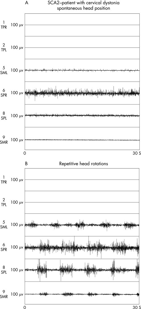
Seven of 11 SCA2 patients with CD had isolated lateroflexion, three had combined lateroflexion and rotation of the head, and one had dystonic head tremor. The severity of dystonia was mild to moderate, with a mean Tsui score of 8 points (range 5–12). A subgroup of six SCA2 patients with clinically diagnosed CD consented to a electromyographic work‐up. Polymyographic recordings revealed involuntary spontaneous muscle activation in at least one recorded neck muscle at rest and disturbed reciprocal inhibition of antagonistic neck muscles (fig 1A, B). Five patients showed a purely tonic muscle activation pattern while CD in one case was characterised by intermittent phasic activity. The polymyography features of the geste revealed tonic activation of one or both muscles that contributed to EMG activity during spontaneous head position. Detailed electromyographic findings are summarised in table 1.
Figure 1 (A, B) Polymyographic recordings of a patient with spinocerebellar ataxia type 2 (SCA2) with cervical dystonia (CD). (A) Spontaneous involuntary muscle activity of the right splenius muscle at rest. (B) Enhanced co‐contraction of the right splenius muscle during repetitive voluntary head movements (disturbed reciprocal innervation) in the same patient. The trapezius muscles (TPR/TPL) have normal EMG activity in both conditions. The x axis (0–30 s) represents 30 s of recording. SML, left sternocleidomastoid muscle; SMR, right sternocleidomastoid muscle; SPL, left splenius capitis muscle; SPR, right splenius capitis muscle; TPL, left trapezius muscle; TPR, right trapezius muscle.
Table 1 Clinical and electrophysiological findings in six SCA2 patients with cervical dystonia.
| Patient no | CAG repeats | Clinical presentation | EMG activity during spontaneous head position | EMG pattern during head rotation | Geste induced EMG changes | |
|---|---|---|---|---|---|---|
| 1 | 40 | Phasic head movements | Tonic/phase activity both SP + SC | Disturbed reciprocal inhibition 4–5 Hz bursts | Tonic both SP | Activation |
| 2 | 43 | Lateroflexion (left) elevation shoulder | Tonic activity both SP + SC | Disturbed reciprocal inhibition | Tonic SCR | Activation |
| 3 | 40 | Rotation (left) and lateroflexion (right) | Tonic activity SPR | Disturbed reciprocal inhibition | Tonic SPL | Activation |
| 4 | 38 | Rotation (left) and lateroflexion (right) | Tonic activity both SP | Disturbed reciprocal inhibition | Tonic SPL | Activation |
| 5 | 37 | Lateroflexion (right) | Tonic activity SPR | Disturbed reciprocal inhibition | Tonic SPL | Activation |
| 6 | 39 | Lateroflexion (right) intermittent head tremor | Tonic–tremulous activation SPR | Disturbed reciprocal inhibition | Tonic both SP | Activation |
SC, sternocleidomastoid muscle; SP, splenius capitis muscle; SPL, left splenius capitis muscle; SPR, right splenius capitis muscle.
CAG repeats represent the length of the expanded allele on chromosome 12. Polymyography of the neck included simultaneous recordings of three muscle pairs.
Discussion
Recent reports indicate a higher frequency of extrapyramidal features in patients with SCA2 than previously suspected, which is in line with post‐mortem studies showing a prominent and consistent basal ganglia pathology in addition to severe atrophy of the cerebellum and brainstem.
The frequency, type and pattern of dystonic symptoms in SCA2 patients with the classic ataxic phenotype, however, has not been assessed in depth. Cancel et al reported on dystonic features in nine of 104 SCA2 patients but did not comment on the type of dystonia in detail.13 We observed CD as the only dystonic feature in 11 of 18 SCA2 patients. Giunti et al found CD to be more frequent in SCA1 and SCA2 than in SCA3, although it was still only found in a minority of their cases.14 Recently, CD as a presenting clinical feature preceding cerebellar ataxia has also been observed in a SCA2 family of Czech origin.15 The mechanisms underlying dystonia in the ataxic SCA2 phenotype are unclear. Dysfunction of the basal ganglia circuitry might account for the frequency of dystonic features in SCA mutations per se. The predilection of the neck in SCA2, however, is unclear. Whether additional dysfunction of the brainstem and/or cerebellum contributes towards abnormal axial posturing and thus to cervical dystonia in SCA2 remains unanswered.16,17
The role of genetic or epigenetic factors on the expression of dystonia in SCA2 are unclear. Cancel et al, studying 32 SCA2 families with the ataxic phenotype, reported that dystonia in general was more frequently present in patients with larger CAG repeat size.13 Giunti et al, in turn, found that extrapyramidal motor symptoms in general correlated with disease duration in SCA2 but not with repeat length.14 In our series, SCA2 patients with CD had identical repeat size to cases without CD (39.6). Disease duration in CD patients was considerably longer (10.3 vs 6.7 years) and severity of cerebellar ataxia was more marked in SCA2 patients with CD (20 vs 14 points). As most of the participants in this study were members of one large family, we cannot exclude overestimation of the frequency because of selection bias due to phenotype variability. However, two SCA2 patients with CD were members of large multigenerational SCA2 families with different genetic backgrounds.
We believe that CD has been an underreported clinical feature in patients with the ataxic SCA2 phenotype. Moreover, CD appears to be frequent in cases with pronounced cerebellar ataxia. As dystonic features such as CD, hemidystonia and writer's cramp have been reported in several different SCA mutations, our findings suggest that a dysfunctional basal ganglia circuitry in addition to dysfunctional pontocerebellar pathways may be important in CD in hereditary ataxias.18,19,20
Abbreviations
CD - cervical dystonia
SCA2 - spinocerebellar ataxia type 2
Footnotes
Competing interests: None declared.
References
- 1.Imbert G, Saudou F, Yvert G.et al Cloning of the gene for spinocerebellar ataxia 2 reveals a locus with high sensitivity to expanded CAG/glutamine repeats. Nat Genet 199614285–291. [DOI] [PubMed] [Google Scholar]
- 2.Durr A, Smadja D, Cancel G.et al Autosomal dominant cerebellar ataxia type I in Martinique (French West Indies). Clinical and neuropathological analysis of 53 patients from three unrelated SCA2 families. Brain 1995118(Pt 6)1573–1581. [DOI] [PubMed] [Google Scholar]
- 3.Estrada R, Galarraga J, Orozco G.et al Spinocerebellar ataxia 2 (SCA2): morphometric analyses in 11 autopsies. Acta Neuropathol (Berl) 199997306–310. [DOI] [PubMed] [Google Scholar]
- 4.Gwinn‐Hardy K, Chen J Y, Liu H C.et al Spinocerebellar ataxia type 2 with parkinsonism in ethnic Chinese. Neurology 200055800–805. [DOI] [PubMed] [Google Scholar]
- 5.Furtado S, Payami H, Lockhart P J.et al Profile of families with parkinsonism‐predominant spinocerebellar ataxia type 2 (SCA2). Mov Disord 200419622–629. [DOI] [PubMed] [Google Scholar]
- 6.Boesch S M, Donnemiller E, Muller J.et al Abnormalities of dopaminergic neurotransmission in SCA2: a combined 123I‐betaCIT and 123I‐IBZM SPECT study. Mov Disord 2004191320–1325. [DOI] [PubMed] [Google Scholar]
- 7.Klockgether T, Schroth G, Diener H C.et al Idiopathic cerebellar ataxia of late onset: natural history and MRI morphology. J Neurol Neurosurg Psychiatry 199053297–305. [DOI] [PMC free article] [PubMed] [Google Scholar]
- 8.Fahn S. Concept and classification of dystonia. Adv Neurol 1988501–8. [PubMed] [Google Scholar]
- 9.Tsui J K, Eisen A, Stoessl A J.et al Double‐blind study of botulinum toxin in spasmodic torticollis. Lancet 19862245–247. [DOI] [PubMed] [Google Scholar]
- 10.Deuschl G, Heinen F, Kleedorfer B.et al Clinical and polymyographic investigation of spasmodic torticollis. J Neurol 19922399–15. [DOI] [PubMed] [Google Scholar]
- 11.Wissel J, Muller J, Ebersbach G.et al Trick maneuvers in cervical dystonia: investigation of movement‐ and touch‐related changes in polymyographic activity. Mov Disord 199914994–999. [DOI] [PubMed] [Google Scholar]
- 12.Muller J, Wissel J, Masuhr F.et al Clinical characteristics of the geste antagoniste in cervical dystonia. J Neurol 2001248478–482. [DOI] [PubMed] [Google Scholar]
- 13.Cancel G, Durr A, Didierjean O.et al Molecular and clinical correlations in spinocerebellar ataxia 2: a study of 32 families. Hum Mol Genet 19976709–715. [DOI] [PubMed] [Google Scholar]
- 14.Giunti P, Sabbadini G, Sweeney M G.et al The role of the SCA2 trinucleotide repeat expansion in 89 autosomal dominant cerebellar ataxia families. Frequency, clinical and genetic correlates. Brain 1998121(Pt 3)459–467. [DOI] [PubMed] [Google Scholar]
- 15.Zaruba K, Ruzicka E, Mazanec R.et al Cervical dystonia in spinocerebellar ataxia type 2. Mov Disord 200419(Suppl 9)S20. [DOI] [PubMed] [Google Scholar]
- 16.Hallett M. The neurophysiology of dystonia. Arch Neurol 199855601–603. [DOI] [PubMed] [Google Scholar]
- 17.LeDoux M S, Brady K A. Secondary cervical dystonia associated with structural lesions of the central nervous system. Mov Disord 20031860–69. [DOI] [PubMed] [Google Scholar]
- 18.Sethi K D, Jankovic J. Dystonia in spinocerebellar ataxia type 6. Mov Disord 200217150–153. [DOI] [PubMed] [Google Scholar]
- 19.Wilder‐Smith E, Tan E K, Law H Y.et al Spinocerebellar ataxia type 3 presenting as an L‐DOPA responsive dystonia phenotype in a Chinese family. J Neurol Sci 200321325–28. [DOI] [PubMed] [Google Scholar]
- 20.Wu Y R, Lee‐Chen G J, Lang A E.et al Dystonia as a presenting sign of spinocerebellar ataxia type 1. Mov Disord 200419586–587. [DOI] [PubMed] [Google Scholar]


