Abstract
Factor XII (FXII), a clotting enzyme that can initiate coagulation in vitro, has long been considered dispensable for normal blood clotting in vivo because hereditary deficiencies in FXII are not associated with spontaneous or excessive bleeding. However, new studies show that mice lacking FXII are protected against arterial thrombosis (obstructive clot formation) and stroke. Thus, FXII could be a unique drug target that could be blocked to prevent thrombosis without the side effect of increased bleeding.
Hemostasis and thrombosis
The hemostatic system has evolved to maintain blood in a fluid state under physiological conditions, but also to react rapidly to vessel injury by sealing defects with fibrin clots. Thrombosis, the primary cause of heart attacks and strokes, is thought to occur when natural anticoagulant mechanisms, which help restrict the clot to the site of injury, are genetically impaired or are overwhelmed by the severity of the initial injury.
Platelets are critical for hemostasis both for the formation of blood clots, as platelet aggregates are an essential constituent of the arterial thrombus, and as a platform for activation of coagulation proteins. However, this commentary will focus predominantly on the coagulation system—a network of sequentially activated plasma serine proteases that trigger the production of fibrin, the protein that forms the meshwork of the clot. Normal blood clotting and thrombosis are thought to occur by the same pathway. In this issue, however, Kleinschnitz et al. (p. 513) show that FXII is required for thrombosis but is dispensable for normal clotting (1), suggesting that the mechanisms regulating these two processes are not identical.
The clotting cascade
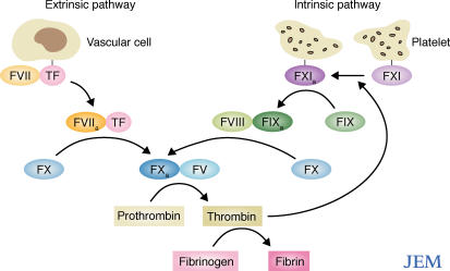
In the current view of physiological blood coagulation, the formation of fibrin can be initiated through either of two converging cascades: the extrinsic pathway and the intrinsic pathway (Fig. 1). The extrinsic pathway is triggered when circulating factor VII (FVII) binds to its membrane-bound receptor tissue factor (TF). TF is expressed on subendothelial layers of blood vessels and on extravascular cells and thus is only exposed to the blood when the normal vasculature is disrupted (2). Once bound to TF, FVII is autoactivated to form FVIIa. The FVIIa–TF complex then cleaves factor X (FX) into its active form (FXa). FXa is a key enzyme common to both the extrinsic and intrinsic coagulation pathways. FXa associates with factor V (FV) to form prothrombinase, which converts prothrombin to thrombin. Thrombin in turn cleaves fibrinogen to form fibrin, thereby initiating the clot.
Figure 1.
Physiological coagulation. The extrinsic pathway is initiated by the binding of FVII to TF which results in the autoactivation of FVII (FVIIa). The activation of this pathway ultimately leads to the formation of thrombin, which feeds back to activate FXI on the surface of platelets. FXIa then triggers the intrinsic pathway generating more thrombin and accelerating fibrin formation.
The intrinsic pathway is triggered by the activation of factor IX (FIX), which is mediated by the TF–VIIa complex (3). Factor XI (FXI), a clotting factor upstream of FIX that is expressed on activated platelets, can be directly activated by thrombin and thus feeds into the intrinsic pathway (4). FXI then activates FIX to FIXa, which in turn activates FX in the presence of factor VIII (FVIII). The end result is a burst in the production of thrombin and the promotion of clot formation.
Hereditary deficiencies in proteins of the extrinsic pathway (FVII), the intrinsic pathway (FXI, FIX, FVIII), or proteins common to both pathways (FX, FV, prothrombin) impair blood clotting in vivo and lead to hemorrhagic disorders, thus demonstrating that both the extrinsic and intrinsic pathways of coagulation are essential for hemostasis.
Protective effects of FXII deficiency
The intrinsic pathway can also be activated in vitro by the autoactivation of FXII, for which the primary substrate is FXI. However, deficiencies in FXII do not cause abnormal or spontaneous bleeding in humans. Thus, FXII, which has been studied for over 50 years, was long considered dispensable for clotting in vivo. But two new studies now challenge this assumption (5). In a recent issue, Renne et al. showed that FXII-deficient mice, like humans, have normal hemostasis and no bleeding abnormalities. However, these mice failed to develop thrombosis in response to vessel injury. Administration of human FXII reversed the protective effect, rapidly inducing thrombus formation. Based on these results, the authors concluded that FXII is required only for pathologic coagulation and not for hemostasis (5). In this issue, a report from the same group strengthens this hypothesis. Kleinschnitz et al. (1) demonstrate that mice lacking FXII, or mice treated with a small molecule inhibitor of FXII, are protected against the development of ischemia-induced brain infarction (stroke) after cerebral artery occlusion (6). Again, the protection was specifically caused by the FXII deficiency, as reconstituting the FXII-deficient mice with human FXII reversed the protective effect. FXI-deficient mice were also protected against stroke and both the FXI- and FXII- deficient mice had reduced ischemia-induced fibrin accumulation in the brain compared with wild-type mice, suggesting that the thrombosis-inducing effects of FXII are mediated through FXI and the intrinsic pathway.
It is interesting to note that some people with hereditary deficiencies in FXI, unlike those lacking FXII, suffer from mild bleeding disorders, suggesting that FXI is required for hemostasis and can be activated independently of FXIIa. As mentioned previously, thrombin can cleave and activate FXI (4), which can then convert FIX to FIXa in the coagulation cascade (7). The ability of thrombin to activate FXI might explain why FXII is not required for physiological hemostasis. But why FXII is strictly required for pathological coagulation is still not completely clear. Although these new data clearly demonstrate a requirement for the activation of FXI, FXII might act on other substrates that also play an essential role in pathological thrombosis.
FXII could induce thrombosis via the extrinsic pathway
In thrombotic conditions, such as sepsis- induced disseminated intravascular coagulation (DIC), bacterial endotoxin or proteoglycans may activate FXII. In addition, polyphosphates that are released upon platelet degranulation can also activate FXII, thus accelerating coagulation (8).
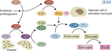
Activated FXII can cleave FVII to an activated form, which is identical to that produced by FXa in the extrinsic pathway. But unlike FXa, which eventually digests and inactivates the FVIIa that it produces, activated FXII produces a stable form of FVIIa (9). The availability of even a small concentration of FVIIa can markedly accelerate the extrinsic pathway by two different mechanisms (Fig. 2). The first is through the normal autoactivation pathway of FVII in the presence of exposed vascular TF, as occurs during physiological blood clotting. The autoactivation of FVII requires a conformational change in FVIIa that occurs upon binding to TF. By cleaving FVII, FXIIa provides the initial few molecules of FVIIa that are needed to trigger for the exponential “explosion” of this autocatalytic cascade. The effectiveness of factor VIIa in triggering this cascade is evident in the successful use of recombinant FVIIa to treat bleeding episodes and during surgery in patients with hemophilia (10–12).
Figure 2.
Pathological thrombosis FXII is activated by exposure to negatively charged molecules to factor XIIa (FXIIa). FXIIa then cleaves FXI to FXIa, thus triggering the intrinsic pathway. Factor XIIa also hydrolyzes FVII to FVIIa, which combines with TF on the surface of activated monocytes or vascular cells to initiate the extrinsic system. Both coagulation pathways are required for pathological thrombosis as occurs during stroke and disseminated intravascular coagulation.
The second mechanism by which FXIIa-mediated activation of FVII can accelerate the extrinsic pathway is through interaction of FVII and TF that is exposed on activated circulating monocytes. One situation that triggers the cellular production of TF occurs during heart bypass surgery. A progressive generation of thrombin during surgery is responsible for the thromboembolic and bleeding complications associated with these operations. During these procedures, circulating monocytes become activated. Monocyte activation triggers an increase in the expression of integrins on the cell surface and the synthesis of TF (13, 14). Substantial concentrations of TF rapidly appear in the pericardial fluid during cardiopulmonary bypass surgery, and the production of TF is accompanied by thrombin formation (15). In the presence of TF, activated monocytes convert all available FVII to FVIIa.
We have recently demonstrated a second mechanism by which soluble TF can generate FVIIa in the presence of FVII and activated monocytes (16). As the velocity of the reaction that produces FVIIa is proportional to the concentration of FVII available, the presence of additional FVIIa, such as that produced by FXIIa, would potentiate the extrinsic pathway. As mentioned previously, FXIIa can also activate FXI to FXIa on the surface of activated platelets in the intrinsic pathway. Both FXIa and FVIIa–TF can convert FIX to FIXa. FIXa, in the presence of FVIII, activates FX—the convergence point in the extrinsic and intrinsic pathways (Fig. 2). Activated monocytes and platelets are present during pathological thrombosis (such as DIC), suggesting that these mechanisms most likely operate in a wide range of inflammatory conditions.
The contact system: an alternative thrombosis inducer?
Activated FXII (FXIIa) also triggers the production of vasoactive peptides known as kinins. This pathway—called the contact system—is triggered by the slow autoactivation of FXII (17) that occurs when FXII is exposed to negatively charged surfaces such as the membranes of activated blood cells. FXIIa activates the serine protease kallikrein from its inactive precursor prekallikrein. Kallikrein then activates FXII (thus amplifying the pathway) and also cleaves high molecular weight kininogen into the potent hypotensive peptide bradykinin (18). Whether the contact system contributes to FXII-driven thrombosis in models of stroke and arteriole injury is not yet known. However, previous work on another model of pathologic coagulation, DIC, suggests that the contact system might not be involved in the hemostatic defect (19).
DIC is a syndrome in which thrombin is produced in excess of antithrombin and exerts its actions systemically rather than locally (20), resulting in the formation of fibrin thrombi in the microvasculature. DIC occurs in humans with bacterial septicemia, a disorder that is often accompanied by hypotensive shock. My group has shown that FXII is activated in these patients, suggesting that this factor might play a role in the development of hypotension associated with septicemia-induced DIC (21). In a baboon model of sepsis, a monoclonal antibody to FXII prevented the development of irreversible hypotension, likely caused by the inhibition of bradykinin release. However, the anti-FXII antibody had no impact on the development of DIC. Thus, the hypotension, but not the hemostatic abnormality, appeared to be related to FXII-dependent activation of the contact system. However, these results do not rule out a role of the contact system in pathologic coagulation because the human FXII-specific antibody only neutralized 60% of baboon FXII. Thus, the residual FXII may have been sufficient to support DIC in this model. Future studies will be required to determine whether the contact pathway contributes to thrombus formation in other models of pathological coagulation.
Therapeutic implications
The selection of anticoagulant therapy—whether for venous thrombembolism or arterial thrombosis (myocardial infarction or stroke)—is based on how well the drug inhibits thrombosis and on the extent of the bleeding side effects. The discovery that FXII deficiency protects against thrombosis without causing spontaneous bleeding makes FXII a unique and ideal target for drug design. Carefully performed experimental studies followed by controlled prospective clinical trials are required to validate this new hypothesis.
References
- 1.Kleinschnitz, C., G. Stoll, M. Bendszus, K. Schuh, H.-U. Pauer, P. Burfeind, C. Renné, D. Gailani, B. Nieswandt, and T. Renné. 2006. Targeting coagulation factor XII provides protection from pathological thrombosis in cerebral ischemia without interfering with hemostasis. J. Exp. Med. 203:513–518. [DOI] [PMC free article] [PubMed] [Google Scholar]
- 2.Mackman, N. 2004. Role of tissue factor in hemostasis, thrombosis, and vascular development. Arterioscler. Thromb. Vasc. Biol. 24:1015–1022. [DOI] [PubMed] [Google Scholar]
- 3.Osterud, B., and S.I. Rapaport. 1977. Activation of factor IX by the reaction product of tissue factor and factor VII: additional pathway for initiating blood coagulation. Proc. Natl. Acad. Sci. USA. 74:5260–5264. [DOI] [PMC free article] [PubMed] [Google Scholar]
- 4.Gailani, D., and G.J. Broze Jr. 1991. factor XI activation in a revised model of blood coagulation. Science. 253:909–912. [DOI] [PubMed] [Google Scholar]
- 5.Renne, T., M. Pozgajova, S. Gruner, K. Schuh, H.U. Pauer, P. Burfeind, D. Gailani, and B. Nieswandt. 2005. Defective thrombus formation in mice lacking coagulation factor XII. J. Exp. Med. 202:271–281. [DOI] [PMC free article] [PubMed] [Google Scholar]
- 6.Choudhri, T.F., B.L. Hoh, H.G. Zerwes, C.J. Prestigiacomo, S.C. Kim, E.S. Connolly Jr., and R.W. Colman. 1998. Reduced microvascular thrombosis and improved outcome in acute murine stroke by inhibiting GP IIb/IIIa receptor-mediated platelet aggregation. J. Clin. Invest. 102:1301–1310. [DOI] [PMC free article] [PubMed] [Google Scholar]
- 7.Davie, E.W., and O.D. Ratnoff. 1964. Waterfall sequence for intrinsic blood clotting. Science. 145:1310–1312. [DOI] [PubMed] [Google Scholar]
- 8.Smith, S.A., N.J. Mutch, D. Baskar, P. Rohloff, R. Docampo, and J.H. Morrissey. 2006. From the cover: Polyphosphate modulates blood coagulation and fibrinolysis. Proc. Natl. Acad. Sci. USA. 103:903–908. [DOI] [PMC free article] [PubMed] [Google Scholar]
- 9.Radcliffe, R., A. Bagdasarian, R. Colman, and Y. Nemerson. 1977. Activation of bovine factor VII by Hageman factor fragments. Blood. 50:611–617. [PubMed] [Google Scholar]
- 10.Veldman, A., M. Hoffman, and S. Ehrenforth. 2003. New insights into the coagulation system and implications for new therapeutic options with recombinant factor VIIa. Curr. Med. Chem. 10:797–811. [DOI] [PubMed] [Google Scholar]
- 11.Habermann, B., K. Hochmuth, L. Hovy, I. Scharrer, and A.H. Kurth. 2004. Management of haemophilic patients with inhibitors in major orthopaedic surgery by immunadsorption, substitution of factor VIII and recombinant factor VIIa (NovoSeven): a single centre experience. Haemophilia. 10:705–712. [DOI] [PubMed] [Google Scholar]
- 12.Levi, M., M. Peters, and H.R. Buller. 2005. Efficacy and safety of recombinant factor VIIa for treatment of severe bleeding: a systematic review. Crit. Care Med. 33:883–890. [DOI] [PubMed] [Google Scholar]
- 13.Kappelmayer, J., A. Bernabei, N. Gikakis, L.H. Edmunds Jr., and R.W. Colman. 1993. Upregulation of Mac-1 surface expression on neutrophils during simulated extracorporeal circulation. J. Lab. Clin. Med. 121:118–126. [PubMed] [Google Scholar]
- 14.Kappelmayer, J., A. Bernabei, L.H. Edmunds Jr., T.S. Edgington, and R.W. Colman. 1993. Tissue factor is expressed on monocytes during simulated extracorporeal circulation. Circ. Res. 72:1075–1081. [DOI] [PubMed] [Google Scholar]
- 15.Hattori, T., M.M. Khan, R.W. Colman, and L.H. Edmunds Jr. 2005. Plasma tissue factor plus activated peripheral mononuclear cells activate factors VII and X in cardiac surgical wounds. J. Am. Coll. Cardiol. 46(4):707–713. [DOI] [PubMed] [Google Scholar]
- 16.Khan, M.M.H., T. Hattori, S. Niewiarowski, L.H. Edmunds Jr., and R.W. Colman. 2006. Truncated and microparticle-free soluble tissue factor bound to peripheral monocytes preferentially activate factor VII. Thromb. Haemost. In press. [DOI] [PubMed]
- 17.Miller, G., M. Silverberg, and A.P. Kaplan. 1980. Autoactivatability of human Hageman factor (factor XII). Biochem. Biophys. Res. Commun. 92:803–810. [DOI] [PubMed] [Google Scholar]
- 18.Cochrane, C.G., S.D. Revak, and K.D. Wuepper. 1973. Activation of Hageman factor in solid and fluid phases. A critical role of kallikrein. J. Exp. Med. 138:1564–1583. [DOI] [PMC free article] [PubMed] [Google Scholar]
- 19.Pixley, R.A., R.A. DeLa Cadena, J.D. Page, N. Kaufman, E.G. Wyshock, A. Chang, F.B. Taylor, Jr., and R.W. Colman. 1993. The contact system contributes to hypotension but not disseminated intravascular coagulation in lethal bacteremia: In vivo use of a monoclonal anti-factor XII antibody to block contact activation in baboons. J. Clin. Invest. 91:61–68. [DOI] [PMC free article] [PubMed] [Google Scholar]
- 20.Colman, R.W., S.J. Robboy, and J.D. Minna. 1972. Disseminated intravascular coagulation (DIC): an approach. Am. J. Med. 52:679–689. [DOI] [PubMed] [Google Scholar]
- 21.Mason, J.W., U. Kleeberg, P. Dolan, and R.W. Colman. 1970. Plasma kallikrein and Hageman factor in Gram-negative bacteremia. Ann. Intern. Med. 73:545–551. [DOI] [PubMed] [Google Scholar]


