Abstract
Calcium and diacylglycerol are critical second messengers that together effect mast cell degranulation after allergen cross-linking of immunoglobulin (Ig)E-bound FcɛRI. Diacylglycerol kinase (DGK)ζ is a negative regulator of diacylglycerol-dependent signaling that acts by converting diacylglycerol to phosphatidic acid. We reported previously that DGKζ−/− mice have enhanced in vivo T cell function. Here, we demonstrate that these mice have diminished in vivo mast cell function, as revealed by impaired local anaphylactic responses. Concordantly, DGKζ−/− bone marrow–derived mast cells (BMMCs) demonstrate impaired degranulation after FcɛRI cross-linking, associated with diminished phospholipase Cγ activity, calcium flux, and protein kinase C–βII membrane recruitment. In contrast, Ras-Erk signals and interleukin-6 production are enhanced, both during IgE sensitization and after antigen cross-linking of FcɛRI. Our data demonstrate dissociation between cytokine production and degranulation in mast cells and reveal the importance of DGK activity during IgE sensitization for proper attenuation of FcɛRI signals.
Mast cells play important roles in both innate and adaptive immune responses. They are central effector cells in immune responses to parasites and in the pathogenesis of diseases such as asthma and allergy (1, 2). The high affinity receptor for IgE (FcɛRI) is one of several cell surface receptors critical for mast cell development and function (3). FcɛRI binds to IgE in the absence of antigen and subsequent cross-linking of IgE-bound FcɛRI by cognate antigen induces a signaling cascade that leads to mast cell degranulation and cytokine secretion, which contribute to both chronic allergic inflammation and acute anaphylaxis. Understanding FcɛRI signaling and mast cell activation is critical to devising new therapies for mast cell–mediated diseases.
Recent studies have greatly improved our understanding of FcɛRI signaling. After FcɛRI engagement, the Src family members Lyn and Fyn and the tyrosine kinase Syk are activated (4, 5). These molecules in turn recruit and activate other kinases such as the Tec family kinase Btk (6), phospholipid modifying enzymes including phosphatidylinositol 4,5-bisphosphate 3-kinase (PI3K) (7), the GTPase-activating molecule Vav1 (8), and adaptor molecules such as linker for activated T cells (LAT) (9), non– T cell activation linker (NTAL/LAB) (10, 11), SH2 domain containing leukocyte phosphoprotein of 76 kD (SLP-76) (12, 13), and Grb2-associated binder protein 2 (Gab2) (14). The formation of a multimolecular signaling complex coordinates activation of various downstream signaling pathways necessary for mast cell effector functions. These pathways include phospholipase Cγ (PLCγ) (15, 16), protein kinase C (PKC) isoforms (17, 18), and mitogen-activated protein kinases (MAPKs) (19). PLCγ hydrolyzes the membrane phospholipid phosphatidylinositol 4,5-bisphosphate (PIP2), leading to the generation of two important second messengers, diacylglycerol (DAG) and inositol 1,4,5-trisphosphate (IP3). IP3 binds to its receptor in the endoplasmic reticulum and induces Ca2+ release into the cytoplasm. DAG recruits to the membrane and activates PKC family members and RasGRPs, which are recently identified guanine nucleotide exchange factors for Ras and Rap (20). Synergistic action of multiple downstream signals, particularly Ca2+ and PKCs, are required to induce mast cell degranulation (18, 21, 22). Activated PKCs and MAPKs together promote transcription of many proinflammatory genes, including cytokines (22–25).
Both in vitro and in vivo evidence suggest a critical role for DAG in the regulation of mast cell function after FcɛRI engagement. Treatment of mast cells with DAG analogues in the presence of a Ca2+ ionophore can mimic FcɛRI engagement and induce mast cells to degranulate and release active mediators (26, 27). Mice lacking PLCγ2, the enzyme that generates IP3 and DAG, have diminished mast cell function (28, 29). Similarly, deficiency in DAG effector molecules alters mast cell function. Multiple PKCs are expressed in mast cells, and activation of both classical and novel isoforms of PKC is regulated by DAG (18, 30). Different PKCs have distinct functions in mast cells. PKCβ−/− mast cells demonstrate decreased IL-6 production and degranulation in response to FcɛRI engagement (22), whereas PKCδ−/− mast cells respond more vigorously to suboptimal FcɛRI stimulation with more sustained Ca2+ mobilization and increased degranulation compared with WT mast cells (31). Thus, proper balance of PKCβ and PKCδ activities appears important for mast cell function.
These observations suggest that DAG levels must be tightly controlled in mast cells. One mechanism for terminating DAG signaling is by phosphorylation catalyzed by the DAG kinase (DGK) family of enzymes. Phosphorylation of DAG by DGKs converts DAG to phosphatidic acid (PA), thus preventing DAG from activating PKCs and RasGRPs (20, 32–34). Additionally, PA itself is a second messenger, and DGK activity could regulate mast cell function by affecting PA accumulation. In vitro, PA is a potent activator of PLC and phosphatidylinositol 4-phosphate 5-kinase (PI5K), enzymes involved in PIP2 degradation and production (35–37). Therefore, through conversion of DAG into PA, DGK enzymes could regulate many aspects of inositol lipid metabolism and mast cell activation after FcɛRI engagement.
We recently described mice deficient in DGKζ and demonstrated that T cells from these animals are hyper-responsive to TCR stimulation. DGKζ−/− mice mount enhanced antiviral immune responses, indicating that DGKζ is an important in vivo negative regulator of TCR signaling and T cell activation (38, 39). We show here that DGKζ also regulates immune receptor signaling in mast cells. To our surprise, in vivo mast cell function is impaired in DGKζ−/− mice as indicated by diminished local anaphylactic responses. To explore the mechanism underlying this finding, we have studied DGKζ−/− bone marrow–derived mast cells (BMMCs). We demonstrate that FcɛRI-induced degranulation of DGKζ−/− BMMCs is diminished, correlating with impaired PLCγ activation and PKCβII membrane translocation. Similar to what we have observed after TCR stimulation, however, FcɛRI-induced activation of the Ras–Erk signaling pathway is enhanced and DGKζ−/− BMMCs produce increased IL-6 during IgE sensitization and after antigen cross-linking of the FcɛRI. Moreover, mast cell survival after growth factor withdrawal is greatly increased by DGKζ deficiency, correlating with maintained phosphorylation of Akt. These findings indicate that DGKζ functions to maintain mast cell responsiveness to antigen stimulation during passive sensitization with IgE and demonstrate separation of cytokine production and degranulation after FcɛRI stimulation of mast cells.
RESULTS
Mast cells develop in DGKζ-deficient mice
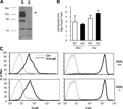
We previously reported that DGKζ plays an important role in T cell activation (38, 39). DGKζ is also expressed in mast cells and, as in T cells, the 115-kD isoform is prominent (Fig. 1 A). There are also smaller species reactive with the anti-DGKζ antibody, which may be products of protein degradation, as mast cells are rich in granules containing proteases. Alternatively, these might represent products of alternate transcripts. Importantly, the three major species reactive with the DGKζ antibody are all absent in BMMCs from targeted mice (Fig. 1 A). Mast cell development is unaffected by DGKζ deficiency for the following reasons: the numbers of mast cells in ear, skin, stomach, and spleen are similar in WT and DGKζ−/− mice (Fig. 1 B and not depicted); electron microscopy did not reveal any differences in cell morphology or number of intracellular granules (not depicted); and KO mast cells express similar levels of c-Kit and FcɛRI on their surfaces (Fig. 1 C). Additionally, DGKζ deficiency does not result in compensatory up-regulation of transcripts for related DGK isoforms (Fig. S1, available at http://www.jem.org/cgi/content/full/jem.20052424/DC1).
Figure 1.
DGKζ expression and surface expression of FcɛRI and c-Kit in BMMCs from WT and DGKζ-deficient mice. (A) DGKζ protein in cell lysates made from WT and DGKζ-deficient BMMCs was detected by Western blot analysis using an antibody specific for the carboxyl-terminus of DGKζ (reference 71). The arrow indicates the 115-kD ζ1 isoform. (B) Mast cell distribution in the skin. Mast cells in the skin (back) and ears were stained with toluidine blue and were counted under a light microscope. Data shown are the means ± standard error of the number of mast cells per high-power field (10 × 100). (C) FcɛRI and c-Kit expression on BMMCs. For FcɛRI expression, WT and DGKζ-deficient BMMCs were incubated with 1 μg/ml anti-DNP IgE for 4 h (solid line) and FcɛRI expression was detected by staining with a FITC-labeled anti-IgE secondary antibody. Cells that had not been incubated with IgE serve as a control (dotted line). For c-Kit expression, WT and DGKζ-deficient BMMCs were either stained with a PE-labeled control antibody (dotted line) or a PE-labeled anti–c-Kit antibody (solid line).
Decreased passive cutaneous anaphylaxis in DGKζ-deficient mice
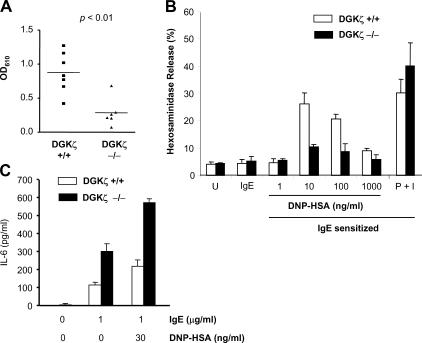
DGKζ−/− T cells have enhanced homeostatic proliferation and antiviral responses in vivo (39). To test the importance of DGKζ in mast cell function, we assessed allergic responses by examining passive cutaneous anaphylaxis (PCA), an in vivo measure of FcɛRI-dependent mast cell function. Unexpectedly, DGKζ−/− mice had significantly impaired localized anaphylactic responses (Fig. 2 A).
Figure 2.
Decreased passive cutaneous anaphylaxis and FcɛRI-induced degranulation but increased IL-6 production as the result of DGKζ deficiency. (A) WT and DGKζ−/− mice were injected subcutaneously with 25 ng IgE in 25 μl PBS in the right ear and with 25 μl PBS in the left ear. 24 h later, DNP-HSA and Evans blue dye were injected intravenously into the mice. 30 min after injection, tissue from the ears was collected, Evans blue was extracted, and the intensity of the dye was measured by absorption at 610 nm (OD610). Each data point is OD610 of IgE-injected ear minus the OD610 of PBS-injected ear from the same mouse. Data shown comprise three experiments. (B) Decreased FcɛRI-induced degranulation in DGKζ−/− BMMCs. IgE-sensitized WT and DGKζ−/− BMMCs were left unstimulated or were stimulated with various concentrations of DNP-HSA at 37°C for 60 min. Cells were spun, and β-hexosaminidase activities in supernatants as well as in whole cell lysates of unstimulated BMMCs were measured. Data were calculated as percentage of β-hexosaminidase activity in the supernatant to the activity in the whole cell lysates of the same genotype. Means ± standard error of triplicates are shown. Data are representative of three experiments. (C) Increased IL-6 production in DGKζ-deficient BMMCs. WT and DGKζ-deficient BMMCs were left unstimulated, were stimulated with 1 μg/ml IgE overnight, or were sensitized with 1 μg/ml IgE for 4 h and cross-linked with 30 ng/ml DNP-HSA overnight. IL-6 in the culture medium was measure by ELISA. Data shown are means ± standard error of triplicates normalized to PMA/Io control and are representative of three experiments.
To explore why DGKζ−/− mice demonstrate impaired PCA responses, we first compared FcɛRI-triggered degranulation in WT versus DGKζ−/− BMMCs by measuring β-hexosaminidase release (Fig. 2 B). FcɛRI-induced degranulation was significantly diminished in DGKζ−/− BMMCs (Fig. 2 B). PMA plus ionomycin (Io) stimulation, however, resulted in similar degranulation in WT and DGKζ−/− BMMCs, indicating that the ability to release granules and total granule content is unaffected by DGKζ deficiency. The impairment of FcɛRI-induced mast cell degranulation likely contributes to the decreased PCA responses in DGKζ−/− mice.
Another consequence of FcɛRI engagement is the production of proinflammatory cytokines such as IL-6. IgE alone can induce a significant amount of IL-6 production in WT BMMCs (Fig. 2 C), which is consistent with previous reports (40). Cross-linking the receptor with antigen induces even more IL-6 production. Under both stimulation conditions, DGKζ−/− BMMCs produce approximately twofold more IL-6 than WT BMMCs. Therefore, DGKζ deficiency impairs FcɛRI-induced degranulation, but increases cytokine production after IgE sensitization and after cross-linking of the receptor with antigen.
Antigen stimulation of IgE-sensitized BMMCs
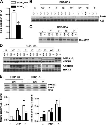
Cross-linking of IgE-bound FcɛRI results in activation of a signaling network that coordinates mast cell effector functions. To investigate why DGKζ−/− BMMCs have impaired degranulation but enhanced cytokine production after FcɛRI stimulation, we assessed how DGKζ deficiency affects antigen-induced FcɛRI signaling in IgE-sensitized BMMCs. We first verified that DGKζ phosphorylates DAG after antigen stimulation of BMMCs by measuring PA production. Addition of DNP-HSA to IgE-sensitized WT BMMCs results in robust PA accumulation beginning 5–10 min after stimulation (Fig. 3 A). As expected, antigen stimulation of DGKζ−/− BMMCs results in significantly less PA accumulation.
Figure 3.
Antigen stimulation of IgE-sensitized BMMCs. (A) DGKζ deficiency impairs PA production by BMMCs after FcɛRI stimulation. BMMCs were sensitized for 4 h with IgE, labeled with 32P-orthorphosphate, and either left unstimulated or stimulated with 10 ng/ml DNP-HSA for 10 min. Lipids were extracted, separated by TLC, and quantified using a phosphorimager. PA, the product of DAG phosphorylation, was identified by comigration with a standard. Data are presented as fold-induction calculated by cpm of stimulated cells divided by cpm of unstimulated cells. Means ± standard error of four experiments are shown; *, P < 0.05. (B–D) WT and DGKζ−/− BMMCs were sensitized with IgE for 4 h and left unstimulated (U) or were stimulated either with 4 ng/ml of DNP-HSA for different times or with 100 ng/ml PMA (P) for 5 min. (B) AKT phosphorylation was assessed by immunoblot analysis using an antibody specific for phosphorylated Ser473 of Akt. Total Akt is shown as a loading control. (C) Active Ras in lysates was measured by affinity precipitation using GST-Raf-RBD agarose beads followed by immunoblot analysis. (D) MEK1/2 and ERK1/2 activity was assessed by immunoblot analysis using phospho-specific antibodies. Total MEK1/2 and ERK1/2 serve as loading controls. (E) Membrane recruitment of PKCβII is diminished in DGKζ−/− BMMCs. IgE-sensitized BMMCs were left unstimulated (−) or were stimulated with 50 ng/ml DNP-HSA (DNP) or PMA (P) for 10 min. Triton-soluble membrane fractions were prepared and membrane translocation of PKC isoforms was determined by Western blot using antibodies to PKCβII, PKCδ, and calnexin (CNX) as a loading control. One representative blot is shown and data from five (PKCβII) or three (PKCδ) experiments were quantified and normalized to CNX; combined data are represented graphically.
We next examined signaling pathways that have been implicated in cytokine production after FcɛRI ligation. Activation of the PI3K/Akt and Ras/Erk pathways is required for cytokine production (23, 25, 41). Low dose stimulation of IgE-sensitized WT BMMCs with DNP-HSA weakly activates Akt, but stimulation of DGKζ−/− BMMCs results in significantly increased Akt activation (Fig. 3 B). FcɛRI activation of Ras is believed to be the result of Grb2-SOS activation (42, 43), but recent reports suggest that DAG might contribute to Ras activation through allosteric activation of the Ras guanine-nucleotide exchange factor RasGRP4 (44–46). As shown in Fig. 3 C, FcɛRI-induced Ras activity is enhanced in DGKζ−/− BMMCs. Consistent with the increased Ras activation, we observed enhanced and prolonged Mek1/2 and Erk1/2 phosphorylation in DGKζ−/− BMMCs compared with WT BMMCs (Fig. 3 D). The effect of DGKζ deficiency is selective, however, as FcɛRI-induced activation of the MAPKs p38 and Jnk is similar in WT and DGKζ−/− BMMCs (unpublished data). These data demonstrate that DGKζ deficiency enhances FcɛRI-induced activation of Akt, Ras, and Erk1/2, likely contributing to enhanced cytokine production.
Activation of PKC family members by DAG and Ca2+ coordinates mast cell degranulation after FcɛRI stimulation (22, 47). As expected, antigen cross-linking induces membrane recruitment of PKCβII and PKCδ in WT BMMCs (Fig. 3 E). However, we find that membrane recruitment of PKCβII is clearly diminished in DGKζ−/− BMMCs, whereas PKCδ recruitment is preserved. Unstimulated IgE-sensitized DGKζ−/− BMMCs also demonstrate diminished levels of membrane-associated PKCβII but normal PKCδ levels. Moreover, treatment with the DAG analogue PMA, which is not a DGK substrate and should be unaffected by DGK deficiency, results in impaired movement of PKCβII to the membrane in DGKζ−/− BMMCs but normal movement of PKCδ (Fig. 3 E, representative blot and graphical summary of replicates). The block in PKCβII recruitment was not the result of diminished protein levels, however, as total levels of PKCβII and PKCδ in whole cell lysates were not decreased in DGKζ−/− BMMCs as compared with WT BMMCs (Fig. S2, available at http://www.jem.org/cgi/content/full/jem.20052424/DC1). These data provide a likely mechanism for the impaired degranulation we observe in DGKζ−/− BMMCs, as FcɛRI cross-linking results in less recruitment of a positive regulator (PKCβII) of mast cell degranulation, but appropriate recruitment of a negative regulator of mast cell degranulation (PKCδ). The data also suggest that DGKζ is essential for FcɛRI activity in addition to its effect on DAG levels, as PMA and PMA/Io (unpublished data) fail to restore PKCβII recruitment in DGKζ−/− BMMCs.
Calcium response and PLCγ activity in DGKζ−/− BMMCs
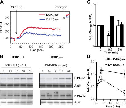
The diminished membrane recruitment of the Ca2+-dependent PKCβII along with the preserved movement of the Ca2+-independent PKCδ led us to question whether DGKζ deficiency affects FcɛRI-induced Ca2+ flux. Compared with WT BMMCs, DGKζ−/− BMMCs have a significantly diminished Ca2+ response after FcɛRI stimulation by antigen (Fig. 4 A). Consistent with the decreased Ca2+ response, phosphorylation of PLCγ1 and PLCγ2 after stimulation of the FcɛRI was decreased in DGKζ−/− BMMCs (Fig. 4 B). As tyrosine phosphorylation of PLCγ does not always correlate with its enzymatic activity (48), we analyzed PLC activity by assessing metabolism of the PLC substrate PIP2 and production of IP3. After receptor cross-linking in WT BMMCs, PIP2 levels quickly decrease (Fig. 4 C), and IP3 levels increase (Fig. 4 D) as a result of PLC activity. Upon stimulation of DGKζ−/− BMMCs, we do not observe a decrease but rather a slight increase in PIP2 levels (Fig. 4 C), and IP3 generation is impaired (Fig. 4 D). These data provide compelling evidence that DGKζ deficiency impairs activation of PLCγ after antigen stimulation of IgE-sensitized mast cells.
Figure 4.
Decreased Ca2+ responses and PLCγ activity in DGKζ−/− mast cells. WT and DGKζ−/− BMMCs were sensitized for 4 h with IgE in cytokine-free media. (A) Deceased Ca2+ flux in DGKζ−/− mast cells after FcɛRI stimulation. WT and DGKζ−/− BMMCs were loaded with Indo-1 and stimulated with 100 ng/ml DNP-HSA to induce Ca2+ responses. Ca2+ flux was determined by flow cytometry based on the change of FL5/FL4 ratio. Data shown are representative of four experiments. (B) Decreased PLCγ phosphorylation in DGKζ−/− BMMCs after FcɛRI stimulation. BMMCs were stimulated with different concentrations of DNP-HSA for 2 min. PLCγ phosphorylation was determined by Western blot with anti–phospho- PLCγ1 and anti–phospho-PLCγ2 antibodies. The blots were probed with an anti-actin antibody as a loading control. (C) Decreased PIP2 hydrolysis in DGKζ−/− BMMCs. Cells were labeled with 32P-orthophosphate and stimulated for the indicated amounts of time with 10 ng/ml DNP-HSA; lipids were analyzed as in Fig. 3. PIP2 was identified by comigration with a standard. Data are means ± standard error of four different experiments. *, P = 0.01, 0.5 min stimulation, WT vs. DGKζ KO. (D) Diminished IP3 generation in DGKζ−/− BMMCs. Cells were left unstimulated or were stimulated with 10 ng/ml DNP-HSA for 20 s or 2 min and lysed using perchloric acid, and IP3 was measured.
IgE signaling and function in the absence of antigen
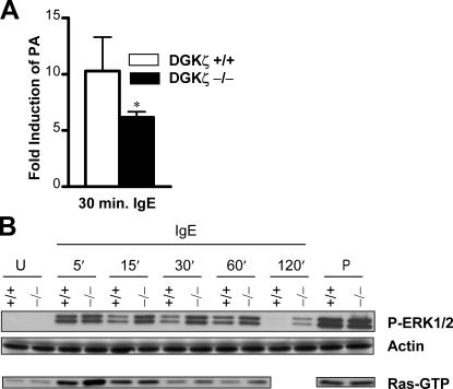
IgE sensitization of DGKζ−/− BMMCs leads to increased cytokine production and impaired PKCβII recruitment to the membrane, so we therefore examined how IgE alone signaling and function is altered in DGKζ deficiency. IgE binding to FcɛRI induces a large increase in PA in WT BMMCs (Fig. 5 A). DGKζ−/− BMMCs, in contrast, have a significant impairment in PA accumulation during IgE sensitization. DAG-dependent signaling is also enhanced in DGKζ−/− BMMCs during IgE sensitization, as Ras and Erk1/2 activity is enhanced (Fig. 5 B).
Figure 5.
IgE stimulation in the absence of antigen cross-linking results in enhanced signaling in DGKζ−/− BMMCs. BMMCs were rested in media without cytokines for at least 4 h and rested for 1 h in Tyrode's buffer before stimulation. (A) Impaired PA production in DGKζ−/− BMMCs. Cells were labeled with 32P-orthophosphate for 1 h and stimulated with 1 μg/ml IgE for 30 min. Lipids were extracted and PA production was quantified as in Fig. 3. Means ± standard error of five experiments are shown. *, P < 0.05. (B) Enhanced Ras/ERK activation in DGKζ−/− BMMCs. BMMCs were stimulated with 1 μg/ml IgE for various amounts of time or with 100 ng/ml PMA for 5 min. Active Ras and ERK1/2 phosphorylation were assessed as in Fig. 3. For the Ras blot, a space is included before the PMA lanes to designate that these samples were run on a separate gel.
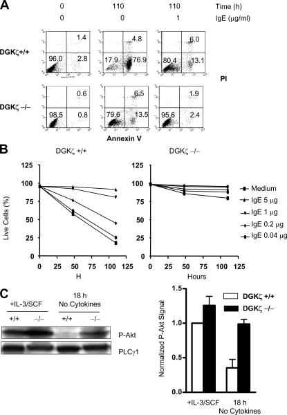
IgE binding to the FcɛRI signals mast cell survival through up-regulation of antiapoptotic proteins (40, 49–52), and ex vivo mast cell survival and expansion requires IL-3 (53, 54). In the presence of IL-3, both WT and DGKζ−/− mast cells survive and expand similarly (unpublished data). When IL-3 is withdrawn, WT BMMCs undergo apoptosis as expected (Fig. 6 A). Surprisingly, DGKζ−/− BMMCs have greatly enhanced survival in the absence of IL-3. IgE enhances survival in both WT and DGKζ−/− BMMCs, but because survival in the absence of cytokines is so much greater in the DGKζ−/− BMMCs, the IgE survival effect is less marked (Fig. 6, A and B).
Figure 6.
Enhanced survival of DGKζ−/− BMMCs after IL-3 withdrawal. WT and DGKζ−/− BMMCs were cultured in media without cytokines in the presence of different concentrations of IgE for 48 or 110 h. Survival of BMMCs was determined by staining with FITC-annexin V and PI and analyzed by flow cytometry. (A) Representative dot-plots. (B) Percentage of live cells (Annexin-V and PI negative) in a full time course and at a range of concentrations of IgE. Data shown are representative of three experiments. (C) Akt phosphorylation during growth in cytokine-replete media (+IL3/SCF) and after 18 h cytokine withdrawal (18 h, no cytokines) was assessed by immunoblot analysis. One representative blot is shown and data from three independent experiments were quantified, normalized to PLCγ1 signal, and represented graphically for comparison. P-AKT signal in WT cells grown in cytokine-replete media was arbitrarily set to 1.
Akt regulates cell survival and is activated by IgE binding to FcɛRI (40). Akt is phosphorylated in WT BMMCs grown in cytokine-replete media, and this activation decreases after withdrawal of cytokines (Fig. 6 C). DGKζ−/− BMMCs, in contrast, have enhanced Akt activity during growth in cytokine-replete media and maintain Akt activity after cytokine removal (Fig. 6 C). As cytokines stimulate inositol metabolism, including PIP2 hydrolysis by PLCγ and phosphorylation by PI3K (55, 56), it is likely that DGKζ regulates signals generated through cytokine receptors in addition to its role in FcɛRI signaling. Future work will explore the biochemical basis of this observation.
DISCUSSION
We have studied mice that lack a key enzyme involved in DAG metabolism, DGKζ, to address the consequences of dysregulated DAG accumulation after immune receptor signaling. We demonstrated recently that DGKζ functions as a key negative regulator of TCR signaling and T cell activation (38, 39). We show here that DGKζ plays an important and unexpected role in regulating signal transduction from the FcɛRI in mast cells. DGKζ−/− BMMCs manifest decreased PA production, enhanced activation of the Ras-Mek-Erk signaling pathway, increased Akt phosphorylation, prolonged survival after cytokine deprivation, and enhanced production of IL-6 as compared with WT BMMCs. Surprisingly, IgE-mediated PCA responses are significantly decreased in DGKζ−/− mice, concordant with impairment of receptor-induced PLCγ activity and degranulation in DGKζ−/− BMMCs. We demonstrate that signals generated by IgE in the absence of antigen cross-linking are enhanced in DGKζ−/− BMMCs. These data demonstrate that DGKζ is important for inactivation of DAG-mediated signaling pathways in mast cells and that DGKζ also plays a critical role in maintaining FcɛRI responsiveness to antigen cross-linking. In addition, we demonstrate a novel FcɛRI signaling alteration in DGKζ−/− BMMCs, in that receptor engagement results in poor PLCγ activity and degranulation but augmented cytokine production.
The observation that DGKζ deficiency impairs PLCγ function after FcɛRI stimulation of mast cells was unexpected, as DGKζ was predicted to function downstream of PLCγ. It is possible that enhanced DAG-dependent signaling during IgE sensitization of DGKζ−/− BMMCs promotes feedback inhibition of PLCγ and Ca2+ responses upon antigen cross-linking of the FcɛRI. Short pretreatment of mast cells with phorbol esters results in feedback inhibition of PLCγ that is mediated by PKCα and PKCɛ (57–59). Prolonged administration of phorbol esters, in contrast, down-regulates PKC isoforms and potentiates PLC activity upon antigen stimulation (59). We do not see changes in PKC protein levels in DGKζ−/− BMMCs, and we report impaired PLC activity. PKC feedback inhibition might be indirect as well, as PKCβ can induce serine-phosphorylation of Btk at an inhibitory site, dampening Btk activation in mast cells (60). Btk phosphorylates and activates PLCγ after FcɛRI stimulation, so feedback inhibition of Btk could contribute to diminished PLCγ activity (61). Alternatively, enhanced Erk activation in DGKζ−/− BMMCs could inhibit PLCγ association with LAT, as has been observed in T cells after TCR engagement (62). However, we have not observed an obvious decrease in PLCγ2-LAT association in DGKζ−/− BMMCs after antigen cross-linking of the FcɛRI (unpublished data).
Alternatively, diminished PLCγ activity and PKCβII membrane recruitment might be a direct consequence of DGKζ deficiency. Although DGK activity terminates DAG signaling, it also creates PA (32, 33, 63). PA is a potent activator of PLCγ enzymatic activity in vitro (35). In addition, a recent report suggests that DGKζ, through production of PA, may increase intracellular PIP2 by activation of PI5K Iα (37). Thus, DGKζ deficiency could lead to decreased PI5K activity, diminishing levels of the PLC substrate PIP2. Our PIP2 measurements did not reveal any differences in 32P incorporation into PIP2 in IgE-sensitized BMMCs (unpublished data), but more direct measures of PIP2 mass should be performed to ensure that steady state levels of 32P-labeled PIP2 reflect total mass. The importance of diminished PA production to the phenotype of DGKζ−/− BMMCs is particularly difficult to assess, as alternative pathways to generate PA exist. These pathways include the hydrolysis of phosphatidylcholine mediated by phospholipase D and the acylation of lyso-PA by lyso-PA acyltransferases (63). phospholipase D–derived PA has been reported to regulate diverse cellular processes, particularly in vesicle transportation and exocytosis (64, 65). It is possible that PA generated by DGKζ performs a similar function to promote mast cell degranulation.
It is intriguing that membrane recruitment of PKCβII is diminished in unstimulated IgE-sensitized DGKζ−/− BMMCs and that PKCβII recruitment is not restored with PMA treatment. PMA is a DAG analogue and is not subject to phosphorylation by DGK, so we predict that if DGKζ acts only by regulating DAG levels, PMA treatment should restore PKCβII membrane recruitment. It is possible that DGKζ has adaptor functions in addition to its role in lipid metabolism, perhaps by participating in a complex with PKCβII or Btk directly (37, 66). Future work will explore this interesting observation.
We have been unable to directly measure an FcɛRI- induced change in DAG mass in WT or DGKζ−/− mast cells using standard biochemical approaches (unpublished data). The likely explanation is that the receptor-induced pool of DAG is a small fraction of the total cellular pool because DAG is important for other aspects of cell biology. We are currently developing imaging approaches to measure DAG localization using a fluorescently labeled DAG probe (67). We also will develop HPLC/MS/MS techniques to quantify particular DAG subspecies, which may be more markedly regulated than total cellular DAG (68). These approaches will allow us to measure dynamic changes in DAG localization after FcɛRI stimulation as well as assess in vivo substrate specificity of DGKζ.
The increased Akt phosphorylation in DGKζ−/− BMMCs stimulated through cytokine or FcɛRs is intriguing. It is possible that increased Akt activity is a consequence of greater PIP3 production in DGKζ−/− BMMCs, and preliminary data support this hypothesis. Decreased PA production in DGKζ−/− BMMCs might increase PI3K activity, as PA has been reported to inhibit this enzyme in vitro (69). Also, perhaps decreased PLCγ activity in DGKζ−/− BMMCs increases the local concentration of PIP2 available as substrate for PI3K (66). The interrelation among different PI metabolites is complex and understanding the exact lipid alterations will require further study.
Our current studies of DGKζ in mast cells reveal important roles for this enzyme in regulating FcɛRI signaling. DGKζ is not only essential for terminating DAG activity but also appears to be critical for maintaining optimal FcɛRI responsiveness to antigen cross-linking. A more complete understanding of the importance of DGK activity to mast cell function requires analysis of other DGK family members. Ongoing biochemical and genetic studies are addressing this question.
MATERIALS AND METHODS
DGKζ-deficient mice.
DGKζ−/− mice have been described previously (39) and were housed in pathogen-free facilities at the University of Pennsylvania and Duke University. All experiments using animals were performed in accordance with regulations of the Institutional Animal Care and Use Committee of the University of Pennsylvania and Duke University.
Microscopic analysis of mast cells.
Skin samples from the back of the trunk and the ears were collected from killed WT and DGKζ−/− mice. Tissues were fixed and mounted in paraffin according to standard histological procedures. Paraffin sections were stained with toluidine blue and mast cells in tissues were evaluated by microscopy. In each tissue, mast cells were counted in 10 contiguous high-power fields (10 × 100).
BMMCs.
BM cells from tibias and femurs from WT and DGKζ−/− mice were harvested and incubated with Isocove's medium (Mediatech, Inc.) supplemented with 15% FBS (Hyclone), 100 U/ml penicillin G, 100 U/ml streptomycin, and 292 μg/ml of l-glutamine, 10 mM Hepes (pH 7.4), 0.1 mM nonessential amino acid, 1 mM sodium pyruvate, and 50 μM 2-mercaptoethanol (IMDM-15) with IL-3–conditioned medium for 2 wk and further expanded for an additional 2–8 wk with the supplement of SCF-conditioned medium made from cell lines engineered to produce these cytokines (70).
Flow cytometry.
BMMCs were analyzed directly or after 4-h sensitization with 1 μg/ml IgE. Cells were stained with fluorescently labeled antibodies in 5% FBS in PBS and were analyzed on a FACSCaliber (Becton Dickinson) with CELLQuest software. Antibodies used were PE-conjugated c-Kit and FITC-conjugated anti-IgE (BD Biosciences). To measure cell survival after cytokine withdrawal, cells were stimulated in IMDM-15 without cytokine at a concentration of 106 cells/ml in a 96-well plate with different concentrations of anti-DNP–IgE (0, 0.04, 0.2, 1, or 5 μg/ml; Sigma-Aldrich). After 48 or 110 h incubation, cells were stained with propidium iodide and annexin V–FITC (BD Biosciences) and were analyzed by flow cytometry.
Measurement of PA and PIP2.
BMMCs were sensitized at 37°C for 4 h in 1 μg/ml IgE in IMDM-15 without IL-3 or SCF. Cells were harvested and rested in Tyrode's buffer (10 mM Hepes, pH 7.4, 130 mM NaCl, 1 mM MgCl2, 5 mM KCl, 1.4 mM CaCl2, 5.6 mM glucose, and 1 mg/ml bovine serum albumin) for 1 h at 37°C. 32P-phosphoric acid (MP Biomedicals) was added to a concentration of 0.25 mCi/ml. Cells were left unstimulated or were stimulated with 10 ng/ml DNP-HSA. Stimulation was terminated by addition of 0.1 volume of ice-cold 2 M HCl and transfer of cells to ice for 10 min. Lipids were extracted with 3 ml of 2:1 methanol:chloroform and phases were separated by addition of 2 ml 1 N NaCl and 2 ml of chloroform. Lipid phase was washed 1× with 3 ml of upper phase buffer (upper phase of 10:10:9 chloroform:methanol:1 M NaCl), dried under nitrogen, and separated by thin-layer chromatography using a basic solvent (9:7:2 chloroform: methanol: 4 M NH4OH). PA and PIP2 were identified by comigration with standards (Sigma-Aldrich) and quantified using a phosphorimager.
Measurement of IP3.
IgE-sensitized BMMCs were rested for 1 h in Tyrode's buffer, then left unstimulated or stimulated for 20 s or 2 min with 10 ng/ml DNP-HSA. Stimulations were terminated by addition of 0.2 vol 20% perchloric acid for 20 min on ice. Proteins were sedimented and supernatants were neutralized to pH 7.5 by titration of 1.5 M KOH 60 mM Hepes containing universal indicator dye (Sigma-Aldrich). KClO4 was precipitated by centrifugation and IP3 was measured in supernatants by radioreceptor assay according to the manufacturer's protocol (PerkinElmer).
Western blot analysis.
For DGKζ expression, BMMCs were lysed in 1% NP-40 lysis buffer (1% NP-40, 150 mM NaCl, 50 mM Tris, pH 7.4) with protease inhibitors. Proteins were resolved by SDS-PAGE, transferred to Trans-Blot Nitrocellulose membrane (Bio-Rad Laboratories), and probed with an anti-DGKζ antibody for DGKζ expression (71). To analyze Ras, Erk, Mek1/2, Jnk, and PLCγ1/2 activation, IgE-sensitized BMMCs were resuspended in Tyrode's buffer. Cells were left unstimulated or stimulated with DNP-HSA for different times or PMA (100 ng/ml) for 5 min. For IgE alone activation of Ras and Erk, cells were not sensitized, but rather were stimulated with 1 μg/ml IgE or PMA. Phosphorylation of these proteins was determined by Western blot with antiphospho-specific antibodies for Erk1/2, Mek1/2, Jnk, Akt, and PLCγ1 and PLCγ2 (Cell Signal Technology). Membranes were stripped and reprobed with antibodies to Erk, Mek1, Jnk, Akt, and actin for loading controls. Activated Ras in cell lysates was determined by GST-Raf-RBD “pull-down” assay as described previously (38).
PKC membrane localization.
BMMCs were sensitized for 4 h with 1 μg/ml IgE in IMDM-15 without cytokines and rested for 1 h at 37°C in Tyrode's buffer. Cells were stimulated with 50 ng/ml DNP-HSA or 100 ng/ml PMA for 10 min, and membrane fractions were isolated as described previously (72). Protein concentration in Triton soluble fraction was quantified using a bicinchonic acid assay (Pierce Chemical Co.) before PAGE analysis. Western blots were probed with antibodies to PKCδ (Cell Signal Technology), PKCβII (Santa Cruz Biotechnology, Inc.), and calnexin (Stressgen Bioreagents).
Detection of IL-6 production.
Measurement of IL-6 production by BMMCs was performed as described with modifications (13). BMMCs were rested overnight in IMDM-15 plus IL-3. For the unstimulated condition, cells were resuspended in IMDM-10 at 2.5 × 10⁵ cells/ml. 200 μl cells were seeded in each well of a 96-well plate in triplicate. For FcɛRI stimulation, cells were washed and sensitized with 1 μg/ml anti-DNP IgE in IMDM-10 at 107 cells/ml for 4 h, pelleted, washed once with IMDM-10, and resuspended at 5 × 10⁵ cells/ml in IMDM-10. 100 μl aliquots of cells were placed into wells of a 96-well plate, followed by addition of 100 μl of IMDM-10 with 2 μg/ml of anti-DNP-IgE, with 60 ng/ml DNP-HSA, or with 40 ng/ml PMA and 200 ng/ml Io. Cells were incubated at 37°C with 5% CO2 for 24 h and IL-6 in supernatants was measured using a murine IL-6 ELISA kit (Pierce Chemical Co.). Data are normalized to PMA/Io control.
β-hexosaminidase release assay.
Hexosaminidase release assay was performed as described previously with modification (13). BMMCs grown in IMDM-15 supplemented with IL-3 were stimulated with IgE at 37°C for 1 h to assess IgE-induced degranulation or were sensitized with 1 μg/ml IgE for 4 h and left unstimulated or stimulated with 10 ng/ml of DNP-HSA or PMA plus Io at 37°C for 1 h. β-Hexosaminidase activities in the medium and in whole cell lysates were determined using p-nitro-phenyl-N-acetyl-β-D-glucosamide as the substrate.
Passive cutaneous anaphylaxis assay.
The passive cutaneous anaphylaxis assay was performed according to published protocols (73). Mice were anesthetized by intraperitoneal injection of 300 μl of 2.5% 2,2,2-tribromoethanol in tert-amyl alcohol/PBS (1/40; Sigma-Aldrich), followed by subcutaneous injection of 25 ng IgE in 25 μl PBS in the right ear and 25 μl PBS in the left ear. 24 h later, mice were anesthetized and 200 μl antigen (100 μg DNP-HSA, 1% Evans blue in PBS) was injected intravenously via the retroorbital sinus. 30 min after the injection, mice were killed and both ears were removed. Ears were incubated in 200 μl formamide at 55°C for 48 h to extract the dye. Tissue debris was removed by centrifugation and the intensities of the dye were measured by absorption at 610 nm. The data are calculated as OD610 of the IgE-injected ear minus the OD610 of the PBS-injected ear from the same mouse.
Calcium flux.
BMMCs were sensitized with 1 μg/mL IgE for 4 h; resuspended at 107 cells/ml in Tyrode's buffer containing 3 μg/ml Indo-1 (Invitrogen) and 4 mM probencid; and incubated for 30 min at 37°C. Cells were washed twice with Tyrode's buffer and resuspended at 2 × 10⁷ cells/ml. 40 μl of cells was added into 460 μl of prewarmed Tyrode's buffer to measure Ca2+ responses by flow cytometry (BD-LSR; Becton Dickinson). After collection of the baseline ratio of FL5 to FL4, 10 μl of 5 μg/ml DNP-HSA was added to stimulate cells. Calcium flux was assessed as the ratio of FL5 to FL4 fluorescence.
Online supplemental material.
For measurement of DGK transcript levels, RNA was isolated from WT and DGKζ−/− BMMCs by TRIzol extraction (Invitrogen) and cDNA generated by reverse transcriptions (CLONTECH Laboratories, Inc.). Transcripts were quantified by SYBR green real-time PCR using the following primers: DGKα: 5′-GATGCAGGCACCCTGTACAAT-3′, 5′-GGACCCATAAGCATAGGCATCT-3′; DGKδ: 5′-GGGACCTCAAGGACCTTGGT-3′, 5′-TCAGCTCCTTGATCCCACAAA; and DGKι: 5′-TTCCCCAGGGCACTCTCA-3′, 5′-CAGACGTTGCATCTAGGAAGCA-3′. No products were amplified from a no reverse transcriptase control sample (unpublished data). Transcripts were normalized to GAPDH signal (5′-GAAGGTACGGAGTCAACGGATTT-3′, 5′-GAATTTGACCATGGGTGGAAT-3′) using the ΔΔCt method. For measurement of total cellular protein levels of PKCβII and PKCδ, WT and DGKζ−/− BMMCs were left unstimulated for 4 h in cytokine-free media with or without IgE (1 μg/ml). Cells were lysed in 1% NP-40 lysis buffer with protease inhibitors, proteins were resolved by SDS-PAGE, and Western blots were probed with antibodies to PKCβII or PKCδ. Blots were stripped and reprobed for actin as a loading control. Online supplemental material is available at http://www.jem.org/cgi/content/full/jem.20052424/DC1.
Supplemental Material
Acknowledgments
We thank J. Stadanlick for help in preparation of the manuscript; M. Jordan for help in the Ca++ experiment; S. Prescott and M. Topham for providing the anti-DGKζ antibody; E. Myers and E. Hainey for technical assistance; and Drs. Q.-C. Yu, H.-W. Yu, and Y. Wang for assistance in histological analysis.
This work was supported by the National Institutes of Health (G.A. Koretzky and X.-P. Zhong), from the Sandler Program for Asthma Research (G.A. Koretzky), and from the National Cancer Institute (B.A. Olenchock).
The authors have no conflicting financial interests.
Abbreviations used: BMMC, bone marrow–derived cell; DAG, diacylglycerol; DGK, DAG kinase; IP3, inositol 1,4,5-trisphosphate; LAT, linker for activated T cells; MAPK, mitogen-activated protein kinase; PA, phosphatidic acid; PCA, passive cutaneous anaphylaxis; PI5K, phosphatidylinositol 4-phosphate 5-kinase; PIP2, phosphatidylinositol 4,5-bisphosphate; PKC, protein kinase C; PLCγ, phospholipase Cγ.
B.A. Olenchock and R. Guo contributed equally to this work.
References
- 1.Benoist, C., and D. Mathis. 2002. Mast cells in autoimmune disease. Nature. 420:875–878. [DOI] [PubMed] [Google Scholar]
- 2.Wedemeyer, J., M. Tsai, and S.J. Galli. 2000. Roles of mast cells and basophils in innate and acquired immunity. Curr. Opin. Immunol. 12:624–631. [DOI] [PubMed] [Google Scholar]
- 3.Kinet, J.P. 1999. The high-affinity IgE receptor (FcɛRI): from physiology to pathology. Annu. Rev. Immunol. 17:931–972. [DOI] [PubMed] [Google Scholar]
- 4.Zhang, J., E. Berenstein, R. Evans, and R. Siraganian. 1996. Transfection of Syk protein tyrosine kinase reconstitutes high affinity IgE receptor-mediated degranulation in a Syk-negative variant of rat basophilic leukemia RBL-2H3 cells. J. Exp. Med. 184:71–79. [DOI] [PMC free article] [PubMed] [Google Scholar]
- 5.Parravicini, V., M. Gadina, M. Kovarova, S. Odom, C. Gonzalez-Espinosa, Y. Furumoto, S. Saitoh, L.E. Samelson, J.J. O'Shea, and J. Rivera. 2002. Fyn kinase initiates complementary signals required for IgE-dependent mast cell degranulation. Nat. Immunol. 3:741–748. [DOI] [PubMed] [Google Scholar]
- 6.Hata, D., Y. Kawakami, N. Inagaki, C.S. Lantz, T. Kitamura, W.N. Khan, M. Maeda-Yamamoto, T. Miura, W. Han, S.E. Hartman, et al. 1998. Involvement of Bruton's tyrosine kinase in FcɛRI-dependent mast cell degranulation and cytokine production. J. Exp. Med. 187:1235–1247. [DOI] [PMC free article] [PubMed] [Google Scholar]
- 7.Fukao, T., Y. Terauchi, T. Kadowaki, and S. Koyasu. 2003. Role of phosphoinositide 3-kinase signaling in mast cells: new insights from knockout mouse studies. J. Mol. Med. 81:524–535. [DOI] [PubMed] [Google Scholar]
- 8.Manetz, T.S., C. Gonzalez-Espinosa, R. Arudchandran, S. Xirasagar, V. Tybulewicz, and J. Rivera. 2001. Vav1 regulates phospholipase cγ activation and calcium responses in mast cells. Mol. Cell. Biol. 21:3763–3774. [DOI] [PMC free article] [PubMed] [Google Scholar]
- 9.Saitoh, S., R. Arudchandran, T.S. Manetz, W. Zhang, C.L. Sommers, P.E. Love, J. Rivera, and L.E. Samelson. 2000. LAT is essential for FcɛRI-mediated mast cell activation. Immunity. 12:525–535. [DOI] [PubMed] [Google Scholar]
- 10.Zhu, M., Y. Liu, S. Koonpaew, O. Granillo, and W. Zhang. 2004. Positive and negative regulation of FcɛRI-mediated signaling by the adaptor protein LAB/NTAL. J. Exp. Med. 200:991–1000. [DOI] [PMC free article] [PubMed] [Google Scholar]
- 11.Volna, P., P. Lebduska, L. Draberova, S. Simova, P. Heneberg, M. Boubelik, V. Bugajev, B. Malissen, B.S. Wilson, V. Horejsi, et al. 2004. Negative regulation of mast cell signaling and function by the adaptor LAB/NTAL. J. Exp. Med. 200:1001–1013. [DOI] [PMC free article] [PubMed] [Google Scholar]
- 12.Pivniouk, V.I., T.R. Martin, J.M. Lu-Kuo, H.R. Katz, H.C. Oettgen, and R.S. Geha. 1999. SLP-76 deficiency impairs signaling via the high-affinity IgE receptor in mast cells. J. Clin. Invest. 103:1737–1743. [PMC free article] [PubMed] [Google Scholar]
- 13.Wu, J.N., M.S. Jordan, M.A. Silverman, E.J. Peterson, and G.A. Koretzky. 2004. Differential requirement for adapter proteins Src homology 2 domain-containing leukocyte phosphoprotein of 76 kDa and adhesion- and degranulation-promoting adapter protein in FcɛRI signaling and mast cell function. J. Immunol. 172:6768–6774. [DOI] [PubMed] [Google Scholar]
- 14.Gu, H., K. Saito, L.D. Klaman, J. Shen, T. Fleming, Y. Wang, J.C. Pratt, G. Lin, B. Lim, J.-P. Kinet, and B.G. Neel. 2001. Essential role for Gab2 in the allergic response. Nature. 412:186–190. [DOI] [PubMed] [Google Scholar]
- 15.Schneider, H., A. Cohen-Dayag, and I. Pecht. 1992. Tyrosine phosphorylation of phospholipase C gamma 1 couples the Fcɛ receptor mediated signal to mast cells secretion. Int. Immunol. 4:447–453. [DOI] [PubMed] [Google Scholar]
- 16.Atkinson, T.P., M.A. Kaliner, and R.J. Hohman. 1992. Phospholipase C-γ 1 is translocated to the membrane of rat basophilic leukemia cells in response to aggregation of IgE receptors. J. Immunol. 148:2194–2200. [PubMed] [Google Scholar]
- 17.Liu, Y., C. Graham, V. Parravicini, M.J. Brown, J. Rivera, and S. Shaw. 2001. Protein kinase Cθ is expressed in mast cells and is functionally involved in Fcɛ receptor I signaling. J. Leukoc. Biol. 69:831–840. [PubMed] [Google Scholar]
- 18.Ozawa, K., Z. Szallasi, M. Kazanietz, P. Blumberg, H. Mischak, J. Mushinski, and M. Beaven. 1993. Ca(2+)-dependent and Ca(2+)-independent isozymes of protein kinase C mediate exocytosis in antigen-stimulated rat basophilic RBL-2H3 cells. Reconstitution of secretory responses with Ca2+ and purified isozymes in washed permeabilized cells. J. Biol. Chem. 268:1749–1756. [PubMed] [Google Scholar]
- 19.Kimata, M., N. Inagaki, T. Kato, T. Miura, I. Serizawa, and H. Nagai. 2000. Roles of mitogen-activated protein kinase pathways for mediator release from human cultured mast cells. Biochem. Pharmacol. 60:589–594. [DOI] [PubMed] [Google Scholar]
- 20.Kazanietz, M.G. 2002. Novel “nonkinase” phorbol ester receptors: the C1 domain connection. Mol. Pharmacol. 61:759–767. [DOI] [PubMed] [Google Scholar]
- 21.Kim, T.D., G.T. Eddlestone, S.F. Mahmoud, J. Kuchtey, and C. Fewtrell. 1997. Correlating Ca2+ responses and secretion in individual RBL-2H3 mucosal mast cells. J. Biol. Chem. 272:31225–31229. [DOI] [PubMed] [Google Scholar]
- 22.Nechushtan, H., M. Leitges, C. Cohen, G. Kay, and E. Razin. 2000. Inhibition of degranulation and interleukin-6 production in mast cells derived from mice deficient in protein kinase Cβ. Blood. 95:1752–1757. [PubMed] [Google Scholar]
- 23.Zhang, C., N. Hirasawa, and M.A. Beaven. 1997. Antigen activation of mitogen-activated protein kinase in mast cells through protein kinase C-dependent and independent pathways. J. Immunol. 158:4968–4975. [PubMed] [Google Scholar]
- 24.Lorentz, A., I. Klopp, T. Gebhardt, M.P. Manns, and S.C. Bischoff. 2003. Role of activator protein 1, nuclear factor-κB, and nuclear factor of activated T cells in IgE receptor-mediated cytokine expression in mature human mast cells. J. Allergy Clin. Immunol. 111:1062–1068. [DOI] [PubMed] [Google Scholar]
- 25.Turner, H., and D.A. Cantrell. 1997. Distinct Ras effector pathways are involved in Fcɛ R1 regulation of the transcriptional activity of Elk-1 and NFAT in mast cells. J. Exp. Med. 185:43–54. [DOI] [PMC free article] [PubMed] [Google Scholar]
- 26.Wolfe, P.C., E.Y. Chang, J. Rivera, and C. Fewtrell. 1996. Differential effects of the protein kinase C activator phorbol 12-myristate 13-acetate on calcium responses and secretion in adherent and suspended RBL-2H3 mucosal mast cells. J. Biol. Chem. 271:6658–6665. [DOI] [PubMed] [Google Scholar]
- 27.Koopmann, W.R., Jr., and R.C. Jackson. 1990. Calcium- and guanine-nucleotide-dependent exocytosis in permeabilized rat mast cells. Modulation by protein kinase C. Biochem. J. 265:365–373. [DOI] [PMC free article] [PubMed] [Google Scholar]
- 28.Wang, D., J. Feng, R. Wen, J.C. Marine, M.Y. Sangster, E. Parganas, A. Hoffmeyer, C.W. Jackson, J.L. Cleveland, P.J. Murray, and J.N. Ihle. 2000. Phospholipase Cγ2 is essential in the functions of B cell and several Fc receptors. Immunity. 13:25–35. [DOI] [PubMed] [Google Scholar]
- 29.Wen, R., S.-T. Jou, Y. Chen, A. Hoffmeyer, and D. Wang. 2002. Phospholipase Cγ2 is essential for specific functions of FcɛR and FcγR. J. Immunol. 169:6743–6752. [DOI] [PubMed] [Google Scholar]
- 30.Chang, E.Y., Z. Szallasi, P. Acs, V. Raizada, P.C. Wolfe, C. Fewtrell, P.M. Blumberg, and J. Rivera. 1997. Functional effects of overexpression of protein kinase C-α, -β, -δ, -ɛ, and -η in the mast cell line RBL-2H3. J. Immunol. 159:2624–2632. [PubMed] [Google Scholar]
- 31.Leitges, M., K. Gimborn, W. Elis, J. Kalesnikoff, M.R. Hughes, G. Krystal, and M. Huber. 2002. Protein kinase C-δ is a negative regulator of antigen-induced mast cell degranulation. Mol. Cell. Biol. 22:3970–3980. [DOI] [PMC free article] [PubMed] [Google Scholar]
- 32.Topham, M.K., and S.M. Prescott. 1999. Mammalian diacylglycerol kinases, a family of lipid kinases with signaling functions. J. Biol. Chem. 274:11447–11450. [DOI] [PubMed] [Google Scholar]
- 33.van Blitterswijk, W.J., and B. Houssa. 2000. Properties and functions of diacylglycerol kinases. Cell. Signal. 12:595–605. [DOI] [PubMed] [Google Scholar]
- 34.English, D., Y. Cui, and R.A. Siddiqui. 1996. Messenger functions of phosphatidic acid. Chem. Phys. Lipids. 80:117–132. [DOI] [PubMed] [Google Scholar]
- 35.Jones, G.A., and G. Carpenter. 1993. The regulation of phospholipase C-γ 1 by phosphatidic acid. Assessment of kinetic parameters. J. Biol. Chem. 268:20845–20850. [PubMed] [Google Scholar]
- 36.Jenkins, G.H., P.L. Fisette, and R.A. Anderson. 1994. Type I phosphatidylinositol 4-phosphate 5-kinase isoforms are specifically stimulated by phosphatidic acid. J. Biol. Chem. 269:11547–11554. [PubMed] [Google Scholar]
- 37.Luo, B., S.M. Prescott, and M.K. Topham. 2004. Diacylglycerol kinase ζ regulates phosphatidylinositol 4-phosphate 5-kinase Iα by a novel mechanism. Cell. Signal. 16:891–897. [DOI] [PubMed] [Google Scholar]
- 38.Zhong, X.P., E.A. Hainey, B.A. Olenchock, H. Zhao, M.K. Topham, and G.A. Koretzky. 2002. Regulation of T cell receptor-induced activation of the Ras-ERK pathway by diacylglycerol kinase ζ. J. Biol. Chem. 277:31089–31098. [DOI] [PubMed] [Google Scholar]
- 39.Zhong, X.P., E.A. Hainey, B.A. Olenchock, M.S. Jordan, J.S. Maltzman, K.E. Nichols, H. Shen, and G.A. Koretzky. 2003. Enhanced T cell responses due to diacylglycerol kinase ζ deficiency. Nat. Immunol. 4:882–890. [DOI] [PubMed] [Google Scholar]
- 40.Kalesnikoff, J., M. Huber, V. Lam, J.E. Damen, J. Zhang, R.P. Siraganian, and G. Krystal. 2001. Monomeric IgE stimulates signaling pathways in mast cells that lead to cytokine production and cell survival. Immunity. 14:801–811. [DOI] [PubMed] [Google Scholar]
- 41.Kitaura, J., K. Asai, M. Maeda-Yamamoto, Y. Kawakami, U. Kikkawa, and T. Kawakami. 2000. Akt-dependent cytokine production in mast cells. J. Exp. Med. 192:729–740. [DOI] [PMC free article] [PubMed] [Google Scholar]
- 42.Jabril-Cuenod, B., C. Zhang, A.M. Scharenberg, R. Paolini, R. Numerof, M.A. Beaven, and J.P. Kinet. 1996. Syk-dependent phosphorylation of Shc. A potential link between FcɛRI and the Ras/mitogen-activated protein kinase signaling pathway through SOS and Grb2. J. Biol. Chem. 271:16268–16272. [DOI] [PubMed] [Google Scholar]
- 43.Turner, H., K. Reif, J. Rivera, and D.A. Cantrell. 1995. Regulation of the adapter molecule Grb2 by the FcɛR1 in the mast cell line RBL2H3. J. Biol. Chem. 270:9500–9506. [DOI] [PubMed] [Google Scholar]
- 44.Li, L., Y. Yang, and R.L. Stevens. 2003. RasGRP4 regulates the expression of prostaglandin D2 in human and rat mast cell lines. J. Biol. Chem. 278:4725–4729. [DOI] [PubMed] [Google Scholar]
- 45.Li, L., Y. Yang, G.W. Wong, and R.L. Stevens. 2003. Mast cells in airway hyporesponsive C3H/HeJ mice express a unique isoform of the signaling protein Ras guanine nucleotide releasing potein 4 that is unresponsive to diacylglycerol and phorbol esters. J. Immunol. 171:390–397. [DOI] [PubMed] [Google Scholar]
- 46.Yang, Y., L. Li, G.W. Wong, S.A. Krilis, M.S. Madhusudhan, A. Sali, and R.L. Stevens. 2002. RasGRP4, a new mast cell-restricted Ras guanine nucleotide-releasing protein with calcium- and diacylglycerol-binding motifs. J. Biol. Chem. 277:25756–25774. [DOI] [PubMed] [Google Scholar]
- 47.Leitges, M., K. Gimborn, W. Elis, J. Kalesnikoff, M.R. Hughes, G. Krystal, and M. Huber. 2002. Protein kinase C-δ is a negative regulator of antigen-induced mast cell degranulation. Mol. Cell. Biol. 22:3970–3980. [DOI] [PMC free article] [PubMed] [Google Scholar]
- 48.Sekiya, F., Y.S. Bae, and S.G. Rhee. 1999. Regulation of phospholipase C isozymes: activation of phospholipase C-γ in the absence of tyrosine-phosphorylation. Chem. Phys. Lipids. 98:3–11. [DOI] [PubMed] [Google Scholar]
- 49.Kawakami, T., and J. Kitaura. 2005. Mast cell survival and activation by IgE in the absence of antigen: a consideration of the biologic mechanisms and relevance. J. Immunol. 175:4167–4173. [DOI] [PMC free article] [PubMed] [Google Scholar]
- 50.Asai, K., J. Kitaura, Y. Kawakami, N. Yamagata, M. Tsai, D.P. Carbone, F.T. Liu, S.J. Galli, and T. Kawakami. 2001. Regulation of mast cell survival by IgE. Immunity. 14:791–800. [DOI] [PubMed] [Google Scholar]
- 51.Kawakami, T., and S.J. Galli. 2002. Regulation of mast-cell and basophil function and survival by IgE. Nat. Rev. Immunol. 2:773–786. [DOI] [PubMed] [Google Scholar]
- 52.Xiang, Z., A.A. Ahmed, C. Moller, K. Nakayama, S. Hatakeyama, and G. Nilsson. 2001. Essential role of the prosurvival bcl-2 homologue A1 in mast cell survival after allergic activation. J. Exp. Med. 194:1561–1569. [DOI] [PMC free article] [PubMed] [Google Scholar]
- 53.Lantz, C.S., J. Boesiger, C.H. Song, N. Mach, T. Kobayashi, R.C. Mulligan, Y. Nawa, G. Dranoff, and S.J. Galli. 1998. Role for interleukin-3 in mast-cell and basophil development and in immunity to parasites. Nature. 392:90–93. [DOI] [PubMed] [Google Scholar]
- 54.Kirshenbaum, A.S., J.P. Goff, S.C. Dreskin, A.M. Irani, L.B. Schwartz, and D.D. Metcalfe. 1989. IL-3-dependent growth of basophil-like cells and mastlike cells from human bone marrow. J. Immunol. 142:2424–2429. [PubMed] [Google Scholar]
- 55.Chaikin, E., H.J. Ziltener, and E. Razin. 1990. Protein kinase C plays an inhibitory role in interleukin 3- and interleukin 4-mediated mast cell proliferation. J. Biol. Chem. 265:22109–22116. [PubMed] [Google Scholar]
- 56.Gold, M.R., V. Duronio, S.P. Saxena, J.W. Schrader, and R. Aebersold. 1994. Multiple cytokines activate phosphatidylinositol 3-kinase in hemopoietic cells. Association of the enzyme with various tyrosine-phosphorylated proteins. J. Biol. Chem. 269:5403–5412. [PubMed] [Google Scholar]
- 57.Park, D.J., H.K. Min, and S.G. Rhee. 1992. Inhibition of CD3-linked phospholipase C by phorbol ester and by cAMP is associated with decreased phosphotyrosine and increased phosphoserine contents of PLC-γ 1. J. Biol. Chem. 267:1496–1501. [PubMed] [Google Scholar]
- 58.Ryu, S.H., U.H. Kim, M.I. Wahl, A.B. Brown, G. Carpenter, K.P. Huang, and S.G. Rhee. 1990. Feedback regulation of phospholipase C-β by protein kinase C. J. Biol. Chem. 265:17941–17945. [PubMed] [Google Scholar]
- 59.Ozawa, K., K. Yamada, M.G. Kazanietz, P.M. Blumberg, and M.A. Beaven. 1993. Different isozymes of protein kinase C mediate feedback inhibition of phospholipase C and stimulatory signals for exocytosis in rat RBL-2H3 cells. J. Biol. Chem. 268:2280–2283. [PubMed] [Google Scholar]
- 60.Kang, S.W., M.I. Wahl, J. Chu, J. Kitaura, Y. Kawakami, R.M. Kato, R. Tabuchi, A. Tarakhovsky, T. Kawakami, C.W. Turck, et al. 2001. PKCβ modulates antigen receptor signaling via regulation of Btk membrane localization. EMBO J. 20:5692–5702. [DOI] [PMC free article] [PubMed] [Google Scholar]
- 61.Kawakami, Y., J. Kitaura, D. Hata, L. Yao, and T. Kawakami. 1999. Functions of Bruton's tyrosine kinase in mast and B cells. J. Leukoc. Biol. 65:286–290. [DOI] [PubMed] [Google Scholar]
- 62.Matsuda, S., Y. Miwa, Y. Hirata, A. Minowa, J. Tanaka, E. Nishida, and S. Koyasu. 2004. Negative feedback loop in T-cell activation through MAPK-catalyzed threonine phosphorylation of LAT. EMBO J. 23:2577–2585. [DOI] [PMC free article] [PubMed] [Google Scholar]
- 63.English, D. 1996. Phosphatidic acid: a lipid messenger involved in intracellular and extracellular signalling. Cell. Signal. 8:341–347. [DOI] [PubMed] [Google Scholar]
- 64.Choi, W.S., Y.M. Kim, C. Combs, M.A. Frohman, and M.A. Beaven. 2002. Phospholipases D1 and D2 regulate different phases of exocytosis in mast cells. J. Immunol. 168:5682–5689. [DOI] [PubMed] [Google Scholar]
- 65.Cockcroft, S., G. Way, N. O'Luanaigh, R. Pardo, E. Sarri, and A. Fensome. 2002. Signalling role for ARF and phospholipase D in mast cell exocytosis stimulated by crosslinking of the high affinity FcɛR1 receptor. Mol. Immunol. 38:1277–1282. [DOI] [PubMed] [Google Scholar]
- 66.Saito, K., K.F. Tolias, A. Saci, H.B. Koon, L.A. Humphries, A. Scharenberg, D.J. Rawlings, J.P. Kinet, and C.L. Carpenter. 2003. BTK regulates PtdIns-4,5-P2 synthesis: importance for calcium signaling and PI3K activity. Immunity. 19:669–678. [DOI] [PubMed] [Google Scholar]
- 67.Oancea, E., and T. Meyer. 1998. Protein kinase C as a molecular machine for decoding calcium and diacylglycerol signals. Cell. 95:307–318. [DOI] [PubMed] [Google Scholar]
- 68.Gronert, K., A. Kantarci, B.D. Levy, C.B. Clish, S. Odparlik, H. Hasturk, J.A. Badwey, S.P. Colgan, T.E. Van Dyke, and C.N. Serhan. 2004. A molecular defect in intracellular lipid signaling in human neutrophils in localized aggressive periodontal tissue damage. J. Immunol. 172:1856–1861. [DOI] [PMC free article] [PubMed] [Google Scholar]
- 69.Lauener, R., Y. Shen, V. Duronio, and H. Salari. 1995. Selective inhibition of phosphatidylinositol 3-kinase by phosphatidic acid and related lipids. Biochem. Biophys. Res. Commun. 215:8–14. [DOI] [PubMed] [Google Scholar]
- 70.Karasuyama, H., and F. Melchers. 1988. Establishment of mouse cell lines which constitutively secrete large quantities of interleukin 2, 3, 4 or 5, using modified cDNA expression vectors. Eur. J. Immunol. 18:97–104. [DOI] [PubMed] [Google Scholar]
- 71.Bunting, M., W. Tang, G.A. Zimmerman, T.M. McIntyre, and S.M. Prescott. 1996. Molecular cloning and characterization of a novel human diacylglycerol kinase ζ. J. Biol. Chem. 271:10230–10236. [PubMed] [Google Scholar]
- 72.Peng, Z., and M.A. Beaven. 2005. An essential role for phospholipase D in the activation of protein kinase C and degranulation in mast cells. J. Immunol. 174:5201–5208. [DOI] [PubMed] [Google Scholar]
- 73.Maekawa, A., K.F. Austen, and Y. Kanaoka. 2002. Targeted gene disruption reveals the role of cysteinyl leukotriene 1 receptor in the enhanced vascular permeability of mice undergoing acute inflammatory responses. J. Biol. Chem. 277:20820–20824. [DOI] [PubMed] [Google Scholar]
Associated Data
This section collects any data citations, data availability statements, or supplementary materials included in this article.


