Abstract
The special elongation factor SelB of Escherichia coli promotes selenocysteine incorporation into formate dehydrogenases. This is thought to be achieved through simultaneous binding to selenocysteyl-tRNASec and, in the case of formate dehydrogenase H, to an fdhF mRNA hairpin structure 3′ adjacent to the UGA selenocysteine codon. By in vitro selection, novel RNA sequences (“aptamers”), which can interact tightly and specifically with SelB, were isolated from an RNA library. The library was comprised of mutagenized variants of the wild-type fdhF mRNA hairpin. One-half of the selected sequences contained the apical stem–loop of the fdhF mRNA hairpin highly conserved. Some of the aptamers showed deviations in the primary sequence within this region of the wild-type fdhF hairpin motif while still binding with high affinity to SelB. Binding studies performed with truncated versions of SelB revealed that aptamers binding to different sites on the protein have been selected. To dissect SelB binding to the fdhF hairpin from the overall biological function of this complex, four selected aptamers were analyzed in vivo for UGA readthrough in a lacZ fusion construct. Among these, one promoted UGA readthrough in vivo. Three of the aptamers, however, were drastically reduced or unable to replace the fdhF mRNA hairpin in vivo, despite the similar secondary structure and binding affinities of these RNAs compared with the wild-type motif. This finding implies functions of the fdhF hairpin that go beyond the mere tethering of selenocysteyl-tRNASec to the UGA codon.
The trace element selenium is essential in several enzymes in eukaryotes as well as prokaryotes (1–4). It is cotranslationally incorporated into selenoproteins in the form of selenocysteine (2). Notable examples of selenoproteins include glutathione peroxidase (5), iodothyronine deiodinase (1, 6), and the mammalian plasma protein selenoprotein P (7). In Escherichia coli, three isoenzymes H, N, and O of the formate dehydrogenases contain a selenocysteine residue in their active sites; its cotranslational incorporation is directed by an in-frame UGA codon (8, 9). Selenocysteyl-tRNASec, unlike all other tRNAs, is recognized by the special elongation factor (EF) SelB (10). SelB shares extensive sequence homology with EF-Tu in its amino terminal and central part and contains a C-terminal extension of 240 amino acids not found in EF-Tu, designated as domain 4 (11).
In addition to the UGA selenocysteine codon, a stable mRNA stem–loop structure 3′ adjacent to the UGA codon is strictly required for selenocysteine incorporation in E. coli (12). SelB binds to selenocysteyl-tRNASec and GTP and forms a quaternary complex with the mRNA hairpin of formate dehydrogenase H (encoded by the fdhF gene) (13). The C-terminal half of domain 4 of the SelB protein is sufficient for binding to this hairpin, whereas the N-terminal part of SelB binds to selenocysteyl-tRNASec (11).
So far it is assumed that by simultaneous binding of SelB to selenocysteyl-tRNASec and to the mRNA hairpin, selenocysteyl-tRNASec is tethered to the UGA codon 5′ to the mRNA hairpin structure (14–16). The molecular mechanism of selenocysteine incorporation and the detailed role of SelB and the fdhF mRNA hairpin in this process, however, is largely unknown. Based on mutational analyses it has been suggested that the binding of the ternary complex to the mRNA results in an increase of the local concentration of the cognate selenocysteyl-tRNASec favoring competition with noncognate aminoacyl-tRNAs (14–16). To test or refine this model experimentally, we wanted to investigate whether we could dissect binding of SelB to the fdhF mRNA hairpin from the overall biological function, i.e., the incorporation of selenocysteine into proteins. We therefore applied in vitro selection (17–19) as a powerful tool to isolate variant RNA sequences, which share as a common phenotype the ability to bind to SelB, and analyzed whether the selected RNAs, which were able to bind to SelB in vitro, still allowed UGA readthrough in vivo. The different SelB-binding RNA aptamers identified in this study should prove useful for further analyzing how RNA sequence and structure contribute to a biological function.
MATERIALS AND METHODS
Pool Construction.
All DNA oligonucleotides were synthezised on a Millipore Expedite DNA syntheziser. Sequence of the pool: 5′-GTC AGG ATG ACT GCT GCG CTC GTG TCN NNC ACG GCC CAT CGG TTG CAG GTC TGC ACC AAT CGG TCG GTA ATG GCG CAA TGA GCA TTA CGG ATT CAA GC-3′ (bold, 30% degenerate insert; italics, completely randomized UGA codon; remainder, primer binding sites). Primers: 5′-TCT AAT ACG ACT CAC TAT AGG TCA GGA TGA CTG CTG CG-3′ and 5′-GCT TGA ATC CGT AAT GCT CA-3′; underlined, T7 promotor. Sequence of wild-type competitor: 5′-CTC GTG TCT GAC ACG GCC CAT CGG TTG CAG GTC TGC ACC AAT CGG TCG GTA ATG GCG CAA-3′; primers: 5′-TCT AAT ACG ACT CAC TAT AGG CTC GTG TCT GAC ACG GCC CA-3′ and 5′-TTG CGC CAT TAC CGA CCG AT-3′. PCR amplification, reverse transcription, and in vitro transcription were performed as described (20). The complexity of the library was 5 × 10¹⁴ different molecules, determined as described (21).
In Vitro Selection.
Pool RNA was incubated with E. coli SelB protein for 1 hr at 37°C in binding buffer [50 mM potassium phosphate, pH 7.0/5.0 mM Mg(OAc)2/0.1 mM EDTA/1.0 mM DTT/0.5 mM GTP/0.02% Tween 20/50 μg 5S rRNA] in the presence of 400 units of RNasin (Promega) in a final volume of 50 μl. The molar ratio of pool RNA to SelB was 20:1. After incubation, the binding reaction was filtered over a 0.45 μm nitrocellulose filter membrane (Millipore). Filters were washed and retained RNA eluted as described (22). The RNA was ethanol precipitated in the presence of 20 μg glycogen, redissolved in TE buffer, and reverse transcribed; cDNA was PCR amplified, followed by in vitro transcription. Cycles 3 and 4 were performed in the presence of a 50-fold excess of wild-type RNA (23). After cycle 4, binding sequences were PCR amplified with primers 5′-TCT AAT ACG ACT CAC TAT AGG GAG GAT CCC GCT CGT GTC-3′ and 5′-GCT TGA ATT CGT AAT GCT CAT TGC-3′, restriction digested with BamHI and EcoRI, cloned, and sequenced.
Competition Studies.
E. coli SelB protein (10 nM) was incubated with pool RNA (40 nM) and wild-type competitor RNA (200 nM) in binding buffer in the presence of 400 units of RNasin. After 1 hr at 37°C, the binding reactions were filtered over nitrocellulose filters, and the bound RNA was eluted and loaded onto a denaturing polyacrylamide gel as modified from ref. 23. The binding ratio of individual aptamers was measured analogously.
Filter Binding and Gel Shift Assays.
Each individual radiolabeled RNA sequence (75 nM) was incubated with SelB protein (150 nM) for 1 hr at 37°C in 50 μl of binding buffer in the presence of 400 units of RNasin. After incubation, the binding reaction was filtered over prewet 0.45 μm nitrocellulose filter membrane (Millipore) to co-retain protein and bound RNA. Filters were washed and the amount of bound RNA determined by scintillation counting. For gel shift assays, RNA sequences (75 nM) were incubated with various concentrations of protein in selection buffer for 1 hr at 37°C and cooled to 0°C. After the addition of 5% glycerol samples were electrophoresed on 4% polyacrylamide gels under native conditions at 4°C.
Chemical and Enzymatic Probing.
Chemical probing of free RNA or RNA–SelB complexes was performed as described (24). Enzymatic probing was carried out with S1 nuclease in 50 mM sodium cacodylate (pH 6.5) or 50 mM Tris⋅HCl (pH 7.2), respectively, and 10 mM MgCl2 for 5 min at 20°C. Reactions were terminated by the addition of 2.5 μg of carrier tRNA and 0.3 M sodium acetate (pH 6.2), followed by two phenol and one chloroform extraction; subsequently, samples were ethanol precipitated and redissolved in 5 μl H2O.
In Vivo β-Galactosidase Assay.
Cloning of PCR fragments into the vector pSKS 106 was performed as described (14). Sequences of all constructs were confirmed by sequencing. Two different transformed E. coli cell lines (FM434 selC+ and FM464 ΔselC) (12) were grown aerobically in TP medium (1% tryptone/0.5% yeast extract/0.5% glycerol/100 mM K2HPO4, pH 7.0/1 mM MgSO4/0.1 mM CaCl2/1 mM isopropyl β-d-thiogalactoside/10 μM Na2MoO4/1 μM Na2SeO3 and micronutrients according to ref. 25. After an OD600 of 0.4 was reached, cells were chilled and assayed for β-galactosidase activity in triplicate as described (26). All activities were reproduced in independent experiments; values are given in Miller units. Values of readthrough (FM434, selC+) were corrected for selC-independent readthrough (FM464, ΔselC) measured in parallel (14).
RESULTS
Selection of SelB-Binding RNA Aptamers.
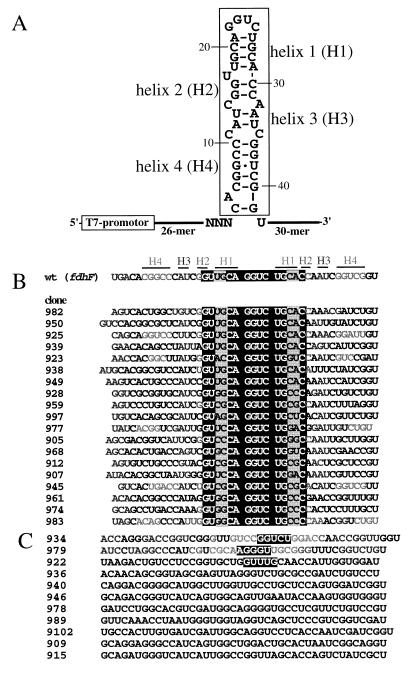
The 39 nucleotide hairpin fragment of the fdhF mRNA was mutagenized at a level of 30% per base position and exhibited a complexity of 5 × 10¹⁴ sequences. In addition, the three bases of the UGA selenocysteine codon were completely randomized to test whether a bias of SelB binding for the UGA codon itself could be selected (Fig. 1A).
Figure 1.
The fdhF mRNA hairpin, pool design, and selected aptamers. (A) Secondary structure of the fdhF mRNA hairpin and pool construction. The boxed region was synthesized at a degeneracy rate of 30% per base position. The sequences corresponding to the UGA selenocysteine codon were completely randomized (NNN). (B) Selected aptamer sequences that contain the apical stem–loop conserved. Only the inserts corresponding to the randomized region are shown. Sequences containing the invariant region are highlighted by the solid box. Within the conserved motif the base pairs U18-A29 and G19-C28 (shaded area) showed extensive Watson–Crick covariations. Aptamer sequences are aligned to the sequence of the wild-type fdhF mRNA hairpin. (C) Sequences that show variations from the consensus motif shown above. Sequence regions in solid boxes represent terminal–loop sequences confirmed by chemical and enzymatic probing analyses.
The SelB-binding RNA molecules of the starting pool were co-retained with SelB on nitrocellulose filters (27, 28). Bound RNA was eluted from the membrane, reverse transcribed to cDNA, and amplified by PCR and in vitro transcription to yield an enriched pool of RNA for the next selection cycle. To increase the stringency of the selection process, a 42 nucleotide sequence corresponding to the wild-type fdhF mRNA hairpin (Fig. 1A) was included as a specific competitor for SelB binding in rounds 3 and 4. The wild-type competitor lacked the primer binding sites of the pool RNA so that it could neither be reverse transcribed nor be amplified in the PCR. After four rounds of selection, RNA aptamers with the ability to compete with wild-type fdhF mRNA for SelB binding were selected.
Selected SelB-Binding Aptamers.
The majority of the selected RNA clones share highly conserved regions that correspond to the apical stem–loop structure of the wild-type fdhF mRNA hairpin (Fig. 1 A and B). Nineteen of the 30 sequences had the secondary structure motif formed by residues G16 to C30 (see solid box in Fig. 1B), highly conserved with only the base pairs U18-A29, and G19-C28 showing Watson–Crick covariations (Fig. 1 A and B). No clone containing the UGA codon was isolated. Furthermore, significant variation from the wild-type sequence in the lower region of the hairpin comprised by helices 3 and 4 was observed in the vast majority of the selected aptamers.
The clones shown in Fig. 1C deviate from the highly conserved apical loop motif found in the sequences of Fig. 1B. For example, the GGUC loop in the wild-type sequence was substituted for a GGUCU loop in clone 934 and an AGGGU loop in 979, whereas the 4-bp helix 1, the bulged U17, and helix 2 are more closely related to the wild type. These clones thus seem to represent classes of sequences toward which we aimed in our study, since they differ in their primary sequence from the SelB-binding apical region of the wild-type fdhF mRNA hairpin but still bind to the protein. Therefore, aptamers were primarily chosen among the sequences shown in Fig. 1C for a more detailed characterization. Clone 945 was chosen as a representative member from the sequences shown in Fig. 1B.
Relative Binding Ratios of Selected RNAs.
Selected monoclonal RNAs were tested for SelB binding in vitro by nitrocellulose binding assays. The ratio of SelB binding of monoclonal RNA aptamers compared with the wild-type was measured in an assay in which radiolabeled aptamer and labeled wild-type RNA of smaller size were allowed to compete for binding to a limited amount of SelB (23) (Table 1). Among the 12 sequences tested, the binding ratio varied between 3-fold better binding (clone 989) and 10-fold weaker binding (clones 909, 936, 946, and 9102) compared with the fdhF mRNA hairpin, which exhibits a Kd of 60 nM to SelB.
Table 1.
Binding activity of individual selected aptamer clones
| Clone | Binding ratio* | Clone | Binding ratio* |
|---|---|---|---|
| 909 | 0.1 | 945 | 2.0 |
| 915 | 0.4 | 946 | 0.1 |
| 922 | 0.5 | 978 | 0.3 |
| 934 | 0.8 | 979 | 0.7 |
| 936 | 0.1 | 989 | 3.0 |
| 940 | 2.7 | 9102 | 0.1 |
The ability of individual selected aptamers to compete with the wild-type fdhF mRNA hairpin is reflected in the “binding ratio”—the ratio of radiolabeled aptamer to radiolabeled wild-type RNA retained on the filter.
In Vitro Characterization of SelB–Aptamer Interaction.
SelB differs from EF-Tu mainly by its larger size due to a prolonged C terminus, designated as domain 4 (10, 11). The mRNA-binding domain of SelB was recently shown to reside within the C-terminal half (domain 4b) of this region (11). To map the SelB-binding region of our selected RNAs, aptamers were analyzed by a gel retardation assay under native conditions (Fig. 2A), as well as by a filter-binding assay (Fig. 2B) for binding to the full-length SelB protein and to a 17-kDa fragment corresponding to domain 4b of SelB. As a control, a fragment of SelB lacking the C terminus was also tested for aptamer binding. Twelve clones sampled from the enriched pool of cycle four were assayed. Ten of these clones were found to interact with the full-length SelB as well as with domain 4b. Clones 922 and 940, however, do not bind to this SelB subdomain. None of the aptamers tested bound to a SelB construct depleted of domain 4 (Fig. 2B).
Figure 2.
Characterization of the aptamer–SelB interaction in vitro. (A) Native gel shift assays of the complex formation of radiolabeled aptamer clones 945 and 922. Each aptamer RNA (75 nM) was assayed with indicated concentrations of full-length SelB protein (SelB), the C-terminal domain 4b, and an N-terminal fragment of SelB without domain 4 (for the sequences of these fragments see ref. 11). (B) Filter binding assays of binding of the wild-type clone 60.2 and individual aptamers to full-length SelB, domain 4b, and SelB without domain 4. Values show the percentage of the total radiolabeled-input RNA co-retained with the protein on nitrocellulose filters after extensive washing with binding buffer. RNA aptamer concentration was 75 nM, concentration of protein was 150 nM. Data are shown representatively for 6 of the 12 clones assayed.
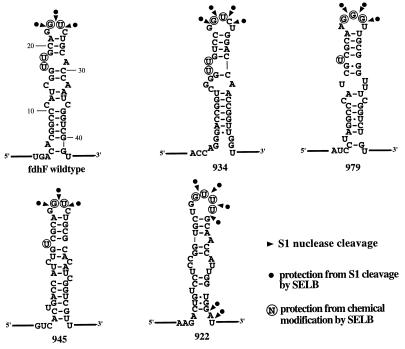
To refine the secondary structure of individual clones for comparison with that of the wild-type fdhF mRNA hairpin (13), chemical and enzymatic probing was performed using base specific (kethoxal, dimethyl sulfate, 1-cyclohexyl-3-(2-morpholinoethyl)-carbodiimide metho-p-toluenesulfonate) or enzymatic probes (Fig. 3). Enzymatic digestion of selected RNA clones with S1 nuclease demonstrated that cleavage or protection from cleavage of proposed loop regions in the presence or absence of SelB is similar, or identical to wild-type fdhF mRNA (13) (Fig. 3). The only exception is clone 922, which shows an additional region of protection at the bottom of the stem possibly reflecting a different binding mode to SelB than the wild-type and the other aptamers tested.
Figure 3.
Secondary structures of the fdhF mRNA hairpin (24) and selected aptamers according to chemical and enzymatic modification analyses. Protection of bases from chemical modification and S1 nuclease cleavage by SelB. ▾, Positions cleaved by S1 nuclease; •, indicates protection from S1 cleavage by SelB. Protection from chemical modification by SelB is shown by circled bases. Chemical and enzymatic probing of free RNA or RNA–SelB complexes was performed as described (24).
In Vivo Function of Selected Aptamers.
We next wanted to analyze whether the selected aptamers that where shown to bind to SelB also function in vivo by promoting UGA readthrough. Therefore, sequences coding for four individual RNA aptamers were cloned in-frame into the lacZ gene of vector pSKS 106 (29); levels of β-galactosidase generated by these constructs were measured as described (12, 14). Because the UGA selenocysteine codon was not selected in these sequences, we reintroduced an in-frame UGA codon on the 5′ side of the stem–loop structures within the selected aptamers. Furthermore, it has been shown previously that the distance of the SelB-binding domain in the upper portion of the fdhF mRNA hairpin from the UGA codon is rather critical and has to be maintained to ensure proper decoding by selenocysteine-tRNASec (14, 30). Therefore, we opted only to analyze those clones for UGA readthrough in which this criterion was fulfilled, i.e., the apical loop that contacts SelB was placed in proper distance from the UGA selenocysteine codon.
The constructs were transformed into E. coli strains FM434 selC+ and, as a negative control, into E. coli strain FM464 ΔselC (12), which lacks tRNASec and is thus unable to incorporate selenocysteine into proteins. As a positive control, the wild-type sequence of the fdhF mRNA hairpin was cloned into the vector pSKS106. Furthermore, as a second negative control, the fdhF hairpin sequence containing a (−1) frameshift by a deletion of one base (14) was analyzed for UGA readthrough (Table 2). UGA readthrough was measured through the generation of an enzymatically active portion of β-galactosidase and calculated in Miller units (Table 2) (26). Clone 945 promoted UGA readthrough to about 87% of the wild-type fdhF mRNA sequence (Table 2). However, aptamers 922, 934, and 979 did not show UGA readthrough in vivo, although they bind SelB in vitro with high affinity (Table 2).
Table 2.
UGA readthrough activity of the pSKS106 plasmid containing undisrupted LacZ gene, wild-type (WT 60.2) positive control, negative (p-1) control, and aptamer constructs, transformed in E. coli strains FM434 (selC+)- and the tRNASec-deficient control strain FM464 (ΔselC) calculated in Miller units
| Clones | FM434 | FM464 | % activity of WT 60.2 |
|---|---|---|---|
| pSKS 106 | 3500 | 3500 | |
| p-1 | 1.6 | 2 | 0 |
| WT 60.2 | 1100 | 10 | 100 |
| 922.1 | 0 | 3 | 0 |
| 922.2 | 0.5 | 4 | 0 |
| 934.1 | 5.5 | 7 | 0 |
| 934.2 | 30 | 4.5 | 3 |
| 945.1 | 970 | 8 | 87 |
| 979.1 | 12 | 12 | 0 |
| 979.2 | 185 | 1.5 | 17 |
Previously it has been proposed that the lower part of the fdhF hairpin structure (helix 4 and flanking regions 5′ and 3′ to the UGA codon) might be involved in promotion of selenocysteine incorporation into proteins, although this region of the hairpin is not required for SelB binding in vitro (12, 24). Thus, it was suggested that the lower part of the fdhF hairpin structure might serve as an anti-determinant for recognition by release factor 2 by entrapping the UGA codon in an intricate tertiary structure (24). Therefore, we tested whether the loss of UGA readthrough in constructs 922, 934, and 979 could be attributed to these findings by replacing the lower parts of the selected aptamer sequences with the corresponding original fdhF mRNA sequence (Fig. 4). These “hybrid” hairpin structures were then tested for UGA readthrough in the in vivo assay. Hybrid clones 922.2 and 934.2 resulted in no (0%) or very limited (3%) UGA readthrough as do the corresponding nonhybrid clones 922.1 and 934.1. Hybrid clone 979.2, however, did promote UGA readthrough to about 17% compared with the fdhF wild-type hairpin (Table 2).
Figure 4.
Constructs for testing in vivo activity of aptamers. (A) Complete selected aptamer sequences are shown in the boxes as comparison with the wild-type fdhF construct 60.2, which was used as the basis for all other constructs. The boxed 60.2 sequence was substituted for aptamer sequences shown in the boxes. (B) Construction of “hybrid” aptamers. The boxed 60.2 sequence was substituted for aptamer sequences shown in the boxes. In clones marked by an asterisk, A43 (solid box) was substituted by a cytosine to avoid the UAA stop codon.
DISCUSSION
Novel SelB-Binding RNA Aptamers.
Previous studies investigating the recognition sites within the fdhF mRNA hairpin motif suggested that only the upper part of the hairpin comprises the major determinants for recognition by the specialized EF SelB (11, 31). By chemical and enzymatic probing analyses only four bases within this region of the apical helix were shown to directly interact with the SelB protein: G23, U24, and U17/U18, respectively (24). Mutational analyses revealed that substitution of G23 within fdhF mRNA for any nucleotide results in almost complete loss of SelB binding and also abolished UGA readthrough (13, 14).
We performed in vitro selection using a partially randomized pool of the fdhF mRNA hairpin to select RNA variants that were able to bind to EF SelB. Since the sequence comprising the fdhF mRNA hairpin has not only evolved to bind to SelB but also has to be translated after selenocysteine incorporation, two sets of constraints are put upon the hairpin sequence in vivo. By in vitro selection of RNA structures, which were only selected for binding to SelB, we attempted to dissect binding from coding features.
Our selection revealed that the most abundant SelB-binding sequences resembled the secondary structure motif formed by residues G16 to C30. Positions within this region were invariant except for the base pairs U18-A29 and G19-C28, which showed an unusually high degree of Watson–Crick covariations (Fig. 1 A and B): The G19-C28 pair changes into a C19-G28 pair in 5 of the 19 selected sequences that contain the apical loop invariant (expected probability Pexp ≤ 0.01; Pfound = 0.26); the U18-A29 pair changes into an alternative base pair (mostly G/C–C/G) in 11 clones (Pexp. = 0.01; Pfound = 0.58). Thus, it appears that the alternative base pairs in helix 1 might be advantageous for SelB binding during the selection, presumably because they impart tighter binding to SelB. Indeed, clone 945, which contains a C18-G29 base pair (Fig. 1A), but is otherwise very similar to the upper portion of the fdhF mRNA hairpin, has a 2-fold higher binding ratio to SelB compared with wild type (Table 1). No examples for covariations were found indicating that U17 can base pair to extend helix 2, suggesting that a bulged residue is required at this position and that the length of helix 1 is critical for SelB binding in vitro. We propose that substitution of the labile U18-A29 base pair by a more stable C/G18-G/C29 base pair might result in a favorable preorganization of the RNA for binding to its complementary binding pocket in the C terminus of the protein, e.g., by “clamping” the bulged U17.
Sequence or secondary structure conservation within the lower part of the hairpin resembling helices 3 and 4 (Fig. 1A) was not observed, consistent with the notion that only the upper portion of the mRNA hairpin serves as a recognition determinant for SelB in vitro. The UGA selenocysteine codon located at the 5′ side of this hairpin was not selected, indicating that the codon itself is not involved in the binding of SelB.
RNA aptamers exhibiting deviations from the consensus motif were also isolated (Fig. 1C). The sequences of these aptamers are defined not by a single, but by several base changes (substitutions, insertions, deletions) within the SelB-binding domain of these hairpins. The wild-type mRNA hairpins cannot tolerate single base substitutions (14); our aptamers, however, show that multiple base changes within the apical hairpin can restore the SelB-binding activity, probably by compensating tertiary structural features providing important SelB–RNA recognition determinants, which are lost by single base substitutions.
Binding of RNA Aptamers to SelB and to Its C-Terminal mRNA Binding Domain.
The aptamers tested revealed binding ratios to SelB ranging between 3-fold better binding and 10-fold weaker binding compared with the wild-type fdhF mRNA hairpin.
We also mapped the SelB-binding site for some of the selected aptamers, since the mRNA-binding domain of SelB was recently shown to reside within a 17-kDa C-terminal region of SelB, designated as domain 4b (11). Among 12 clones, 10 bound domain 4b in the same way as they bind full-length SelB (Fig. 2B) and did not interact with the N-terminal SelB protein lacking domain 4. This result is important with respect to the comparison of aptamer clones 934 and 979 and the wild-type fdhF mRNA in their UGA readthrough activity in vivo (see below): These aptamers, while differing from the wild-type fdhF sequence in the apical loop exhibit (i) similar secondary structural features compared with wild type, (ii) similar binding affinities in vitro, and (iii) use the same domain within SelB for binding.
The selected pool also contained aptamers that use binding sites on the protein that differ from those of the wild-type. Such sequences are represented by clones 922 and 940, which only bind to the full-length protein and not to the truncated version of SelB or to domain 4b (Fig. 2). In these clones, secondary structure refinements by chemical modification and enzymatic probing analyses revealed no obvious homology to natural SelB-binding RNAs, such as the fdhF or fdnG mRNAs or to tRNASec (Fig. 3).
In Vivo Activity of Selected Aptamers.
To investigate whether our selected RNA aptamers were not only capable of binding to SelB in vitro but also promoted readthrough of the UGA selenocysteine codon in vivo, we cloned four individual aptamers in-frame into the lacZ gene and measured the levels of β-galactosidase activity. Special care had to be taken in designing these fusion constructs. First, since in none of the sequences the UGA codon was selected, it had to be re-introduced at its original position in the RNA hairpins. Second, it was shown previously that the distance between the UGA codon and the apical SelB-binding domain of the hairpin is important for selenocysteine incorporation (14, 30); therefore, we selected only those aptamers for the in vivo analysis in which the UGA codon was placed at a proper distance from the SelB-binding domain of the RNA aptamers. Finally, we only chose those clones that did not contain a termination codon when inserted into the lacZ gene.
Clone 945 promoted UGA readthrough similar to wild-type levels. This result shows that in vitro selected RNA aptamers can replace the natural fdhF mRNA hairpin in promoting UGA readthrough in vivo. It provides the second general example showing that an aptamer can adopt the in vivo function of a natural RNA. Previously, RNA aptamers selected for binding the HIV-1 Rev protein were shown to be Rev responsive in vivo (32). Of the four clones investigated, however, three did not result in UGA readthrough (922, 934, and 979), although they bind to SELB at levels similar to wild type. This might be due to the lower part of the wild-type fdhF hairpin being important for selenocysteine incorporation (24). To test this, only the upper, SelB-binding part of the fdhF RNA hairpin was replaced by the corresponding upper portion of the selected RNA aptamers, leaving the lower portion of the hairpin as wild type (Fig. 4). Again, hybrid clone 934.2 did not result in significant UGA readthrough (3%). Hybrid clone 979.2 promoted UGA readthrough to about 17% compared with wild type. This activity, although rather moderate, indicates that in this clone the lower part of the hairpin might directly or indirectly contribute to UGA readthrough to some extent, although not being involved in SelB binding. Further investigations will have to show which features within the lower region of the hairpin contribute to selenocysteine incorporation and how this is achieved.
The Biological Role of the mRNA Hairpin Goes Beyond Mere Tethering of SelB to the UGA Codon.
It has been suggested that the biological function of the fdhF mRNA hairpin might be to tether SelB in close proximity to the UGA selenocysteine codon, thereby enhancing the local concentration of the selenocysteyl-tRNASec bound to the SelB protein on the ribosome (14–16). If this model is correct, it would require that SelB-binding RNAs that contain mutations in the upper portion of the hairpin but still bind to the C terminus of SelB not differ significantly from the wild type in UGA readthrough activity. In this study, however, we obtained RNA sequences (e.g., clones 934 and 979) that fulfill the aforementioned criteria but nevertheless are devoid or drastically reduced in their UGA readthrough activity in vivo. In these aptamers, binding of SelB and its overall biological function are clearly dissected. These results therefore seem to imply functions of the mRNA stem–loop structure that go beyond the mere tethering of selenocysteyl-tRNASec to the UGA codon by simultaneous binding of SelB to selenocysteyl-tRNASec and the mRNA hairpin.
Our results therefore argue for alternative or additional functions of the mRNA hairpin during decoding. For example, the spatial arrangement of SelB/Sec-tRNASec, with respect to the ribosomal decoding site, might be different in the aptamer mRNAs compared with the wild-type fdhF mRNA hairpin. This could result in a loss of UGA readthrough. On the other hand, chemical and enzymatic probing data point to a similar recognition mode of these hairpins by SelB compared with wild-type, which would argue against this model.
Alternatively, binding of SelB to the fdhF mRNA hairpin might trigger a conformational change within SelB—only upon which the EF is able to deliver tRNASec to the ribosome and thereby promote selenocysteine incorporation. What would be the advantage of such a mechanism? Because SelB is homologous to EF-Tu in its N-terminal and central region (11), delivery of selenocysteyl-tRNASec to the ribosomal A site by SelB might proceed in a similar manner to the EF-Tu ternary complex. However, this would involve a major risk for the cell, since it would enable the selenocysteine-inserting machinery to decode regular UGA stop codons as UGA selenocysteine codons. Because the proposed conformational switch within SelB could only be induced if SelB is present in the very vicinity of a UGA selenocysteine codon, this would prevent readthrough of regular UGA stop codons. This model therefore goes beyond the mere enhancement of SelB concentration on the ribosome, but rather implicates a conformational switch within the quaternary complex. Thus aptamers 934 and 979, which contain mutations within the stem–loop structure of the hairpin but are still able to bind tightly to SelB, might fail to lead to UGA readthrough because of their inability to trigger the necessary conformational switch. The failure of aptamer 922 and the hybrid clone 922.2 to promote UGA readthrough is most likely explained by the result that clone 922 is not binding to the SelB domain 4b, but rather exhibits a different binding mode for the SelB protein.
In conclusion, our in vitro selection study provides for the first time, to the best of our knowledge, SelB-binding variants of the SelB-responsive wild-type fdhF mRNA hairpin, which allow for the dissection of SelB binding from the overall biological function of this mRNA secondary structure.
Acknowledgments
We dedicate this paper to Prof. August Böck on the occasion of his 60th birthday. We thank A. Böck for the generous gift of SelB and cell lines FM434 selC+ and FM464 ΔselC. We are also grateful to A. Böck for critical reading of the manuscript, to A. Böck, S. Müller, and M. Blind for helpful discussions, and to E.-L. Winnacker for support. This work was supported by Grant Fa275/2–1 from the Deutsche Forschungsgemeinschaft to M.F.
ABBREVIATION
- EF
elongation factor
References
- 1.Berry M J, Banu L, Larsen P R. Nature (London) 1991;349:438–440. doi: 10.1038/349438a0. [DOI] [PubMed] [Google Scholar]
- 2.Böck A, Forchhammer K, Heider J, Leinfelder W, Baron C. Trends Biochem Sci. 1991;16:463–467. doi: 10.1016/0968-0004(91)90180-4. [DOI] [PubMed] [Google Scholar]
- 3.Sturchler C, Carbon P, Krol A. Medicine/Sciences. 1995;11:1081–1088. [Google Scholar]
- 4.Walczak R, Westhof E, Carbon P, Krol A. RNA. 1996;2:367–379. [PMC free article] [PubMed] [Google Scholar]
- 5.Chambers I, Goldfarb P, Hampton J, Affara N, McBain W, Harrison P. EMBO J. 1986;5:1121–1127. doi: 10.1002/j.1460-2075.1986.tb04350.x. [DOI] [PMC free article] [PubMed] [Google Scholar]
- 6.Berry M J, Banu L, Chen Y Y, Mandel S J, Kieffer J D, Harney J W, Larsen P R. Nature (London) 1991;353:273–276. doi: 10.1038/353273a0. [DOI] [PubMed] [Google Scholar]
- 7.Hill K E, Lloyd R S, Yang J G, Read R, Burk R F. J Biol Chem. 1991;266:10050–10053. [PubMed] [Google Scholar]
- 8.Zinoni F, Birkmann A, Stadtman T, Böck A. Proc Natl Acad Sci USA. 1986;83:4560–4564. doi: 10.1073/pnas.83.13.4650. [DOI] [PMC free article] [PubMed] [Google Scholar]
- 9.Leinfelder W, Zehelein E, Mandrand-Berthelot M A, Böck A. Nature (London) 1988;331:723–725. doi: 10.1038/331723a0. [DOI] [PubMed] [Google Scholar]
- 10.Forchhammer K, Leinfelder W, Böck A. Nature (London) 1989;342:453–456. doi: 10.1038/342453a0. [DOI] [PubMed] [Google Scholar]
- 11.Kromayer M, Wilting R, Tormay P, Böck A. J Mol Biol. 1996;262:413–420. doi: 10.1006/jmbi.1996.0525. [DOI] [PubMed] [Google Scholar]
- 12.Zinoni F, Heider J, Böck A. Proc Natl Acad Sci USA. 1990;87:4660–4664. doi: 10.1073/pnas.87.12.4660. [DOI] [PMC free article] [PubMed] [Google Scholar]
- 13.Baron C, Heider J, Böck A. Proc Natl Acad Sci USA. 1993;90:4181–4185. doi: 10.1073/pnas.90.9.4181. [DOI] [PMC free article] [PubMed] [Google Scholar]
- 14.Heider J, Baron C, Böck A. EMBO J. 1992;11:3759–3766. doi: 10.1002/j.1460-2075.1992.tb05461.x. [DOI] [PMC free article] [PubMed] [Google Scholar]
- 15.Baron C, Böck A. In: The Selenocysteine-Inserting tRNA Species: Structure and Function. Söll D, RayBhandary U, editors. Washington, DC: ASM; 1995. pp. 529–544. [Google Scholar]
- 16.Gesteland R F, Atkins J. Annu Rev Biochem. 1996;65:741–768. doi: 10.1146/annurev.bi.65.070196.003521. [DOI] [PubMed] [Google Scholar]
- 17.Gold L, Polisky B, Uhlenbeck O, Yarus M. Annu Rev Biochem. 1995;64:763–797. doi: 10.1146/annurev.bi.64.070195.003555. [DOI] [PubMed] [Google Scholar]
- 18.Joyce G F. Curr Opin Struct Biol. 1994;4:331–336. doi: 10.1016/s0959-440x(94)90100-7. [DOI] [PubMed] [Google Scholar]
- 19.Klug S J, Famulok M. Mol Biol Rep. 1994;20:97–107. doi: 10.1007/BF00996358. [DOI] [PubMed] [Google Scholar]
- 20.Famulok M. J Am Chem Soc. 1994;116:1698–1706. [Google Scholar]
- 21.Geiger A, Burgstaller P, Von der Eltz H, Roeder A, Famulok M. Nucleic Acids Res. 1996;24:1029–1036. doi: 10.1093/nar/24.6.1029. [DOI] [PMC free article] [PubMed] [Google Scholar]
- 22.Tuerk C, Gold L. Science. 1990;249:505–510. doi: 10.1126/science.2200121. [DOI] [PubMed] [Google Scholar]
- 23.Bartel D P, Zapp M L, Green M R, Szostak J W. Cell. 1991;67:529–536. doi: 10.1016/0092-8674(91)90527-6. [DOI] [PubMed] [Google Scholar]
- 24.Hüttenhofer A, Westhof E, Böck A. RNA. 1996;2:354–366. [PMC free article] [PubMed] [Google Scholar]
- 25.Neidhardt F C, Bloch P L, Smith D F. J Bacteriol. 1974;119:736–747. doi: 10.1128/jb.119.3.736-747.1974. [DOI] [PMC free article] [PubMed] [Google Scholar]
- 26.Miller J F. Experiments in Molecular Genetics. Plainview, NY: Cold Spring Harbor Lab. Press; 1972. [Google Scholar]
- 27.Carey J, Uhlenbeck O C. Biochemistry. 1983;22:2601–2610. doi: 10.1021/bi00280a002. [DOI] [PubMed] [Google Scholar]
- 28.Fitzwater T, Polisky B. Methods Enzymol. 1996;267:275–301. doi: 10.1016/s0076-6879(96)67019-0. [DOI] [PubMed] [Google Scholar]
- 29.Shapira S K, Chou J, Richaud F V, Casadaban M C. Gene. 1983;25:71–82. doi: 10.1016/0378-1119(83)90169-5. [DOI] [PubMed] [Google Scholar]
- 30.Chen G F, Fang L, Inouye M. J Biol Chem. 1993;268:23128–23131. [PubMed] [Google Scholar]
- 31.Ringquist S, Schneider D, Gibson T, Baron C, Böck A, Gold L. Genes Dev. 1994;8:376–385. doi: 10.1101/gad.8.3.376. [DOI] [PubMed] [Google Scholar]
- 32.Symensma T L, Giver L, Zapp M, Takle G B, Ellington A D. J Virol. 1996;70:179–187. doi: 10.1128/jvi.70.1.179-187.1996. [DOI] [PMC free article] [PubMed] [Google Scholar]


