Abstract
Aphthoviruses use a conserved Arg-Gly-Asp triplet for attachment to host cells and this motif is believed to be essential for virus viability. Here we report that this triplet—which is also a widespread motif involved in cell-to-cell adhesion—can become dispensable upon short-term evolution of the virus harboring it. Foot-and-mouth disease virus (FMDV), which was multiply passaged in cell culture, showed an altered repertoire of antigenic variants resistant to a neutralizing monoclonal antibody. The altered repertoire includes variants with substitutions at the Arg-Gly-Asp motif. Mutants lacking this sequence replicated normally in cell culture and were indistinguishable from the parental virus. Studies with individual FMDV clones indicate that amino acid replacements on the capsid surface located around the loop harboring the Arg-Gly-Asp triplet may mediate in the dispensability of this motif. The results show that FMDV quasispecies evolving in a constant biological environment have the capability of rendering totally dispensable a receptor recognition motif previously invariant, and to ensure an alternative pathway for normal viral replication. Thus, variability of highly conserved motifs, even those that viruses have adapted from functional cellular motifs, can contribute to phenotypic flexibility of RNA viruses in nature.
Replication fidelity for RNA viruses is several-thousand-fold lower than for DNA based organisms (1–3). This is the consequence of a lack of proofreading activities associated with RNA polymerases and retroviral transcriptases. The outcome of the highly mutable viral genomes is that RNA viral populations form complex distributions of mutant genomes termed viral quasispecies (4–7). The evolutionary potential of such systems is largely unexplored. It is often assumed that nucleotide sequence conservation is linked to functionally essential domains of proteins or of regulatory genomic regions. This is often extended to RNA viruses, and statements correlating sequence conservation with functional essentiality abound in the literature of RNA viruses. But even this rule, which is very rooted in DNA genetics, may fail for RNA viruses, as documented in this report.
Work with the important animal pathogen foot-and-mouth disease virus (FMDV) by several groups over the last decade has permitted identification of a highly conserved, essential Arg-Gly-Asp domain on capsid protein VP1, which is involved in recognition of cellular receptors for this virus, including αvβ3 (8–13¶). Structural and functional work with rhinovirus and poliovirus indicated that the receptor recognition domain of these viruses was located at a depression or canyon inside the viral capsid (14, 15). This separated the receptor recognition site, conveniently hidden from immune attack, from variable surface residues corresponding to antigenic sites. The need to conserve critical residues was again equated with prohibition to change. It was work with FMDV that changed this picture dramatically. First, the FMDV structures determined by Stuart and colleagues (16–19) revealed that, contrary to other picornaviruses, no canyon or pit was present on the smooth FMDV surface. Furthermore, the critical receptor recognition Arg-Gly-Asp domain was located on a highly mobile loop (the G–H loop of VP1), which was also a major antigenic site for the virus (20–22). Additional structural work by Fita and colleagues (23) further indicated that the Arg-Gly-Asp motif is not only part of the receptor recognition site but it also interacts directly with some anti-viral neutralizing antibodies. Recent work with poliovirus and rhinovirus has also suggested that, interestingly, there is significant overlap between the subset of amino acid residues involved in receptor recognition and those belonging to antigenic sites (24‖). In addition to implications for coevolution of antigenicity and cell tropism, these observations emphasize one of the major evolutionary mechanisms used by RNA viruses: extremely active mutant production with continuous action of negative selection (3, 25). The Arg-Gly-Asp triplet is conserved not because it is not subjected to antibody attack but because the viruses that incorporate variations at this site cannot survive and, thus, they cannot be isolated and analyzed.
Here we document that the evolutionary capability of FMDV embraces the possibility that upon replication of the virus in a constant cell culture environment, even the Arg-Gly-Asp (RGD) motif may become dispensable.
MATERIALS AND METHODS
Virus and Monoclonal Antibody (mAb).
FMDV C-S8c1 is a plaque-purified derivative of the vaccine strain FMDV C-S8 (Santa Pau, Spain) (26). The passage history of FMDV C-S8c1 in cell culture involved three successive plaque isolations and an amplification from about 105 to 108 plaque-forming units (pfu) in BHK-21 cells. FMDV C-S8c1p100 is the C-S8c1 virus propagated 100 times in cell culture. Serial passages of FMDV C-S8c1 were performed by infecting a monolayer of about 2 × 10⁶ BHK-21 cells with 4 × 10⁶ to 8 × 10⁶ pfu (multiplicity of infection 2–4 pfu per cell) (27). The progeny of each infection was used to infect a fresh cell monolayer at the same multiplicity of infection. It must be emphasized that contrary to persistent infections in which the BHK-21 cells coevolved with FMDV (28), in the present experiments BHK-21 cells did not evolve, since a fresh monolayer from the same clonal stock of cells was used for each viral passage. mAb SD6 recognizes an extensively characterized epitope within the G–H loop of VP1 (29).
Selection of FMDV Mutants Resistant to mAb SD6.
Mutants resistant to neutralization by mAb SD6 (MARM) were selected either from FMDV C-S8c1 and FMDV C-S8c1p100 or from plaque-purified viral clones derived from the same viral populations. Selection of plaque-purified viruses ensured that each MARM analyzed originated from an independent mutational event. About 105-106 pfu of virus were incubated for 2 hr at 4°C with a 1:1 dilution of the mAb (supernatant of the SD6 hybridoma culture), and plated with a 1:50 dilution of the same mAb in the agar overlay, as previously described (29). Virus from a single plaque (about 105 pfu) was isolated and its resistance to the antibody (no detectable reduction in the number of plaques) tested in the neutralization assay (29). The virus was amplified to about 108 pfu for nucleotide sequencing.
To determine the MARM frequency, 105 to 106 viral pfu were incubated either in the presence or the absence of antibodies and adsorbed onto BHK-21 monolayers following described procedures (27). A 1:1 dilution of the supernatant of an SD6 hybridoma culture suppressed plaque formation of 100 pfu of FMDV C-S8c1 without any significant decrease of plaque numbers of resistant mutants previously selected with SD6 (29).
Virus Growth Curves.
Confluent BHK-21 cell monolayers (2 × 10⁶ cells) were infected at a multiplicity of infection of 10 pfu per cell with either FMDV C-S8c1 or FMDV C-S8c1p100 or mutants resistant to mAb SD6. After a 1 hr absorption period at 37°C, monolayers were washed, and infection allowed to proceed at 37°C as described. Samples (20 μl) were taken at different times after infection for titration of infectivity (26).
Nucleotide Sequencing.
The viral RNA encoding the FMDV capsid (P1) region was retrotranscribed into cDNA with primer 1: 5′-GAAGGGCCCAGGGTTGGACT-3′ (complementary to the RNA region encoding the 2A–2B junction, residues 3869–3888). The cDNA was PCR amplified with primer 1 and primer 2: 5′-GCTGTGGTAAACGCCATCA-3′ [complementary to the RNA region encoding the L (leader) protease, residues 1063–1081]. For sequencing the VP1-coding region primer 1 and primer 3: 5′-GCACGCTTCATGCGCAC-3′ (complementary to residues 3727–3743) were used. The complete capsid (P1) region was sequenced with primers 1, 3, and 4 (5′-GACCTTCACAAACCGGTCA-3′, residues 3315–3333), 5 (5′-GTTTCAGCCTCGTGGGACGC-3′, residues 3058–3077), 6 (5′-GCCGGCCAAGTAGGTGTTTGA-3′, residues 2807–2828), 7 (5′-GGTTGGGGCTATGTTGGC-3′, residues 2497–2514), 8 (5′-CCAGCCATTGCGCATGTACG-3′, residues 2192–2211), 9 (5′-CTAGAGACGCGCGTTC-3′, residues 2047–2062), 10 (5′-ACTGATGGCGTTGTC-3′, residues 1759–1773), and 11 (5′-CACTGGCAGCATAATTAAC-3′, residues 1692–1710). PCRs were sequenced using thermosequenase (United States Biochemical/Amersham).
Molecular Modeling.
Using the crystallographic coordinates of FMDV C-S8c1 (17), the substitutions found in the capsid of FMDV C-S8c1p100, mutant (Asp-143-Gly) (see Table 2), have been modeled with the program turbo (32) by placing the side chain atoms of the substituted amino acids in their standard conformations. The model was then optimized by energy minimization using the program x-plor (33).
Table 2.
Capsid amino acids found in FMDV C-S8c1p100c10 and FMDV C-S8c1p100 MARM (Asp-143-Gly), which differ from FMDV C-S8c1
| Secondary structure* | Position† | Amino acid substitution |
|---|---|---|
| VP1 βB strand | 41 | Lys → Glu |
| VP1 B–C loop | 46 | Asp → Glu |
| VP1 C terminus | 197 | His → Arg |
| VP3 N terminus | 25 | Ala → Val |
| VP3 G–H loop | 173 | Glu → Lys |
| VP3 C terminus | 218 | Gln → Lys |
Secondary structure assignments are as described in ref. 36.
Only positions different from those in FMDV C-S8c1 are shown.
RESULTS
Overcoming Restrictions to Variation: A New Repertoire of Amino Acid Substitutions Among Antibody-Resistant Mutants of FMDV.
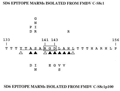
mAb SD6, which recognizes a continuous epitope located within amino acids 136–147 of VP1 of FMDV C-S8c1 (23, 29, 32, 33), was used to isolate resistant mutants. Viruses C-S8c1 and its derivative C-S8c1p100, obtained by propagating C-S8c1 in cell culture (see Materials and Methods) were incubated with mAb SD6 and resistant mutants were selected. To identify the mutations responsible for the resistance phenotype, the sequence of the VP1-coding region was determined. Sequences of 26 mutants isolated from FMDV C-S8c1 and 31 mutants isolated from FMDV C-S8c1p100 were compared with 56 additional mutants previously isolated from FMDV C-S8c1 (29, 34) (Table 1). The amino acid substitutions found in mutants derived from FMDV C-S8c1 affected only VP1 residues Ala-138, Ser-139, and His-146 (Table 1 and Fig. 1). Both immunochemical (refs. 29 and 35, M.G.M. and E.D., unpublished work) and structural (23) results have shown that, in addition to residues 138, 139, and 146, the highly conserved VP1 amino acids 141–145 and 147 were critically involved in recognition by mAb SD6 (Fig. 1). The finding of 86 antigenic variants substituted only in 3 different residues out of the 10 that interact with the antibody paratope clearly demonstrates the restrictions to variation that must be operating to keep residues 141–145 of FMDV C-S8c1, which include the Arg-Gly-Asp triplet, invariant (24, 35). In contrast, the sequences of 31 SD6-resistant mutants isolated from FMDV C-S8c1p100 revealed a completely different, expanded repertoire of antigenic variants (Table 1), with most substitutions affecting the highly conserved residues 142–145 (Fig. 1). Thus, the restrictions to variation in this antigenic region were largely lost after serial passages of FMDV C-S8c1 in BHK-21 cells.
Table 1.
SD6 epitope MARMs isolated from FMDV C-S8c1 and FMDV C-S8c1p100
| Mutation* | Amino acid substitution† | No. of mutants
|
|
|---|---|---|---|
| C-S8c1 | C-S8c1p100 | ||
| G(412) → C | Ala-138 → Pro | 1 | 0 |
| C(413) → A | Ala-138 → Asp | 3 | 1 |
| A(415) → C | Ser-139 → Arg | 2 | 0 |
| A(415) → G | Ser-139 → Gly | 2 | 0 |
| G(416) → A | Ser-139 → Asn | 8 | 2 |
| G(416) → T | Ser-139 → Ile | 17 | 8 |
| T(417) → A | Ser-139 → Arg | 2 | 0 |
| T(417) → G | Ser-139 → Arg | 16 | 0 |
| G(425) → A | Gly-142 → Glu | 0 | 4 |
| A(428) → G | Asp-143 → Gly | 0 | 1 |
| T(430) → G | Leu-144 → Val | 0 | 5 |
| T(431) → C | Leu-144 → Ser | 0 | 8 |
| C(434) → T | Ala-145 → Val | 0 | 2 |
| A(437) → G | His-146 → Arg | 31 | 0 |
| A(437) → G | His-146 → Arg | ||
| C(446) → T | Thr-149 → Met | 1 | 0 |
The first letter corresponds to the wild-type nucleotide; the number gives nucleotide position in the VP1-coding region; the last letter corresponds to the mutant nucleotide.
The first amino acid is the one found in the wild-type virus; the number gives the amino acid position in VP1; the second amino acid is the one found in the different MARMs. The vertical line indicates the isolation of a double mutant.
Figure 1.
Alignment of the amino acid sequences of the central region of the VP1 G–H loop of FMDV C-S8c1 and SD6 epitope MARMs. The single-letter amino acid code is used. Numbering is according to that for FMDV C-S8c1. Antigenic site A of type C viruses involves residues 136–150. The continuous epitope recognized by mAb SD6 is underlined. The RGD triplet is boxed. Solid triangles correspond to amino acid residues that cannot be replaced by most other amino acids with regard to binding to SD6; open triangles indicate residues that can be replaced by some other residues without having a drastic effect on SD6 recognition; residues 137 and 140 were generally replaceable without affecting SD6 recognition (ref. 33 and M.G.M. and E.D., unpublished work). These results are also consistent with structural data regarding the mAb SD6–G–H loop peptide interaction (23).
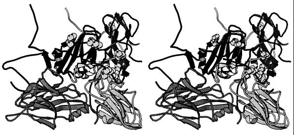
To determine whether the FMDV C-S8c1p100 population incorporated additional amino acid substitutions during the 100 passages in BHK-21 cells, the sequence of the entire capsid coding region was determined for one FMDV C-S8c1p100-derived mutant (Asp-143-Gly, see Table 1), and for a plaque-purified wild-type clone (c10) isolated from population FMDV C-S8c1p100 in the absence of mAb SD6. Apart from the mutation responsible for resistance to SD6 in the mutant clone, both clones differed from C-S8c1 in six capsid amino acids (Table 2). Three of them were located in capsid protein VP1 and three in VP3. With the exception of Ala-25-Val in VP3, all substitutions were located at the virion surface and they clustered around the position found for the G–H loop of VP1 in C-S8c1 (Fig. 2), suggesting they could have some influence on the conformation of the G–H loop (see Discussion).
Figure 2.
Location of the amino acid substitutions found in FMDV C-S8c1p100c10 on the virus capsid. Stereoview of a ribbon protein diagram of a crystallographic protomer of C-S8c1 in which the relevant substitutions have been modeled (see Materials and Methods). The capsid proteins VP1, VP2, and VP3 are indicated as dark, medium, and light grey, respectively. VP4 is entirely internal and has not been included for clarity. VP1 from a neighboring protomer is shown on the left side (dark grey). The substituted residues are depicted in CPK representation. The position of the G–H loop of VP1 in C-S8c1 complexed with mAb SD6 is indicated in light grey (37).
Selection and Frequencies of FMDVs Resistant to Neutralization by mAb SD6.
To determine the frequencies of MARMs, a MARM phenotypic assay with mAb SD6 was carried out as described. The MARM frequencies obtained were (8.5 ± 1) × 10−4 for FMDV C-S8c1 and (4.1 ± 0.5) × 10−3 for FMDV C-S8c1p100 (Table 3). Similar frequencies were obtained with a MARM genotypic assay (mAb SD6 added only to the agar overlay after cell adsorption of viruses) (data not shown). The large MARM frequencies obtained with FMDV C-S8c1p100 could reflect either a special trait of the complex quasispecies generated after many passages in BHK-21 cells or a feature of the viral capsid irrespective of the complexity of the viral population. To distinguish between these two possibilities, the MARM frequencies of two plaque-purified clones derived from population C-S8c1p100 were obtained (Table 3). Although such biological clones develop also into a quasispecies distribution, their complexity should be lower than that of the parental C-S8c1 p100 population. The large MARM frequencies found for both clones (Table 3) indicate that the high MARM frequencies depended on a feature of the viral particles and not on population complexity. The substitutions found in C-S8p100c10 (Table 2) suggest the relevance of the additional capsid amino acid substitutions in determining the increased MARM frequencies of the multiply passaged FMDV C-S8c1p100 population.
Table 3.
Frequencies of SD6 epitope MARMs in populations FMDV C-S8c1 and FMDV C-S8c1p100, and some clonal derivatives
| Viruses* | No. of assays | Frequency of MARM |
|---|---|---|
| C-S8c1 | 3 | (8.5 ± 1.0) × 10−4 |
| C-S8c1p100 | 3 | (4.1 ± 0.5) × 10−3 |
| C-S8c1p100c1 | 3 | (8.7 ± 0.3) × 10−3 |
| C-S8c1p100c10 | 3 | (8.5 ± 2.0) × 10−3 |
C-S8c1p100 is the initial FMDV C-S8c1 propagated 100 times in cell culture. C-S8c1p100c1 and C-S8c1p100c10 are two independent clones isolated from the FMDV C-S8c1p100 population.
Replication Kinetics of Selected SD6 MARMs.
The finding of MARMs with substitutions at the previously thought invariant RGD sequence (Fig. 1 and Table 1) begged the question as to their replication capacity. RGD is an essential cell-binding motif, and in contrast to the present findings, previous studies showed that BHK cells transfected with FMDV RNAs containing changes within this sequence produced noninfectious virus (10). To determine whether the substitutions found in SD6 MARMs influenced viral production, one-step growth analysis was carried out with several MARMs. The replication kinetics analysis included mutants with substitutions at the RGD sequence, as well as the MARM repertoire from FMDV C-S8c1p100 (Fig. 1). No differences were found among the mutants and the parental virus C-S8c1p100, demonstrating that substitutions at the RGD sequence or in surrounding residues did not affect the virus replication capacity (Fig. 3). Furthermore, the RNA from viruses recovered at 20 hr after infection (Fig. 3) were PCR amplified and their nucleotide sequence showed no signs of reversion at the relevant mutant positions, nor the presence of additional substitutions (data not shown). Likewise, no detectable reduction in the number of plaques was observed in the progeny mutants in a neutralization assay with mAb SD6 indicating maintenance of the MARM phenotype. As expected, the parental virus C-S8c1, with a limited passage history in BHK-21 cells (27), shows a delay in its replication kinetics relative to C-S8c1p100 and to all the mutants tested (Fig. 3).
Figure 3.
Replication of SD6 epitope MARMs. BHK-21 cells were infected with SD6 MARMs isolated from FMDV C-S8c1p100 and wild-type viruses C-S8c1 and C-S8c1p100 at a multiplicity of infection of 10 pfu per cell. Virus titer at different times after infection was determined by plaque assay in BHK-21 cells. Each time point is the average of duplicate samples.
DISCUSSION
The high mutation rates operating during RNA virus replication create complex quasispecies. Because such a complexity is generated in a highly probabilistic fashion due to the stochastic process of mutagenesis, the behavior of RNA virus populations is often unpredictable (6). Here we show that large FMDV populations serially propagated in cell culture, with constant environmental conditions, generate a different antigenic repertoire. Interestingly, this new antigenic repertoire includes variants with substitutions at the RGD motif (Gly-142-Glu and Asp-143-Gly) (Fig. 1), previously proposed to be essential for the viability of FMDV. The 86 mutants isolated from FMDV C-S8c1 (Table 1), together with many field viruses and laboratory variants sequenced (29, 36, 38–42) should be sufficient to consider FMDV of serotype C highly intolerant to amino acid substitutions at the RGD motif and several contiguous residues. Work with peptides indicates that, in addition to the RGD triplet, neighboring residues 144, 145, and 147 are also involved in receptor recognition of C-S8c1 (13), thus explaining their high degree of conservation. The relevance of the RGD motif in cell recognition of FMDV has been confirmed by studies of infectivity with cells transfected with the relevant integrin genes, and by site-directed mutagenesis of the critical loop residues (10, 11, 14, 43). The mAb SD6 MARM repertoire found in the FMDV C-S8c1p100 population, with substitutions in VP1 residues 141–145 (Table 1 and Fig. 1), clearly demonstrates the loss of restrictions to variation in this highly conserved region. Structural and immunochemical work have shown that all the residues found substituted in the SD6 MARMs (Fig. 1) interact with mAb SD6 (refs. 23 and 33 and references therein).
Using the atomic coordinates of FMDV C-S8c1 obtained by x-ray crystallography (18), the substitutions found in the capsid of FMDV C-S8c1p100 mutant Asp-143-Gly and of a plaque-purified wild-type clone (c10) isolated from C-S8c1p100 (Table 2) have been modeled on the capsid structure (Fig. 2). The G–H loop of VP1 is disordered in the crystal structures determined for native FMDVs. However, the structure of C-S8c1 complexed with the Fab fragment of mAb SD6 has been recently determined by cryoelectron microscopy (37); this allowed the positioning of the G–H loop on the capsid (Fig. 2). It may be significant that five of the six amino acid replacements found in the C-S8c1p100 clones relative to C-S8c1 cluster around the position found for the G–H loop. Substitutions at the B–C loop of VP1 of FMDV type O have been proposed to affect antibody recognition of the G–H loop by forcing this flexible loop to a different position (44). In addition, the conformation adopted by the RGD triplet within the G–H loop is probably critical for receptor binding (13). Furthermore, recent evidence suggests that FMDV of serotype O uses also heparan sulfate to interact with the cell (45). Thus, it is tempting to propose that some of the mutations in C-S8c1p100 may alter cell recognition of FMDV. Dispensability of the RGD could reflect either relaxation of the structural features needed to recognize a cellular integrin or the acquisition by FMDV of the ability to recognize an entirely new receptor molecule. It is noteworthy that the virus harbors, within the mutant spectrum of the evolving quasispecies, the evolutionary potential to render dispensable a receptor–recognition motif and to ensure an alternative pathway for normal viral replication. This implies a close relationship between antigenic variation and changes in cell tropism attained in the absence of immune selection and in a constant cell culture environment.
Wimmer and coworkers (24, 46**) have suggested that the involvement of some poliovirus capsid residues in antigenic determinants and in receptor binding could lead to the emergence of new enterovirus species with altered tissue tropism. Experiments are in progress to try to elucidate whether C-S8c1p100 and the mAb SD6 MARMs derived from it, especially those with substitutions in the RGD motif, have an altered cell tropism. The results reported here emphasize the great adaptive potential of RNA viruses. Indeed their genetic flexibility extends to rendering dispensable highly conserved functional domains due to variations occurring in a constant biological environment. This has clear implications for the emergence of new viruses in nature.
Acknowledgments
We acknowledge E. Hewat, D. Stuart, A. King, and I. Fita for providing unpublished data obtained in collaboration with us. The work in Madrid was supported by Grant PB94-0034-CO2-01 from Dirección General de Investigación Cientifica y Ténica (DGICYT), and Fundación Ramón Areces, and in Barcelona by Grant PB92–0707 from DGICYT.
ABBREVIATIONS
- FMDV
foot-and-mouth disease virus
- MARM
mutants resistant to neutralization by mAb SD6
- pfu
plaque-forming unit(s)
Footnotes
Carvalho, D., Rieder, E., Neff, S., Baxt, B., Rodarte, R., Tanuri, A. & Mason, P. (1996) Ninth Meeting of the European Study Group on the Molecular Biology of Picornaviruses, May 18–24, Gmunden, Austria (abstr.).
Mosser, A. G., Smith, T. J., Leippe, D. M., Olson, N., Baker, T. & Rueckert, R. R. (1996) Ninth Meeting of the European Study Group on the Molecular Biology of Picornaviruses, May 18–24, Gmunden, Austria (abstr.).
Wimmer, E., Harber, J., Bernhardt, G., Lu, H.-H., Sgro, J.-Y. & Gromeier, M. (1996) Ninth Meeting of the European Study Group on the Molecular Biology of Picornaviruses, May 18–24, Gmunden, Austria (abstr.).
References
- 1.Batschelet E, Domingo E, Weissmann C. Gene. 1976;1:27–32. doi: 10.1016/0378-1119(76)90004-4. [DOI] [PubMed] [Google Scholar]
- 2.Drake J W. Proc Natl Acad Sci USA. 1993;90:4171–4175. doi: 10.1073/pnas.90.9.4171. [DOI] [PMC free article] [PubMed] [Google Scholar]
- 3.Domingo E, Holland J J. In: The Evolutionary Biology of Viruses. Morse S, editor. New York: Raven; 1994. pp. 161–183. [Google Scholar]
- 4.Eigen M. Naturwissenschaften. 1971;58:465–523. doi: 10.1007/BF00623322. [DOI] [PubMed] [Google Scholar]
- 5.Eigen M, Schuster P. The Hypercicle: A Principle of Natural Self-Organization. Berlin: Springer; 1979. [DOI] [PubMed] [Google Scholar]
- 6.Holland J J, de la Torre J C, Steinhauer D A. Curr Top Microbiol Immunol. 1992;176:1–20. doi: 10.1007/978-3-642-77011-1_1. [DOI] [PubMed] [Google Scholar]
- 7.Domingo E, Holland J J. In: The Evolutionary Biology of Viruses. Morse S, editor. New York: Raven; 1994. pp. 161–184. [Google Scholar]
- 8.Fox G, Parry N R, Barnett P V, McGinn B, Rowlands D J, Brown F. J Gen Virol. 1989;70:625–637. doi: 10.1099/0022-1317-70-3-625. [DOI] [PubMed] [Google Scholar]
- 9.Baxt B, Becker Y. Virus Genes. 1990;4:73–83. doi: 10.1007/BF00308567. [DOI] [PubMed] [Google Scholar]
- 10.Mason P W, Rieder E, Baxt B. Proc Natl Acad Sci USA. 1994;91:1932–1936. doi: 10.1073/pnas.91.5.1932. [DOI] [PMC free article] [PubMed] [Google Scholar]
- 11.Berinstein A, Roivainen M, Hovi T, Mason P W, Baxt B. J Virol. 1995;69:2664–2666. doi: 10.1128/jvi.69.4.2664-2666.1995. [DOI] [PMC free article] [PubMed] [Google Scholar]
- 12.Hernández J, Valero M L, Andreu D, Domingo E, Mateu M G. J Gen Virol. 1996;77:257–264. doi: 10.1099/0022-1317-77-2-257. [DOI] [PubMed] [Google Scholar]
- 13.Mateu M G, Valero M L, Andreu D, Domingo E. J Biol Chem. 1996;271:12814–12819. doi: 10.1074/jbc.271.22.12814. [DOI] [PubMed] [Google Scholar]
- 14.Rossmann M G, Johnson J E. Annu Rev Biochem. 1989;58:533–573. doi: 10.1146/annurev.bi.58.070189.002533. [DOI] [PubMed] [Google Scholar]
- 15.Rossmann M G. J Biol Chem. 1989;264:14587–14590. [PubMed] [Google Scholar]
- 16.Acharya R, Fry E, Stuart D, Fox G, Rowlands D, Brown F. Nature (London) 1989;337:709–716. doi: 10.1038/337709a0. [DOI] [PubMed] [Google Scholar]
- 17.Lea S, Hernández J, Blakemore W, Brocchi E, Curry S, Domingo E, Fry E, Abu-Ghazaleh R, King A M Q, Newman J, Stuart D, Mateu M G. Structure (London) 1994;2:123–139. doi: 10.1016/s0969-2126(00)00014-9. [DOI] [PubMed] [Google Scholar]
- 18.Lea S, Abu-Ghazaleh R, Blakemore W, Curry S, Fry E, Jackson T, King A, Logan D, Newman J, Stuart D. Structure (London) 1995;3:571–580. doi: 10.1016/s0969-2126(01)00191-5. [DOI] [PubMed] [Google Scholar]
- 19.Curry S, Fry E, Blakemore W, Abu-Ghazaleh R, Jackson T, King A M Q, Lea S, Newman J, Rowlands D, Stuart D. Structure (London) 1996;4:135–145. doi: 10.1016/s0969-2126(96)00017-2. [DOI] [PubMed] [Google Scholar]
- 20.Bittle J L, Houghten R A, Alexander H, Shinnick T M, Sutcliffe J G, Lerner R A, Rowlands D, Brown F. Nature (London) 1982;298:30–33. doi: 10.1038/298030a0. [DOI] [PubMed] [Google Scholar]
- 21.Pfaff E, Mussgay M, Böhm H O, Schulz G E, Schaller H. EMBO J. 1982;1:869–874. doi: 10.1002/j.1460-2075.1982.tb01262.x. [DOI] [PMC free article] [PubMed] [Google Scholar]
- 22.Strohmaier K, Franze R, Adam K H. J Gen Virol. 1992;59:295–306. doi: 10.1099/0022-1317-59-2-295. [DOI] [PubMed] [Google Scholar]
- 23.Verdaguer N, Mateu M G, Andreu D, Giralt E, Domingo E, Fita I. EMBO J. 1995;14:1690–1696. doi: 10.1002/j.1460-2075.1995.tb07158.x. [DOI] [PMC free article] [PubMed] [Google Scholar]
- 24.Harber J, Bernhardt G, Lu H-H, Sgro J-Y, Wimmer E. Virology. 1995;214:559–570. doi: 10.1006/viro.1995.0067. [DOI] [PubMed] [Google Scholar]
- 25.Domingo E, Mateu M G, Escarmís C, Martínez-Salas E, Andreu D, Giralt E, Verdaguer N, Fita I. Virus Genes. 1996;11:125–135. doi: 10.1007/BF01728659. [DOI] [PubMed] [Google Scholar]
- 26.Sobrino F, Dávila M, Ortín J, Domingo E. Virology. 1983;128:310–318. doi: 10.1016/0042-6822(83)90258-1. [DOI] [PubMed] [Google Scholar]
- 27.Martínez M A, Carrillo C, González-Candelas F, Moya A, Domingo E, Sobrino F. J Virol. 1991;65:3954–3957. doi: 10.1128/jvi.65.7.3954-3957.1991. [DOI] [PMC free article] [PubMed] [Google Scholar]
- 28.de la Torre J C, Martínez-Salas E, Díez J, Villaverde A, Gebauer F, Rocha E, Dávila M, Domingo E. J Virol. 1988;62:2050–2058. doi: 10.1128/jvi.62.6.2050-2058.1988. [DOI] [PMC free article] [PubMed] [Google Scholar]
- 29.Mateu M G, Martínez M A, Rocha E, Andreu D, Parejo J, Giralt E, Sobrino F, Domingo E. Proc Natl Acad Sci USA. 1989;86:5883–5887. doi: 10.1073/pnas.86.15.5883. [DOI] [PMC free article] [PubMed] [Google Scholar]
- 30.Roussel A, Cambillau C. turbo-frodo in Silicon Graphics Geometry Partners Directory. Mountain View, CA: Silicon Graphics; 1989. pp. 77–78. [Google Scholar]
- 31.Brünger A T. x-plor Version 3.1: A System for X-Ray Crystallography and NMR. New Haven, CT: Yale Univ. Press; 1992. [Google Scholar]
- 32.Mateu M G, Rocha E, Vicente O, Vayreda F, Navalpotro C, Andreu D, Pedroso E, Giralt E, Enjuanes L, Domingo E. Virus Res. 1987;8:261–274. doi: 10.1016/0168-1702(87)90020-7. [DOI] [PubMed] [Google Scholar]
- 33.Mateu M G. Virus Res. 1995;38:1–24. doi: 10.1016/0168-1702(95)00048-u. [DOI] [PubMed] [Google Scholar]
- 34.Carrillo C, Plana J, Mascarella R, Bergadá J, Sobrino F. Virology. 1990;179:890–892. doi: 10.1016/0042-6822(90)90162-k. [DOI] [PubMed] [Google Scholar]
- 35.Novella I S, Borrego B, Mateu M G, Domingo E, Giralt E, Andreu D. FEBS Lett. 1993;330:253–259. doi: 10.1016/0014-5793(93)80883-v. [DOI] [PubMed] [Google Scholar]
- 36.Mateu M G, Hernández J, Martínez M A, Feigelstock D, Lea S, Peréz J J, Giralt E, Stuart D, Palma E L, Domingo E. J Virol. 1994;68:1407–1417. doi: 10.1128/jvi.68.3.1407-1417.1994. [DOI] [PMC free article] [PubMed] [Google Scholar]
- 37.Hewat E A, Verdaguer N, Fita I, Blakemore W, Brookes S, King A, Newman J, Domingo E, Mateu M G, Stuart D. EMBO J. 1997;16:1492–1500. doi: 10.1093/emboj/16.7.1492. [DOI] [PMC free article] [PubMed] [Google Scholar]
- 38.Mateu M G, Martínez M A, Capucci L, Andreu D, Giralt E, Sobrino F, Brocchi E, Domingo E. J Gen Virol. 1990;71:629–637. doi: 10.1099/0022-1317-71-3-629. [DOI] [PubMed] [Google Scholar]
- 39.Martínez M A, Hernández J, Piccone M E, Palma E, Domingo E, Knowles N, Mateu M G. Virology. 1991;184:695–706. doi: 10.1016/0042-6822(91)90439-i. [DOI] [PubMed] [Google Scholar]
- 40.Martínez M A, Dopazo J, Hernández J, Mateu M G, Sobrino F, Domingo E, Knowles N. J Virol. 1992;66:3557–3565. doi: 10.1128/jvi.66.6.3557-3565.1992. [DOI] [PMC free article] [PubMed] [Google Scholar]
- 41.Hernández J, Martínez M A, Rocha E, Domingo E, Mateu M G. J Gen Virol. 1992;73:213–216. doi: 10.1099/0022-1317-73-1-213. [DOI] [PubMed] [Google Scholar]
- 42.Borrego B, Novella I S, Giralt E, Andreu D, Domingo E. J Virol. 1993;67:6071–6079. doi: 10.1128/jvi.67.10.6071-6079.1993. [DOI] [PMC free article] [PubMed] [Google Scholar]
- 43.Rieder E, Baxt B, Mason P W. J Virol. 1994;68:5296–5299. doi: 10.1128/jvi.68.8.5296-5299.1994. [DOI] [PMC free article] [PubMed] [Google Scholar]
- 44.Parry N, Fox G, Rowlands D, Brown F, Fry E, Acharya R, Logan D, Stuart D. Nature (London) 1990;347:569–572. doi: 10.1038/347569a0. [DOI] [PubMed] [Google Scholar]
- 45.Jackson T, Ellard F M, Abu-Ghazaleh R, Brookes S M, Blakemore W E, Corteyn A H, Stuart D I, Newman J W I, King A M Q. J Virol. 1996;70:5282–5287. doi: 10.1128/jvi.70.8.5282-5287.1996. [DOI] [PMC free article] [PubMed] [Google Scholar]
- 46.Wimmer E, Hellen C U T, Cao X. Annu Rev Genet. 1993;27:353–436. doi: 10.1146/annurev.ge.27.120193.002033. [DOI] [PubMed] [Google Scholar]


