Abstract
cAMP/cAMP-dependent protein kinase (PKA) signaling pathway has been recently proposed to participate in both the late phase of long term potentiation in the hippocampus and in the late, protein synthesis-dependent phase of memory formation. Here we report that a late memory consolidation phase of an inhibitory avoidance learning is regulated by an hippocampal cAMP signaling pathway that is activated, at least in part, by D1/D5 receptors. Bilateral infusion of SKF 38393 (7.5 μg/side), a D1/D5 receptor agonist, into the CA1 region of the dorsal hippocampus, enhanced retention of a step-down inhibitory avoidance when given 3 or 6 h, but not immediately (0 h) or 9 h, after training. In contrast, full retrograde amnesia was obtained when SCH 23390 (0.5 μg/side), a D1/D5 receptor antagonist, was infused into the hippocampus 3 or 6 h after training. Intrahippocampal infusion of 8Br-cAMP (1.25 μg/side), or forskolin (0.5 μg/side), an activator of adenylyl cyclase, enhanced memory when given 3 or 6 h after training. KT5720 (0.5 μg/side), a specific inhibitor of PKA, hindered memory consolidation when given immediately or 3 or 6 h posttraining. Rats submitted to the avoidance task showed learning-specific increases in hippocampal 3H-SCH 23390 binding and in the endogenous levels of cAMP 3 and 6 h after training. In addition, PKA activity and P-CREB (phosphorylated form of cAMP responsive element binding protein) immunoreactivity increased in the hippocampus immediately and 3 and 6 h after training. Together, these findings suggest that the late phase of memory consolidation of an inhibitory avoidance is modulated cAMP/PKA signaling pathways in the hippocampus.
Keywords: cAMP signaling pathway, D1/D5 receptors, long term potentiation, hippocampus, PKA
Memory is a temporally graded process during which new information becomes consolidated and stored (1–3). From mollusks to mammals, memory can be divided into at least two phases: a protein and RNA synthesis-independent phase that lasts minutes to 1–3 h (short term memory) and a protein and RNA synthesis-dependent component [long term memory (LTM)] that lasts several hours to days, weeks, or even longer periods (4–7).
Recent work in Aplysia (8–10), Drosophila (11, 12), and mouse (13) has clearly demonstrated that cAMP-responsive transcription, mediated by the CREB (cAMP responsive element binding protein) family of proteins, is a crucial step for the establishment of LTM. cAMP-responsive genes participate in the long term facilitation of neurotransmitter release in Aplysia neurons, a model of nonassociative learning (8–10); the overexpression of a CREB repressor isoform blocks the formation of LTM for a Pavlovian odor avoidance task in Drosophila (11) whereas it does not affect a consolidated, protein synthesis-independent form of memory (7), and, more importantly, transgenic flies expressing an activating isoform of CREB showed an enhancement of LTM (12); in CREB knockout mice, LTM of fear conditioning and spatial learning is disrupted whereas acquisition and short term memory, which lasts 30–60 minutes, are normal (13). Taken together, these findings strongly suggest that cAMP-regulated pathways, via phosphorylation of CREB isoforms, are important for the long term consolidation of memories. CREB appears to act as a molecular switch to control the formation of LTM. This molecular switch depends on the functional ratio of CREB activators to blockers (14). In this context, by using a one trial step-down inhibitory avoidance task in rats, we recently demonstrated that hippocampal cAMP appears to be crucial for consolidating the late phase of an aversive memory (15).
In remarkable parallel with memory, long term potentiation (LTP), a long lasting activity-dependent mechanism of synaptic plasticity (16, 17), has been shown to possess also two components: an early transient phase that lasts less than 2–3 h and is resistant to inhibitors of protein synthesis, and a late, persistent component that requires both transcription and protein synthesis during a critical time window (17–20). This late protein synthesis-dependent phase of LTP is mediated, at least in part, by cAMP signaling pathways (19, 21).
Recently, Kandel and coworkers found that D1/D5 receptors, which activate adenylyl cyclase, are involved in the late phase of LTP in the CA1 region of the hippocampus (22). Bath application of D1/D5 receptor agonists induced a long-lasting synaptic potentiation that reached a peak 3–4 h after drug application, simulating the late phase of LTP. This effect requires new protein synthesis, is occluded by the potentiation produced by an activator of the cAMP-dependent protein kinase (PKA), and is blocked by the specific D1/D5 dopamine receptor antagonist SCH 23390 (22).
Given that LTP has been proposed repeatedly to be a model for information storage in the brain (16, 17) and that hippocampal LTP may underlie memory consolidation of an aversively motivated learning task in rats (23–26), we decided to explore the role of hippocampal D1/D5 receptor–cAMP signaling pathways in memory consolidation of a step down inhibitory avoidance learning.
MATERIALS AND METHODS
Surgery.
Under thionembutal anesthesia (30 mg/kg, i.p.), 647 male Wistar rats (3–4 months old; body weight 240–310 g) were bilaterally implanted with a 30-g cannula aimed 1 mm above area CA1 of the dorsal hippocampus at coordinates A 4.3, L 4.0, V 2.0 of the atlas of Paxinos and Watson (27). Postmortem histological controls of the cannula placements (28–30) showed that the injection tips were within 1.5 mm2 of the intended sites in 598 of the 647 animals. Only the behavioral data from animals with the cannula located in the intended sites were used.
Step Down Inhibitory Avoidance Task.
Once recovered from surgery, animals were trained in a one-trial step down inhibitory avoidance task and tested for retention 24 h later (28, 29). Rats were placed on a 2.5-cm high, 7 × 25 cm platform facing a 42 × 25 cm grid of parallel 0.1-cm stainless steel bars spaced 1 cm apart. Their latency to step down, placing their four paws on the grid, was measured. In training sessions, immediately upon stepping down, the rats received a 0.3-mA, 2-s, scrambled foot shock. No foot shock was given in test sessions. Test minus training session step down latencies (to a ceiling of 180 s) were taken as a measure of retention.
Drug Infusion Procedures.
At the time of infusion, 27-g cannulae were fitted into the guide cannula. The animals were gently withdrawn from the training apparatus, wrapped in a soft cloth left with their head out, and subjected to the infusion procedure. Handling was kept as gentle as possible to minimize stress. It is to be noted, however, that this was the same for all animals, vehicle or drug-treated. The time between withdrawal of the animals from the training apparatus and the start of the first infusion was between 20 and 60 s. The entire infusion procedure took ≈2 min, including 45 s for the infusions themselves, first on one side and then on the other, and the handling. Infusions were performed immediately (0 h), 3, 6, or 9 h after training. The infusion cannula was connected to a microsyringe by a polyethylene tube. The tip of the infusion cannula protruded 1 mm beyond that of the guide cannulae and was therefore aimed at CA1 in the dorsal hippocampus. The animals received bilateral 0.5-μl infusions of vehicle (saline or 20% DMSO in saline) or of different drugs dissolved in the vehicle.
Statistical analysis of the behavioral data was by a Kruskal–Wallis ANOVA followed by individual Mann–Whitney U tests, two-tailed. Differences in training session latency among groups were not significant (H = 1.09, P > 0.1). In control groups, training test latency differences were significant at P < 0.001 in a nonparametric Mann–Whitney U test (two-tailed).
Biochemical Measurements.
For all of the biochemical measurements, nonimplanted male Wistar rats weighing 220–250 g were divided into three groups: naive controls, killed immediately after withdrawal from their home cages; shocked animals, placed directly over the electrified grid, given a 2-s, 0.3-mA foot shock, and immediately returned to their home cage; and a group trained in the step down inhibitory avoidance as described above. Animals from the three groups were killed by decapitation at different time intervals after training.
3H-SCH 23390 Binding Assay.
For the autoradiographic assay, brains were rapidly frozen (−70°C) after removal. Sagittal sections 12-μm thick were cut in a cryostat, thaw-mounted onto chrome-alum gelatin-coated slides, and kept at −70°C until use. 3H-SCH 23390 [72.5 Ci/mmol (1 Ci = 37 GBq); NEN] autoradiography was carried out as described (32). Sections were incubated in buffer Tris⋅HCl (50 mM) with a saturating concentration of 3H-SCH 23390 (4 nM) at room temperature for 1 h. Incubations were terminated by washing the sections three times in buffer Tris⋅HCl, followed by two washes in ice-cold distilled water, and then the sections were rapidly air-dried
Autoradiograms were generated by exposing a 3H-sensitive film (Hyperfilm, Amersham) to the slices at 4°C for 13 days before developing. The autoradiograms were analyzed using a computerized microdensitometer (mcid 4.02, Imaging Research, Toronto, Canada), and ODs were converted to equivalent disintegrations per minute per square millimeter (autoradiographic 3H-microscales standards, Amersham; ref. 26). Statistical analysis was performed using statistical software (instat, Graph Pad, San Diego). Differences among groups were determined by one-way ANOVA, followed by Neuman–Keuls test.
cAMP Assay.
The hippocampi were rapidly dissected out, placed in buffer sodium acetate 0.5 mM with 1-methyl-3-isobutylxantine for 1 min at 60°C, homogenized, and centrifuged at 12,500 rpm for 10 min. The pellet was discarded, and a radioimmunoassay was performed using the ligand 125I-cAMP (25,000–28,000 cpm) and an anti-cAMP antibody (Sigma), according to the method of Domino et al. (31), with slight modifications as described elsewhere (15). After overnight incubation of triplicate tubes at 4°C, 50 μl of 2% BSA and 2 ml of 96% ethanol were added to each tube and mixed. Then, the tubes were centrifuged at 3500 rpm for 15 min, the supernatant was discarded, and the radioactivity in the pellet was counted. Hippocampal cAMP levels were determined by log-logit analysis and were expressed as pmol/mg tissue. Statistical significance was determined by two-way ANOVA followed by a Tukey–Kramer multiple comparison test.
cAMP-Dependent Phosphodiesterase Activity.
cAMP-dependent phosphodiesterase was assayed according to the method of Thompson et al. (33). In brief, hippocampal samples of naive, shocked, and trained animals were incubated in 80 mM buffer Tris (10 mM Cl2Mg/16 mM β-mercaptoethanol/2 mM AMP/200 μM cAMP) with the addition of 70,000 cpm of 3H-cAMP at 37°C for 10 min. Then, the samples were boiled during 1 min and incubated with 50 μl of snake venom containing 5′-nucleotidase (1 mg/ml) at 30°C for 10 min. After this, 1 ml of a mixture of Dowex W50 resin/methanol (1/3) was added and centrifuged at 3000 rpm for 15 min. The resulting supernatants were collected and counted in liquid scintillator.
PKA Activity Assay.
PKA activity was assayed using Kemptide (CALBIOCHEM), a selective substrate for PKA (34, 35). In brief, hippocampi were homogenized in 20 mM Tris⋅HCl buffer containing 0.5 mM 3-isobutylmethylxanthine, 10 mM DTT, 5 mM NaF, 10 mM EDTA, 10 mM EGTA, and a mixture of protease inhibitors. After centrifugation at 2.800 rpm for 10 min, the supernatants were collected. Ten microliters of tissue [2 mg protein/ml, as determined by the method of Bradford (36)] was incubated at 30°C for 5 min in buffer containing 30 μM Kemptide, 10 mM cAMP, and 200 μM [γ-32P]ATP (200–300 cpm/pmol; NEN). The reaction was stopped onto phosphocellulose strips (Whatman P81), and these were washed 3 times in 75 mM phosphoric acid (8 ml/sample). Filters were dried and then counted by liquid scintillation.
P-CREB Immunocytochemistry.
P-CREB immunocytochemistry was performed according to the method of Ginty et al. (37). Naive controls and shocked and trained animals were anesthetized with chloral hydrate (28%) and perfused with 4% paraformaldehyde in PBS for 5 min. Then, the brains were removed and fixed for 3 h at 4°C. Cryostat sagittal sections (20 μm) were incubated with anti-P-CREB (0.5 μg/ml) for 16 h at 4°C, and specific immune complexes were visualized with an avidin–biotin detection system (Vector Laboratories). Quantitative densitometric analysis was performed using a computerized microdensitometer (mcid 4.02).
RESULTS
Modulation of Memory Consolidation by Hippocampal D1/D5 Receptors.
To examine whether hippocampal D1/D5 receptors are involved in the early and late stages of memory formation, we investigated the effects on memory consolidation of the bilateral intrahippocampal infusion of a specific D1/D5 receptor agonist (SKF 38393, 7.5 μg/side) or a specific D1/D5 receptor antagonist (SCH 23390, 0.5 μg/side) at different time intervals after training.
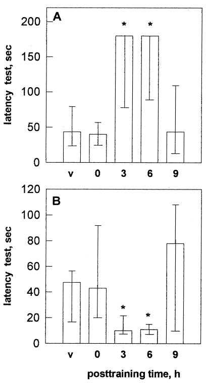
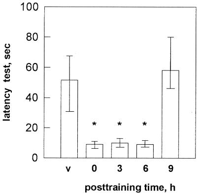
Test session step down latency in the inhibitory avoidance, when either SKF 38393 or SCH 23390 was infused into the CA1 region of the dorsal hippocampus, is depicted in Fig. 1 A and B, respectively. SKF 38393 facilitated memory consolidation when given 3 or 6 h (P < 0.002), but not 0 or 9 h, after training (Fig. 1A). In contrast, the intrahippocampal infusion of SCH 23390 caused full retrograde amnesia when given 3 or 6 h posttraining (P < 0.002) but not 0 or 9 h after training (Fig. 1B). These findings suggest that hippocampal D1/D5 receptors participate in the late, but not in the early, phase of memory consolidation of a one-trial inhibitory avoidance learning. Earlier work has shown that the early phase of memory formation of a one-trial stepdown avoidance task is associated with a learning-specific, time-dependent increase in both 3H-AMPA binding to AMPA glutamate receptors and 3H-phorbol dibutyrate binding to membrane-bound PKC in the hippocampus (26, 38).
Figure 1.
Effects of bilateral intrahippocampal infusion of 7.5 μg of SKF 38393 (A) or 0.5 μg of SCH 23390 (B) at different time intervals after training, on test session step down latency. Data are expressed as median (interquartile range). ∗, Significant difference from saline at P < 0.002. Number of animals ranged between 10 and 12 per group.
To evaluate whether the inhibitory avoidance training also is accompanied by changes in hippocampal D1/D5 receptors, we used a quantitative autoradiographic method (32, 38) to study the binding of 3H-SCH 23390 at different time intervals after training. As can be seen in Table 1, avoidance learning is associated with a 90% increase in CA1 hippocampal 3H-SCH 23390 binding 3 h after training (P < 0.01). Also, there was a smaller, but significant, increase (47%; P < 0.05) 6 h after training.
Table 1.
Quantitative densitometric analysis of 3H-SCH 23390 binding to D1/D5 receptors in the CA1 region of the hippocampus
| Group | n | 3H-SCH binding, dpm/mm2 |
|---|---|---|
| Naive | 6 | 42 ± 8.1 |
| Shocked | 6 | 50 ± 9.3 |
| Trained 0 h | 6 | 52 ± 9.9 |
| Trained 1 h | 6 | 52 ± 7.5 |
| Trained 3 h | 6 | 98 ± 8.9† |
| Trained 6 h | 6 | 74 ± 10.5* |
| Trained 9 h | 6 | 51 ± 9.3 |
Data are expressed as mean ± SEM.
P < 0.05;
P < 0.01 in Newman–Keuls test after ANOVA.
Modulation of Memory Consolidation by Hippocampal cAMP Signaling Pathways.
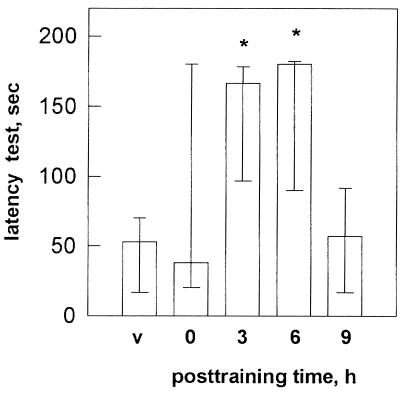
Confirming and extending recent findings from our laboratories (15), the bilateral intrahippocampal infusion of the membrane-permeable analog of cAMP (8Br-cAMP; 1.25 μg/side) enhanced retention scores when given 3 or 6 h after training (P < 0.002) (Fig. 2). No changes in memory retention scores were observed when 8Br-cAMP was administered immediately or 9 h after training.
Figure 2.
Effects of 8Br-CAMP (1.25 μg/side) administered by bilateral infusion into the CA1 region on step down latency. ∗, Significant difference from test session values of saline group at P < 0.002. All training test latency differences were significant at P < 0.001. Number of animals ranged between 10 and 12 per group.
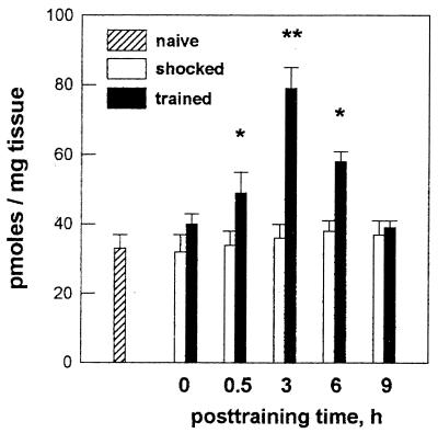
In addition, we found that rats submitted to the avoidance learning showed a learning-specific, time-dependent increase in the endogenous levels of cAMP in the hippocampus (Fig. 3). This increase was maximal 3 h after training (+135% in comparison with naive animals; P < 0.001); a marginal increment in the hippocampal endogenous cAMP level also was evident 6 h posttraining (P < 0.05). These results suggest that cAMP signaling pathways in the hippocampus are important for the late, but not the early, posttraining phase of memory consolidation of this task.
Figure 3.
Endogenous cAMP levels in the hippocampus of naive, shocked, or trained animals killed at different time intervals after training. Data are expressed as mean ± SE pmol/mg tissue of six to nine independent experiments done by triplicate. ∗, P < 0.01; ∗∗, P < 0.001 compared with naive controls, Tukey–Kramer test after ANOVA.
To test whether these changes in hippocampal cAMP levels might be related to an increased breakdown of the cyclic nucleotide, we determined the activity of cAMP-dependent phosphodiesterase in hippocampal samples from naive, shocked, or trained rats (33). No alterations in hippocampal phosphodiesterase activity were observed in trained animals in comparison with naive or shocked controls (Table 2).
Table 2.
Hippocampal cAMP-dependent PDE activity
| Group | n | PDE, pmol/mg tissue |
|---|---|---|
| Naive | 8 | 20.3 ± 0.88 |
| Shocked | 6 | 20.2 ± 1.15 |
| Trained 0 h | 6 | 20.9 ± 1.46 |
| Trained 3 h | 6 | 23.7 ± 1.63 |
| Trained 6 h | 6 | 21.1 ± 1.71 |
| Trained 9 h | 6 | 19.4 ± 1.53 |
Data are expressed as mean ± SEM.
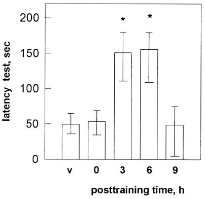
To explore further the possibility that hippocampal cAMP signaling pathways are involved in the late phase of memory formation, we next examined the effect on inhibitory avoidance memory of forskolin, an activator of adenylyl cyclase, given into the CA1 region at different time intervals after training (Fig. 4). Forskolin (0.5 μg/side) caused a clear-cut facilitation of memory when infused 3 (P < 0.002) or 6 h (P < 0.02) posttraining.
Figure 4.
Effects of bilateral intrahippocampal infusion of vehicle or forskolin (0.5 μg/side) on step down latency. Data are expressed as in Fig. 1. Number of rats ranged between 9 and 12 per group. Training test differences were significant at P < 0.001 by Mann–Withney U tests (two tailed). ∗, P < 0.02; ∗∗, P < 0.002 in comparison with test session values of vehicle group (Mann–Whitney U test).
The effect of forskolin on memory consolidation was quite similar in both the nature and time course to those observed with SKF 38393 (Fig. 1A) and 8Br-cAMP (Fig. 3), reinforcing the assumption that cAMP-regulated mechanisms in the hippocampus are important for the consolidation of an aversively motivated learning task.
The activation of adenylyl cyclases leads to the dissociation of regulatory and catalytic subunits of PKA. Only the free catalytic subunit is active. Therefore, to determine more directly the role of hippocampal PKA in memory of an avoidance learning, we next examined the effects of KT5720, a specific inhibitor of the catalytic subunit of PKA, on memory consolidation. The bilateral infusion of KT5720 (0.5 μg/side) into the CA1 region produced full amnesia when given 0, 3, or 6 h posttraining (P < 0.002) (Fig. 5). No changes in test session latencies were observed when KT5720 was administered 9 h after training. These results are consistent with the effects of 8BrcAMP and forskolin on the late posttraining phase of memory consolidation. However, PKA activation in the hippocampus also appears to be required for the early phase of memory formation.
Figure 5.
Effects of KT5720 on memory of step down inhibitory avoidance learning in rats that received a bilateral infusion of vehicle or KT5720 (0.5 μg/side) into the CA1 region 0, 3, 6, or 9 h after training. Data are expressed as in Fig. 1; n = 10–11 animals per group. Training test latency differences significant at P < 0.001. ∗, P < 0.002 compared with vehicle values (Mann–Whitney U tests).
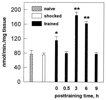
If activation of hippocampal PKA were involved in memory consolidation, training rats in the avoidance task should probably cause an increase in PKA activity. We therefore carried out biochemical assays to measure directly the activity of PKA in hippocampal preparations from naive, shocked, and trained animals. PKA activity increased 49% at 0 h (P < 0.01), 139% at 3 h (P < 0.001), and 110% at 6 h (P < 0.001) posttraining (Fig. 6). Shocked animals had similar values of PKA activity as naive controls.
Figure 6.
Effect of an inhibitory avoidance learning on hippocampal PKA activity. Data are expressed as mean ± SE nmol/min/mg tissue of seven independent experiments done by triplicate. ∗, P < 0.01; ∗∗, P < 0.001 in comparison with naive control values in Newman–Keuls test after ANOVA.
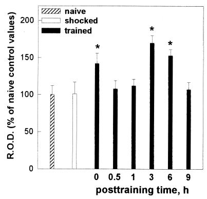
Given that cAMP regulates the transcription of numerous genes via the PKA-mediated phosphorylation of the CREB family of proteins, we next examined the effect of the inhibitory avoidance task on CREB phosphorylation. We have used a specific antibody against the activated, phosphorylated form of CREB (37) to show that acquisition and consolidation of an aversively motivated learning task induced the phosphorylation of CREB in CA1 hippocampal neurons (Fig. 7). Quantitative densitometric analysis revealed two peaks of increased immunoreactivity in the hippocampus of trained rats at 0 h and 3 and 6 h after training (0 h: +40%, P < 0.01; 3 h: +75%, P < 0.01; 6 h: + 56%, P < 0.01). This effect appears to be learning-specific because no alteration in P-CREB immunoreactivity was found in shocked animals.
Figure 7.
Quantitative densitometric analysis of the immunocytochemistry of P-CREB in CA1 region of the dorsal hippocampus of naive, shocked, or trained rats. Data are expressed as percentage of control values [mean ± SE of relative OD (R.O.D.) per mm2]. n = five independent experiments done by triplicate. ∗, P < 0.01, in comparison with naive control values, in Newman–Keuls test after ANOVA.
DISCUSSION
Memories are not acquired directly in their definitive form. They are labile and can be modified in the period that follows acquisition. This is due to the influence of modulatory systems (1–3, 24, 25, 39, 40). These modulatory systems regulate a process of consolidation, whereby memories are transformed from their initial labile into a later stable state. Consolidation leads to storage: What becomes consolidated is stored. The hippocampus and related brain areas are mainly involved in the acquisition and consolidation of an inhibitory avoidance learning but apparently do not participate in the long term storage of memory (23–25).The role of the hippocampus in the acquisition and consolidation of this task has been confirmed recently using reversible inactivation with tetrodotoxin (41).
We have shown that hippocampal ionotropic and metabotropic glutamate receptors (24), CAMK II (30), protein kinase C (26, 28), and retrograde messengers (25, 29) are critically involved in the early (within 1 h) neural events responsible for the acquisition and consolidation of an inhibitory avoidance learning.
The main finding of the present study is that cAMP signaling pathways in the hippocampus, activated at least in part by D1/D5 receptors, are crucial for the late phase of memory consolidation of an aversively motivated learning paradigm in rats. This is based on five series of data. First, the activation of hippocampal D1/D5 receptors or adenylyl cyclases 3 or 6 h after training produced facilitation of memory consolidation. Second, the intrahippocampal infusion of 8Br-cAMP provoked enhancement of memory only when given 3 or 6 h posttraining (Fig. 2). Third, the blockade of D1/D5 receptors or PKA in the hippocampus caused full retrograde amnesia when SCH 23390 or KT 5720 were infused into the CA1 region at the same time intervals after training (Figs. 1B and 5, respectively). Fourth, rats submitted to the avoidance learning showed significant increases in hippocampal cAMP levels and PKA activity at the same time window observed in the behavioral experiments (Fig. 3 and 6). Fifth, there is a peak of increased P-CREB immunoreactivity in CA1 neurons 3 h after training (Fig. 7). Taken together, these results strongly suggest that cAMP signal transduction pathways in the hippocampus are mainly involved in the late phase of memory consolidation.
Also, our findings endorse and extend the assumption that LTM requires a functional cAMP signaling pathway regulating the transcription of several genes via the PKA-mediated phosphorylation of the CREB family of proteins. Nonassociative learning in Aplysia, odor avoidance learning in Drosophila, and Pavlovian conditioning and spatial learning in mice have provided strong evidence that the activation of the CREB family of transcriptional factors is a necessary step for the establishment of LTM (8–14). As a corollary, these data, taken together with those found in the present study, suggest that, even though different forms of learning probably activate different distributed neuronal pathways, the molecular mechanism for the storage of LTM may be conserved.
Our results indicate also that the late phase of memory consolidation of an inhibitory avoidance is modulated by hippocampal D1/D5 receptors. A mesolimbic dopaminergic input to the CA1 region acting on D1/D5 receptors has been demonstrated (42, 43). Both D1 and D5 receptors stimulate adenylyl cyclase (44). Therefore, the D1/D5 receptor modulation of memory consolidation seems to result from the functional coupling with adenylyl cyclase and cAMP. This is support by the finding that SKF 38393, forskolin, and 8Br-cAMP have a similar effect on memory. However, the involvement of other hippocampal modulatory neurotransmission that might contribute to the late phase of memory consolidation cannot be ruled out (40). In fact, recent pharmacological data reveal that hippocampal β adrenergic receptors and 5-HT1A receptors also modulate the late phase of memory formation (L.B., et al., unpublished observations).
Earlier work has shown that the dopaminergic system participates in learning and memory. Using a foot shock-motivated brightness discrimination task in rats, Grekch and Matthies (45) found that the intrahippocampal infusion of the nonspecific dopaminergic agonist apomorphine improves memory, whereas haloperidol, a nonspecific dopamine receptor antagonist, impairs memory consolidation. Moreover, posttraining i.p administration of SKF 38393 facilitated retention of an inhibitory avoidance in mice whereas SCH 23390 impaired memory (46). SKF 38393 (3 mg/kg, i.p.) also improved memory of aged rats subjected to a spatial learning (33), and local injection of SCH 23390 into the prefrontal cortex hindered working memory in monkeys (47). Therefore, our present results confirm and extend the above mentioned findings, showing that D1/D5 receptors in the hippocampus participate in the late consolidation phase of an avoidance learning.
Recently, it has been demonstrated that chicks submitted to an inhibitory avoidance training showed a selective increase in D1/D5, but not in D2, binding in selected brain regions (32). In accordance, we found that inhibitory avoidance in rats provoked a learning-specific, time-dependent increase in 3H-SCH 23390 binding in the hippocampus. This learning-induced change occurred 3 and 6 h posttraining, the same critical period when SKF 38393 and SCH 23390 modulate memory consolidation.
Consistent with Hebbian models of learning and memory (16, 17, 23) and in remarkable parallel with the present findings, cAMP signaling pathways participate in the late phase of LTP in several regions of the hippocampus (18, 21). Thus, our results may be taken to further endorse the hypothesis that LTP in the hippocampus underlies memory processes (24, 25).
A dopamine-independent, PKA-mediated mechanism may be also required in the immediate early phase of memory formation in the hippocampus. The amnestic effect of the PKA inhibitor when given 0 h posttraining (Fig. 5), the lack of effect of SCH 23390, SKF 38393, 8Br-cAMP, or forskolin when given at this early time (Figs. 1, 2, and 4), and the peak of P-CREB immunoreactivity observed in CA1 region immediately posttraining (Fig. 7) indicate that there is also an early, PKA-mediated modulation of memory, apparently independent of D1/D5 receptors or of the endogenous levels of cAMP. These findings are consistent with previous results in Drosophila, showing that cAMP signaling pathways also are important for the acquisition and the early phase of memory processing (14, 48). Furthermore, a PKA-dependent early phase of LTP has been observed (19–21, 49). In this context, it has been postulated recently that, in the early phase of LTP, the cAMP signaling pathway functions as a gate to modulate the establishment of LTP (49).
The early peak of increased P-CREB immunoreactivity coincides with a small but significant increase in PKA activity observed immediately after training. However, it must be borne in mind that CREB is also phosphorylated by CAMK II (50). In this connection, it is important to stress that KN 62, a specific inhibitor of CAMK II, caused full retrograde amnesia only when infused immediately, but not 2 or 4 h, after training into the hippocampus (30).
In conclusion, the present behavioral and biochemical results indicate that cAMP signaling pathways in the hippocampus, activated at least in part by D1/D5 receptors, are mainly involved in the modulation of a late consolidation phase of an inhibitory avoidance learning.
Acknowledgments
This work was supported by Fundacao de Amparo a Pesquisa do Estado do Rio Grande do Sul and Financiadora de Estudos e Projetos, Brasil, and the University of Buenos Aires and Fundacion Antorchas, Argentina.
ABBREVIATIONS
- LTP
long term potentiation
- LTM
long term memory
- PKA
cAMP-dependent protein kinase
- P-CREB
phosphorylated form of cAMP-responsive element binding protein
References
- 1.McGaugh J L. Science. 1966;153:1351–1358. doi: 10.1126/science.153.3742.1351. [DOI] [PubMed] [Google Scholar]
- 2.Mc Gaugh J L. In: Perspectives of Memory Research. Solomon P R, Goethals G R, Kelley C M, Stephens B R, editors. New York: Springer; 1988. pp. 33–66. [Google Scholar]
- 3.Izquierdo I. Behav Neural Biol. 1989;59:171–202. doi: 10.1016/s0163-1047(89)90812-1. [DOI] [PubMed] [Google Scholar]
- 4.Davis H P, Squire L R. Psychol Bull. 1984;96:518–559. [PubMed] [Google Scholar]
- 5.Castellucci V, Blumenfeld H, Goelet P, Kandel E R. J Neurobiol. 1989;20:1–9. doi: 10.1002/neu.480200102. [DOI] [PubMed] [Google Scholar]
- 6.Frank D A, Greenberg M E. Cell. 1994;79:5–8. doi: 10.1016/0092-8674(94)90394-8. [DOI] [PubMed] [Google Scholar]
- 7.Tully T, Preat T, Boynton S C, Del Vecchio M. Cell. 1994;79:37–48. doi: 10.1016/0092-8674(94)90398-0. [DOI] [PubMed] [Google Scholar]
- 8.Dash P K, Hochner B, Kandel E R. J Neurosci. 1990;7:2232–2238. [Google Scholar]
- 9.Kaang B-K, Kandel E R, Grant S G N. Neuron. 1993;10:427–435. doi: 10.1016/0896-6273(93)90331-k. [DOI] [PubMed] [Google Scholar]
- 10.Alberini C M, Ghirardi M, Metz R, Kandel E R. Cell. 1994;85:1099–1114. doi: 10.1016/0092-8674(94)90386-7. [DOI] [PubMed] [Google Scholar]
- 11.Yin J C P, Wallach J S, Del Vecchio M, Wilder E L, Zhou H, Quinn W G, Tully T. Cell. 1994;79:49–58. doi: 10.1016/0092-8674(94)90399-9. [DOI] [PubMed] [Google Scholar]
- 12.Yin J C P, Del Vecchio M, Zhou H, Tully T. Cell. 1995;81:107–115. doi: 10.1016/0092-8674(95)90375-5. [DOI] [PubMed] [Google Scholar]
- 13.Bourtchuladze R, Frenguelli B, Cioffi D, Blendy J, Schutz G, Silva A. Cell. 1994;79:59–66. doi: 10.1016/0092-8674(94)90400-6. [DOI] [PubMed] [Google Scholar]
- 14.Yin J C P, Tully T. Curre Opin Neurobiol. 1996;6:204–208. doi: 10.1016/s0959-4388(96)80082-1. [DOI] [PubMed] [Google Scholar]
- 15.Bernabeu R, Schmitz P, Faillace M P, Izquierdo I, Medina J H. NeuroReport. 1996;7:585–588. doi: 10.1097/00001756-199601310-00050. [DOI] [PubMed] [Google Scholar]
- 16.Bliss T V, Collingridge G L. Nature (London) 1993;361:31–39. doi: 10.1038/361031a0. [DOI] [PubMed] [Google Scholar]
- 17.Nicoll R A, Malenka R C. Nature (London) 1995;377:115–118. doi: 10.1038/377115a0. [DOI] [PubMed] [Google Scholar]
- 18.Nguyen P V, Abel T, Kandel E R. Science. 1994;265:1104–1107. doi: 10.1126/science.8066450. [DOI] [PubMed] [Google Scholar]
- 19.Huang Y-Y, Li X C, Kandel E R. Cell. 1994;79:69–79. doi: 10.1016/0092-8674(94)90401-4. [DOI] [PubMed] [Google Scholar]
- 20.Nguyen P V, Kandel E R. J Neurosci. 1996;16:3189–3198. doi: 10.1523/JNEUROSCI.16-10-03189.1996. [DOI] [PMC free article] [PubMed] [Google Scholar]
- 21.Frey U, Huang Y-Y, Kandel E R. Science. 1993;260:1661–1664. doi: 10.1126/science.8389057. [DOI] [PubMed] [Google Scholar]
- 22.Huang Y-Y, Kandel E R. Proc Natl Acad Sci, USA. 1995;92:2446–2450. doi: 10.1073/pnas.92.7.2446. [DOI] [PMC free article] [PubMed] [Google Scholar]
- 23.Izquierdo I. FASEB J. 1994;8:1139–1145. [PubMed] [Google Scholar]
- 24.Izquierdo I, Medina J H. Neurobiol Learn Mem. 1995;63:19–32. doi: 10.1006/nlme.1995.1002. [DOI] [PubMed] [Google Scholar]
- 25.Medina J H, Izquierdo I. Brain Res Rev. 1995;21:185–194. doi: 10.1016/0165-0173(95)00013-5. [DOI] [PubMed] [Google Scholar]
- 26.Bernabeu R, Izquierdo I, Cammarota M, Jerusalinsky D, Medina J H. Brain Res. 1995;685:163–168. doi: 10.1016/0006-8993(95)00310-m. [DOI] [PubMed] [Google Scholar]
- 27.Paxinos G, Watson C. The Rat Brain in Stereotaxic Coordinates. San Diego: Academic; 1986. [DOI] [PubMed] [Google Scholar]
- 28.Jerusalinsky D, Quillfeltd J A, Walz R, Da Silva R C, Medina J H, Izquierdo I. Behav Neural Biol. 1994;61:107–109. doi: 10.1016/s0163-1047(05)80063-9. [DOI] [PubMed] [Google Scholar]
- 29.Izquierdo I, Fin C, Schmitz P, Da Siva R, Jerusalinsky D, Quillfeltd J A, Ferreira M B, Medina J H, Bazan N G. Proc Natl Acad Sci USA. 1995;92:5047–5051. doi: 10.1073/pnas.92.11.5047. [DOI] [PMC free article] [PubMed] [Google Scholar]
- 30.Wolfman C, Fin C, Dias M, Bianchin M, Da Silva R, Schmitz P, Medina J H, Izquierdo I. Behav Neural Biol. 1994;61:203–205. doi: 10.1016/s0163-1047(05)80001-9. [DOI] [PubMed] [Google Scholar]
- 31.Domino SE, Tubb D J, Garbers D L. Methods Enzymol. 1991;195:345–355. doi: 10.1016/0076-6879(91)95179-n. [DOI] [PubMed] [Google Scholar]
- 32.Stewart M G, Kabai P, Harrison E, Steele R J, Kossut M, Gierdalski M, Csillag A. Neuroscience. 1996;70:7–14. doi: 10.1016/0306-4522(95)00345-j. [DOI] [PubMed] [Google Scholar]
- 33.Thompson W J, Broker G, Appleman M M. Methods Enzymol. 1974;38:205–212. doi: 10.1016/0076-6879(74)38033-0. [DOI] [PubMed] [Google Scholar]
- 34.Roskoski R., Jr Methods Enzymol. 1983;99:3–6. doi: 10.1016/0076-6879(83)99034-1. [DOI] [PubMed] [Google Scholar]
- 35.Clegg C H, Correll L A, Cadd G G, McKnight G S. J Biol Chem. 1987;262:13111–13119. [PubMed] [Google Scholar]
- 36.Bradford M M. Anal Biochem. 1976;72:248–260. doi: 10.1016/0003-2697(76)90527-3. [DOI] [PubMed] [Google Scholar]
- 37.Ginty D D, Kornhauser J M, Thompson M A, Bading H, Mayo K E, Takahashi J S, Greenberg M E. Science. 1993;260:238–241. doi: 10.1126/science.8097062. [DOI] [PubMed] [Google Scholar]
- 38.Cammarota M, Izquierdo I, Wofman C, Levi de Stein M, Bernabeu R, Jerusalinsky D, Medina J H. Neurobiol Learn Mem. 1995;64:257–264. doi: 10.1006/nlme.1995.0008. [DOI] [PubMed] [Google Scholar]
- 39.Izquierdo I, Medina J H. Trends Pharmacol Sci. 1991;12:260–265. doi: 10.1016/0165-6147(91)90567-c. [DOI] [PubMed] [Google Scholar]
- 40.Izquierdo I, Medina J H. In: Brain Processes and Memory. McGaugh J L, Ishikawa K, editors. Amsterdam: Elsevier–North Holland; 1996. pp. 25–38. [Google Scholar]
- 41.Lorenzini C A, Baldi E, Bucherelli C, Sacchetti B, Tassoni G. Brain Res. 1996;730:32–39. doi: 10.1016/0006-8993(96)00427-1. [DOI] [PubMed] [Google Scholar]
- 42.Huang Q, Zhuo D, Chase K, Gusella J, Aronon N, Di Figlia M. Proc Natl Acad Sci USA. 1992;89:11988–11992. doi: 10.1073/pnas.89.24.11988. [DOI] [PMC free article] [PubMed] [Google Scholar]
- 43.Monsma F J, Jr, Mahan L C, McVittie L D, Gerefen C R, Sibley D R. Proc Natl Acad Sci USA. 1990;87:6723–6727. doi: 10.1073/pnas.87.17.6723. [DOI] [PMC free article] [PubMed] [Google Scholar]
- 44.Nestler E J. Cell. 1994;79:923–926. doi: 10.1016/0092-8674(94)90022-1. [DOI] [PubMed] [Google Scholar]
- 45.Grecksch G, Matthies H. In: Neuronal Plasticity and Memory Formation. Ajmone Marsan C, Matthies H, editors. New York: Raven; 1982. pp. 353–357. [Google Scholar]
- 46.Castellano C, Cestari V, Cabib S, Puglisi-Allegra S. Behav Neural Biol. 1991;56:283–291. doi: 10.1016/0163-1047(91)90439-w. [DOI] [PubMed] [Google Scholar]
- 47.Sagawuchi T, Goldman-Rakic P S. Science. 1991;251:947–950. doi: 10.1126/science.1825731. [DOI] [PubMed] [Google Scholar]
- 48.Levin L R, Han P L, Hwang P M, Feinstein P G, Davis R L, Reed R R. Cell. 1992;68:479–489. doi: 10.1016/0092-8674(92)90185-f. [DOI] [PubMed] [Google Scholar]
- 49.Blitzer R D, Wong T, Nouranifar R, Iyengar R, Landau E H. Neuron. 1995;15:1403–1414. doi: 10.1016/0896-6273(95)90018-7. [DOI] [PubMed] [Google Scholar]
- 50.Sheng M, McFadden G, Greenberg M E. Neuron. 1990;4:571–582. doi: 10.1016/0896-6273(90)90115-v. [DOI] [PubMed] [Google Scholar]


