

Abstract
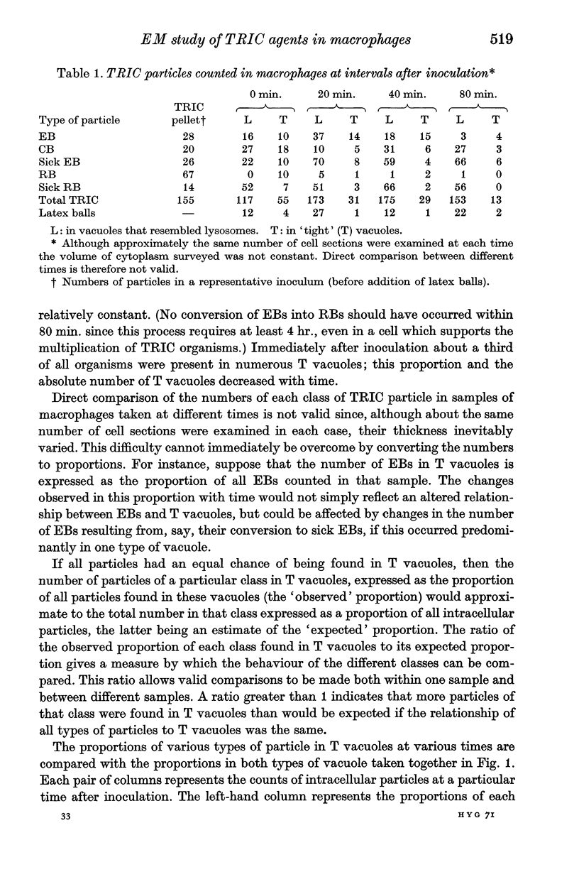
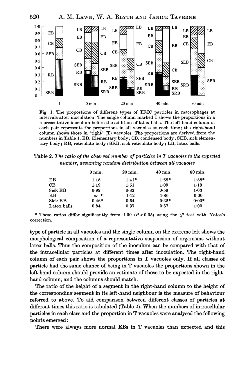
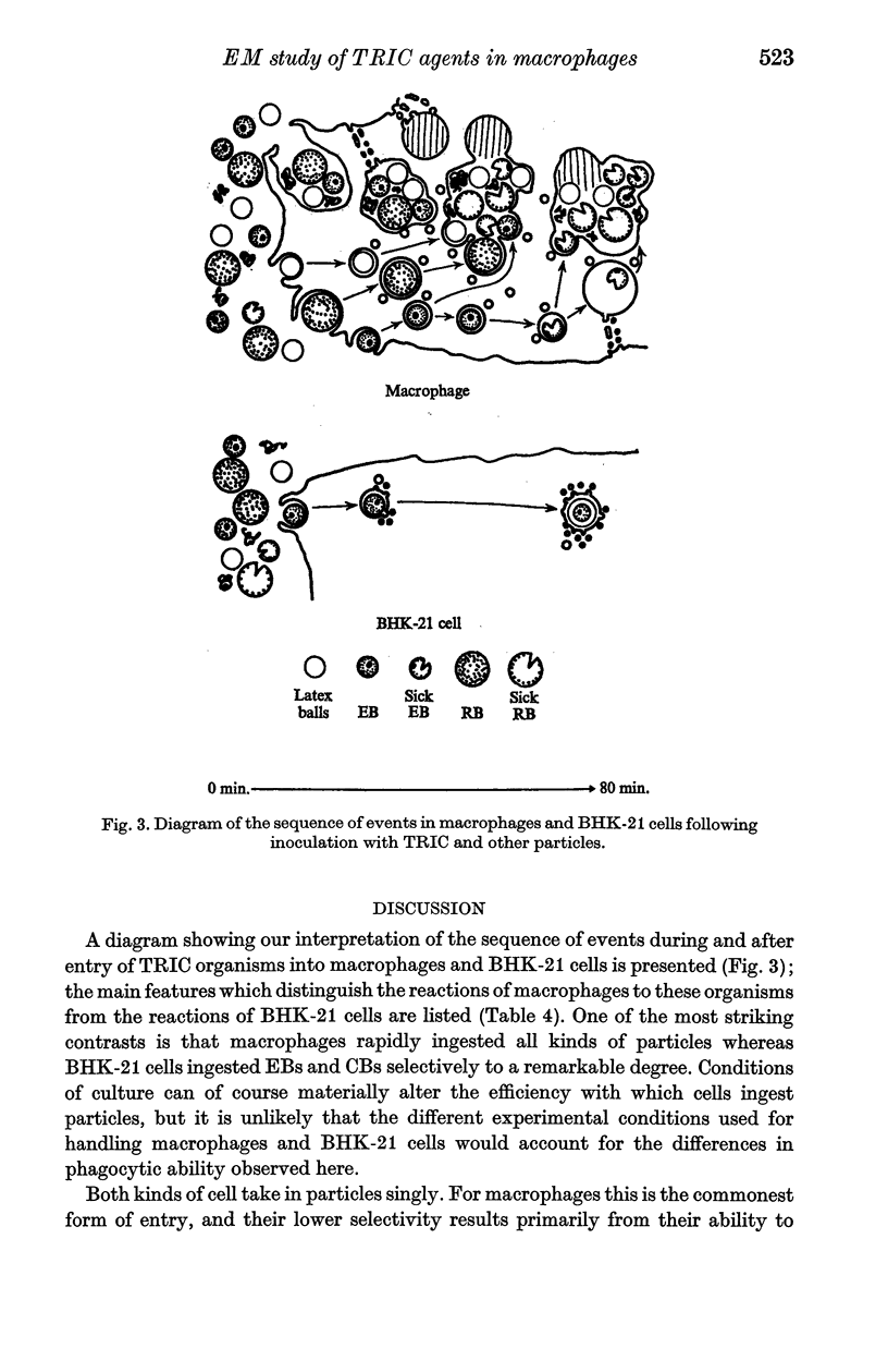
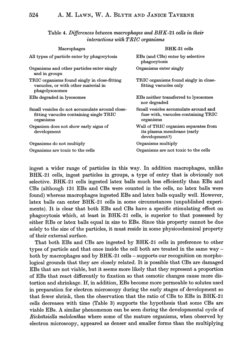
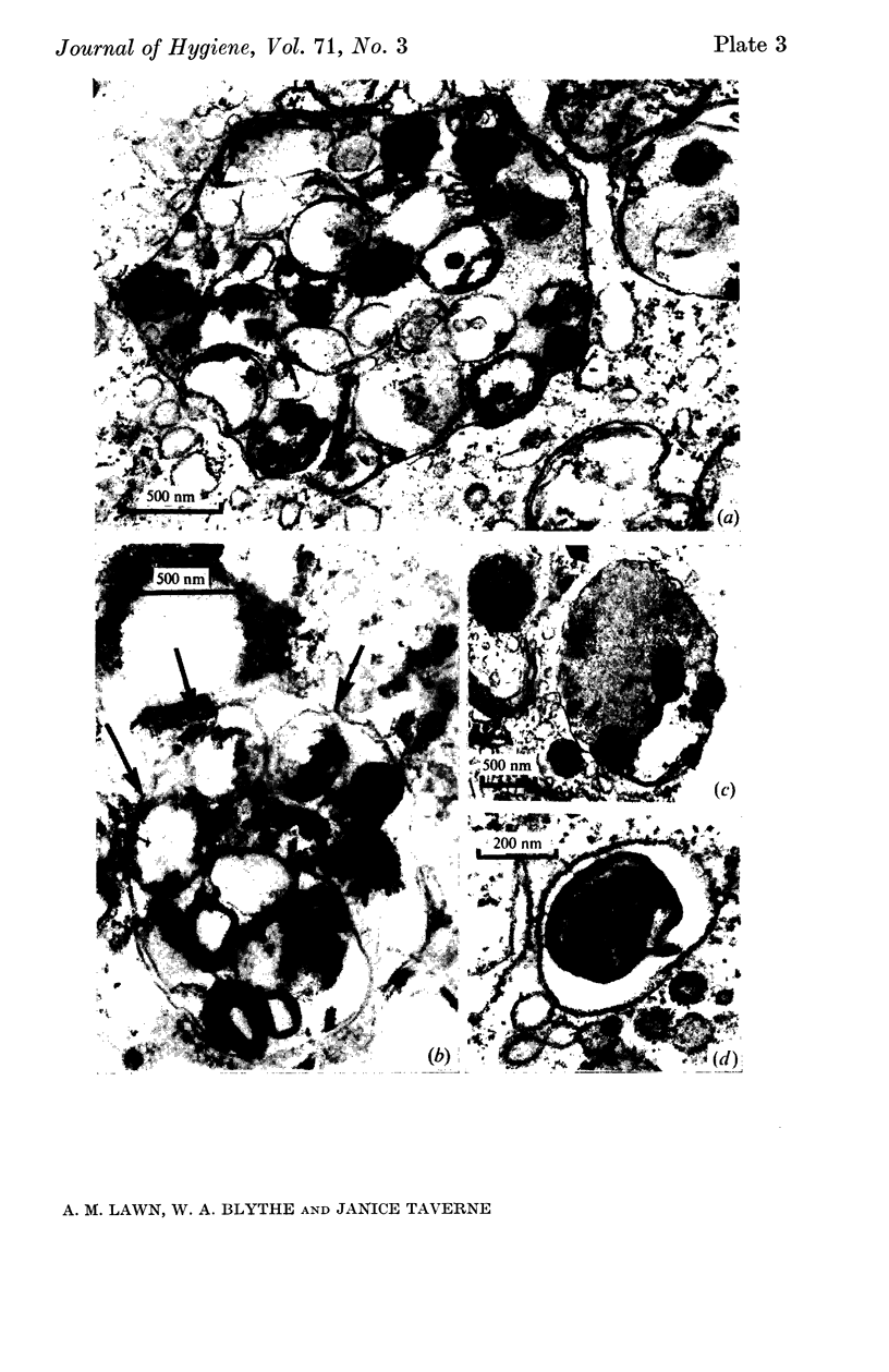
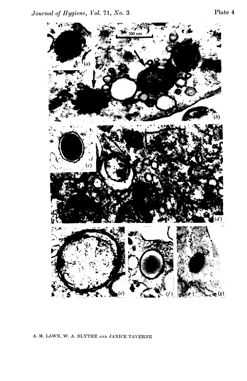
TRIC agents do not multiply in mouse peritoneal macrophages in culture but have a toxic effect on them, whereas they multiply readily in BHK-21 cells. Sections of macrophages and of BHK-21 cells fixed during the first 90 min after inoculation were examined by electron microscopy. Macrophages ingested all forms of the organism, which were eventually degraded in lysosomes. However, elementary bodies were distinguished from other TRIC particles by the delay in their transfer to lysosomes. BHK-21 cells ingested elementary bodies selectively, and in these cells the organisms were neither found in lysosomes nor degraded. Instead they showed morphological changes that probably represented an early stage of development.
Full text
PDF
Images in this article
Selected References
These references are in PubMed. This may not be the complete list of references from this article.
- Anderson D. R., Hopps H. E., Barile M. F., Bernheim B. C. Comparison of the ultrastructure of several rickettsiae, ornithosis virus, and Mycoplasma in tissue culture. J Bacteriol. 1965 Nov;90(5):1387–1404. doi: 10.1128/jb.90.5.1387-1404.1965. [DOI] [PMC free article] [PubMed] [Google Scholar]
- Armstrong J. A., Hart P. D. Response of cultured macrophages to Mycobacterium tuberculosis, with observations on fusion of lysosomes with phagosomes. J Exp Med. 1971 Sep 1;134(3 Pt 1):713–740. doi: 10.1084/jem.134.3.713. [DOI] [PMC free article] [PubMed] [Google Scholar]
- Brown C. A., Draper P. An electron-microscope study of rat fibroblasts infected with Mycobacterium lepraemurium. J Pathol. 1970 Sep;102(1):21–26. doi: 10.1002/path.1711020105. [DOI] [PubMed] [Google Scholar]
- Devauchelle G., Meynadier G., Vago C. Etude ultrastructurale du cycle de multiplication de Rickettsiella melolonthae (Krieg), Philip, dans les hémocytes de son hôte. J Ultrastruct Res. 1972 Jan;38(1):134–148. doi: 10.1016/s0022-5320(72)90088-3. [DOI] [PubMed] [Google Scholar]
- Dumont A., Robert A. Electron microscopic study of phagocytosis of Histoplasma capsulatum by hamster peritoneal macrophages. Lab Invest. 1970 Sep;23(3):278–286. [PubMed] [Google Scholar]
- Friis R. R. Interaction of L cells and Chlamydia psittaci: entry of the parasite and host responses to its development. J Bacteriol. 1972 May;110(2):706–721. doi: 10.1128/jb.110.2.706-721.1972. [DOI] [PMC free article] [PubMed] [Google Scholar]
- Hirsch J. G., Fedorko M. E. Ultrastructure of human leukocytes after simultaneous fixation with glutaraldehyde and osmium tetroxide and "postfixation" in uranyl acetate. J Cell Biol. 1968 Sep;38(3):615–627. doi: 10.1083/jcb.38.3.615. [DOI] [PMC free article] [PubMed] [Google Scholar]
- Jones T. C., Hirsch J. G. The interaction between Toxoplasma gondii and mammalian cells. II. The absence of lysosomal fusion with phagocytic vacuoles containing living parasites. J Exp Med. 1972 Nov 1;136(5):1173–1194. doi: 10.1084/jem.136.5.1173. [DOI] [PMC free article] [PubMed] [Google Scholar]
- Novikoff P. M., Novikoff A. B., Quintana N., Hauw J. J. Golgi apparatus, GERL, and lysosomes of neurons in rat dorsal root ganglia, studied by thick section and thin section cytochemistry. J Cell Biol. 1971 Sep;50(3):859–886. doi: 10.1083/jcb.50.3.859. [DOI] [PMC free article] [PubMed] [Google Scholar]
- REEVE P., TAVERNE J. A simple method for total particle counts of trachoma and inclusion blennorrhoea viruses. Nature. 1962 Sep 1;195:923–924. doi: 10.1038/195923a0. [DOI] [PubMed] [Google Scholar]
- TAVERNE J., BLYTH W. A., REEVE P. TOXICITY OF THE AGENTS OF TRACHOMA AND INCLUSION CONJUNCTIVITIS. J Gen Microbiol. 1964 Nov;37:271–275. doi: 10.1099/00221287-37-2-271. [DOI] [PubMed] [Google Scholar]
- VENABLE J. H., COGGESHALL R. A SIMPLIFIED LEAD CITRATE STAIN FOR USE IN ELECTRON MICROSCOPY. J Cell Biol. 1965 May;25:407–408. doi: 10.1083/jcb.25.2.407. [DOI] [PMC free article] [PubMed] [Google Scholar]