Abstract
TRPC3 (or Htrp3) is a human member of the trp family of Ca2+-permeable cation channels. Since expression of TRPC3 cDNA results in markedly enhanced Ca2+ influx in response to stimulation of membrane receptors linked to phospholipase C (Zhu, X., J. Meisheng, M. Peyton, G. Bouley, R. Hurst, E. Stefani, and L. Birnbaumer. 1996. Cell. 85:661–671), we tested whether TRPC3 might represent a Ca2+ entry pathway activated as a consequence of depletion of intracellular calcium stores. CHO cells expressing TRPC3 after intranuclear injection of cDNA coding for TRPC3 were identified by fluorescence from green fluorescent protein. Expression of TRPC3 produced cation currents with little selectivity for Ca2+ over Na+. These currents were constitutively active, not enhanced by depletion of calcium stores with inositol-1,4,5-trisphosphate or thapsigargin, and attenuated by strong intracellular Ca2+ buffering. Ionomycin led to profound increases of currents, but this effect was strictly dependent on the presence of extracellular Ca2+. Likewise, infusion of Ca2+ into cell through the patch pipette increased TRPC3 currents. Therefore, TRPC3 is stimulated by a Ca2+-dependent mechanism. Studies on TRPC3 in inside-out patches showed cation-selective channels with 60-pS conductance and short (<2 ms) mean open times. Application of ionomycin to cells increased channel activity in cell-attached patches. Increasing the Ca2+ concentration on the cytosolic side of inside-out patches (from 0 to 1 and 30 μM), however, failed to stimulate channel activity, even in the presence of calmodulin (0.2 μM). We conclude that TRPC3 codes for a Ca2+-permeable channel that supports Ca2+-induced Ca2+-entry but should not be considered store operated.
Various mechanisms exist by which Ca2+ influx may be evoked in response to stimulation of membrane receptors that are coupled to phospholipase C and mediate production of inositol-1,4,5-trisphosphate (InsP3).1 Ca2+ entry pathways have been described that are activated by intracellular Ca2+ (von Tscharner et al., 1986) or by second messengers such as InsP3 (Kuno and Gardner, 1987; Restrepo et al., 1990; Mozhayeva et al., 1991), inositol-1,3,4,5-tetrakisphosphate (Lückhoff and Clapham, 1992), or cGMP (Bahnson et al., 1993), but these mechanisms appear to be confined to a few specialized cells. One other Ca2+ entry mechanism, however, appears to be of particular importance in a broad range of different cell types that is named “capacitative” or “store-operated” (Putney, 1990; Berridge, 1995; Clapham, 1995). The term describes the phenomenon that depletion of intracellular calcium stores generates an unknown signal that is communicated to Ca2+-permeable channels in the plasma membrane and activates these channels. Recently, several cDNAs have been found in different species, including human (Petersen et al., 1995; Wes et al., 1995; Zhu et al., 1996; Philipp et al., 1996), which are homologous to the trp gene in Drosophila (Montell and Rubin, 1989). Like trp, all functionally expressed members of the new trp gene family code for Ca2+-permeable cation channels. We have reported that expression of one of these trp homologues named TRPC1A, a shortened variant of the previously cloned cDNAs TRPC1 (Wes et al., 1995) or Htrp1 (Zhu et al., 1995), results in currents with the typical characteristics of store-operated Ca2+ entry pathways (Zitt et al., 1996). Specifically, these currents were stimulated by store-depletion with InsP3 or thapsigargin, but were inhibited by Ca2+. In terms of ion permeability and predicted single-channel conductance, however, TRPC1A showed properties sharply distinct from previously described store-operated Ca2+ currents (Hoth and Penner, 1992; Zweifach and Lewis, 1993; Parekh et al., 1993; Lückhoff and Clapham, 1994). Therefore, other members of the trp family may exist that are store-operated as well, but may more closely resemble the other known pathways. Evidence for this possibility has been provided by the study of Zhu et al. (1996), which demonstrates that capacitative Ca2+ entry in murine Ltk− cells was virtually abolished by expression of a mixture of antisense RNAs directed against six murine members of the trp family. In the same study, Zhu et al. expressed the cDNA Htrp3 (a full-length cDNA of the PCR fragment TRPC3 described by Wes et al., 1995) and found that Htrp3 induced a significant and sustained enhancement of receptor-mediated increases in [Ca2+]i. Since expression of Htrp1 resulted in less pronounced increases in Ca2+ influx than that of Htrp3 (Zhu et al., 1996), one might consider Htrp3 (or TRPC3) a promising candidate as a channel responsible for store-operated Ca2+ influx (Birnbaumer et al., 1996) and an important tool to study this elusive mechanism in greater detail.
Independently of Wes et al. (1995) and Zhu et al. (1996), we have cloned TRPC3 and report here its functional characterization. With fluorometric methods, we confirmed that expression of TRPC3 in CHO cells enhanced receptor-activated Ca2+ influx. More detailed studies with the patch-clamp method revealed that TRPC3 did not exhibit any of the properties expected for store-operated channels. In contrast, TRPC3 currents were stimulated by Ca2+. This finding can explain the effects of TRPC3 expression on [Ca2+]i, which may be interpreted as Ca2+-induced Ca2+ entry. Thus, TRPC3 represents the prototype of a Ca2+ channel activated by a Ca2+-dependent mechanism.
Materials and Methods
Cell Culture and Microinjection of Expression Plasmids
CHO cells (subclone CHO-K1) were cultured in Ham's F12 medium supplemented with 10% FCS, 60 U/ml penicillin, 60 μg/ml streptomycin, and 1 mM glutamine. Cells were seeded at a density of ∼103 cells/mm2 on coverslips imprinted with squares for localization of injected cells. Intranuclear microinjection was performed with a manual injection system (Eppendorf, Hamburg, Germany). The aqueous injection solution contained 0.25 μg/μl of the reporter plasmid pS65T-C1 (Clontech Laboratories, Palo Alto, CA) that codes for a modified green fluorescent protein from Aequora victoria and either 1.2 μg/μl of the eukaryotic expression plasmid pcDNA3 (Invitrogen, Leek, Netherlands) (control) or 1.2 μg/μl of the plasmid carrying the TRPC3 cDNA insert. Cells that coexpressed the angiotensin II receptor AT1A were injected with a solution that also contained the cDNA of the receptor in the plasmid pcDNA3 (1.2 μg/μl). Approximately 10–20 fl were injected with microcapillaries with an outlet diameter of ∼0.5 μm. The pressure was 20–40 hPa, and the injection time was 0.3 s. After injection, the cells were kept in culture medium for 15–24 h.
Measurement of [Ca2+]i in CHO Cells
Measurement of [Ca2+]i in single CHO cells loaded with fura-2 by incubation in fura-2/acetoxymethylester (MoBiTec, Göttingen, Germany) was performed with a digital imaging system (T.I.L.L. Photonics, München, Germany) as described, including the calibration procedure (Dippel et al., 1996). The standard bath solution contained (mM): NaCl 138, KCl 6, MgCl2 1, CaCl2 1, glucose 5.5, Hepes 20, pH 7.4.
Electrophysiology
Whole-cell currents in CHO cells were measured with the patch-clamp technique (Hamill et al., 1981). The standard intracellular solution contained (mM): CsCl 140, MgCl2 2, EGTA 1 or 10, ATP 0.3, GTP 0.03, Hepes 10, pH 7.2. Thapsigargin (3 μM) or InsP3 (10 μM) was added as indicated. In some experiments, the Ca2+ concentration in the pipette solution was set to a calculated value (Schubert, 1996) of 10 μM with a mixture of EGTA (10 mM) and CaCl2 (9.85 mM). The standard bath solution contained (mM): NaCl 140, MgCl2 1.2, CaCl2 1.2, glucose 10, Hepes 11.5, pH 7.4. In some experiments, EGTA (5 or 10 mM) was added, or NaCl was substituted with N-methyl-d-glucamine (NMDG)/HCl, or CaCl2 was changed to 0 or to 10 mM. The standard holding potential was −70 mV. Currents were normally filtered with 1 kHz. The procedure for noise analysis has been described (Zitt et al., 1996).
Single-channel analysis was performed in the cell-attached or inside-out mode. Cell-attached patches were studied with the following solutions (mM): bath, CsCl 140, MgCl2 1, CaCl2 1.8, glucose 10, Hepes 10, pH 7.4; EGTA 1 was present instead of CaCl2 when a Ca2+-free bath was desired. The pipette contained (mM): CsCl 140, MgCl2 1, CaCl2 1.8, glucose 10, Hepes 10, pH 7.4.
For inside-out experiments, the bath solution facing the cytosolic side of the patches contained (mM): Na gluconate 140, Mg gluconate 1, EGTA 1, glucose 10, Hepes 10, pH 7.4, or NMDG 140, Mg gluconate 1, EGTA 1, glucose 10, Hepes 10, pH 7.4 (adjusted with methanesulfonic acid). Various Ca2+ concentrations in the first solutions were obtained by the addition of Ca gluconate, according to a computer program (Schubert, 1996). If calmodulin (0.2 μM) was added to the bath, it contained also ATP (0.3 mM, taken into account as Ca2+ chelator for the calculation of [Ca2+]i). The pipette solution contained (mM): CsCl 140, MgCl2 1, CaCl2 1.8 (or sometimes EGTA 1), glucose 10, Hepes 10, pH 7.4. Data were sampled at 50 kHz and filtered at 5 kHz. Analysis was performed with the Pclamp 6 software (Axon Instruments, Foster City, CA). Channel activity is expressed as NPo, the product of the number (N) of channels in the patch and the open probability. All experiments were performed at room temperature (21–26°C). Data are presented as mean ± SEM.
Results
Using a DNA fragment of the expressed sequence tag R34716 amplified by the PCR as a probe, we isolated a full-length cDNA of TRPC3 from human brain cDNA libraries by plaque hybridization. In comparison to the sequence reported by Zhu et al. (1996), we found different bases at five positions within the coding region (data not shown), one of them resulting in a change in the amino acid sequence (A 820 E). The cDNA of TRPC3 was cloned into the eukaryotic expression plasmid pcDNA3 and injected into the nucleus of CHO cells for transient expression of TRPC3.
Under the assumption that TRPC3 codes for a store-operated cation channel, we first tried to demonstrate expression of TRPC3 under the same conditions we previously used for the characterization of the store-operated channel TRPC1A. For patch-clamp measurements, we kept the cells in Ca2+-free solution for 10–30 min and then measured the whole-cell currents with pipette solutions containing InsP3 (10 μM), thapsigargin (3 μM), and low Ca2+/high Ca2+ buffer capacity (10 mM EGTA). In our previous study (Zitt et al., 1996), cation currents were observed in 73% of cells injected with cDNA coding for TRPC1A. In the present study with TRPC3, however, only 3 out of more than 50 cells exhibited cation currents markedly larger than control cells (injected with pcDNA3 alone). Addition of Ca2+ to the bath (1.2 or 10 mM) failed to elicit Ca2+-selective currents in any cell (data not shown).
We then changed the conditions by keeping the cells in Ca2+-containing (1.2 or 10 mM) solution throughout, lowering the Ca2+ buffer capacity of the intracellular solution (only 1 mM EGTA), and omitting InsP3 and thapsigargin from the pipette solution. Furthermore, the cells were injected with the cDNA of TRPC3 and, additionally, the reporter plasmid pS65T-C1 that codes for a modified green fluorescent protein (GFP) from A. victoria (Cubitt et al., 1995). Only cells exhibiting marked GFP fluorescence were measured 1 d after injection. Under the new conditions, most of the cells showed inward currents at a holding potential of −70 mV that were considerably larger than in control cells (intranuclear injection of GFP and pcDNA3 only). Specifically, 10 out of 16 cells exceeded the range of currents in control cells (Fig. 1 A). The median of the peak cation current density in cells expressing TRPC3 was about 15 times higher than in control cells (29.5 vs. 2.05 pA/pF). It should be noted that the peak inward current density in control cells was usually smaller than 6 pA/pF (20 out of 22 cells), and currents below this limit were considered for analysis without further test for their ionic nature. In all cells revealing higher current densities (2 control cells, 13 TRPC3-injected cells), the relevant cation currents are given as the difference between the peak inward current and the current remaining after application of NMDG, which discriminates cation currents from chloride and leak currents.
Figure 1.
Cation currents expressed in CHO cells after intranuclear injection of TRPC3. (A) Comparison of current densities (peak inward currents at −70 mV divided by the cell capacity) in TRPC3-expressing cells and control cells (injected with pcDNA3). All cells additionally expressed GFP and were identified by their marked fluorescence. (B) Current– voltage relation of TRPC3 currents obtained during voltage ramps from −90 to +60 mV in three different bath solutions with the following cation concentrations (mM): Na+ 140, Ca2+ 1.2 (1); Na+ 131, Ca2+ 10 (2); and NMDG 140 (3). The insert shows a magnified view of the currents at potentials around 0 mV.
TRPC3 currents were characterized by a reversal potential close to 0 mV, with a slight shift in the reversal potential to the right (by 3–6 mV, n = 4) when the Ca2+ concentration in the bath was increased from 1.2 to 10 mM, with a corresponding reduction of the Na+ concentration (Fig. 1 B). Inward currents were almost completely abolished when NMDG was the only extracellular cation. Thus, expression of TRPC3 results in nonselective cation currents permeant to Na+, Cs+, and Ca2+, with little selectivity for Ca2+ over Na+.
As soon as we had demonstrated expression of TRPC3, we wished to obtain evidence in our expression system that TRPC3 leads to enhanced increases in [Ca2+]i after stimulation with an agonist. Therefore, we coinjected cells with cDNAs for GFP, the angiotensin II receptor AT1A, and either TRPC3 or pcDNA3 (control) and measured [Ca2+]i responses with the fura-2 method. Fig. 2 shows that control cells responded to angiotensin II (1 μM) with a transient rise of [Ca2+]i. [Ca2+]i returned to baseline after less than 1 min. TRPC3-expressing cells, in contrast, exhibited sustained increases in [Ca2+]i. Although already the peak [Ca2+]i values were higher than in control cells, the most striking difference was that [Ca2+]i remained elevated for more than 2 min. 100 s after stimulation with angiotensin II, the mean [Ca2+]i exceeded the baseline by 525 ± 174 nM (n = 12 cells in three separate experiments). In controls, the corresponding value was −20 ± 8 nM (n = 12; P < 0.01, rank sum test). Thus, expression of TRPC3 markedly enhances Ca2+ influx in response to receptor stimulation. Therefore, we used electrophysiological techniques to study the mechanisms of TRPC3 activation in detail. Specifically, we tested whether TRPC3 is store-operated or activated by some other mechanisms.
Figure 2.

Effects of TRPC3 on [Ca2+]i in CHO cells. [Ca2+]i was measured with the fura-2 method in cells coinjected with cDNAs for GFP, angiotensin II receptor AT1A, and either TRPC3 or pcDNA3 (control). Angiotensin II (1 μM) was added 10 s after the beginning of the measurements (arrow). Each trace represents the mean value of five cells obtained during one experiment; two further experiments showed similar results.
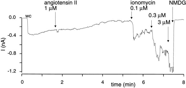
Whole-cell currents were observed as soon as the whole-cell configuration was obtained, as if the currents were constitutively active. A steady decline of the current was observed in the course of the experiments (Fig. 3 A). This decline was not prevented when the pipette solution contained either InsP3 (10 μM, n = 4), thapsigargin (3 μM, n = 3), or both (n = 2) (data not shown). In cells additionally expressing the angiotensin II receptor, angiotensin II (1 μM) induced increases in the current (see Fig. 3 A, n = 4 out of 6 cells, mean increase 36 ± 20 pA), but these increases were always transient. No such current increases were observed in controls (expressing angiotensin II receptor and GFP, n = 6, Fig. 3 B). The calcium ionophore ionomycin induced strong current increases (n = 8) in a concentration-dependent manner (Fig. 4). Specifically, ionomycin (0.1 μM) enhanced currents by 120 ± 45 pA (n = 7) and at a higher concentration (1 μM) by 580 ± 166 pA (n = 8). In controls, ionomycin (1 μM) did not enhance NMDG-blockable currents by more than 10 pA in any cell (n = 8).
Figure 3.
Stimulation of TRPC3 currents by angiotensin II. Shown is the tracing of the whole-cell current in a cell expressing TRPC3, GFP, and the angiotensin II receptor (A) and in a control cell (expressing GFP and the angiotensin II receptor, B). The whole-cell configuration was obtained at the time indicated (wc). Angiotensin II (1 μM) was applied at the time point indicated by the arrow. At the end of the experiment, the bath was exchanged (arrow) to a solution containing NMDG instead of Na+ and Ca2+. The holding potential was −70 mV.
Figure 4.
Stimulation of TRPC3 currents by ionomycin in the presence of extracellular Ca2+. To a cell expressing TRPC3 and the angiotensin II receptor, angiotensin II and ionomycin at increasing concentrations were cumulatively added. See also the legend to Fig. 3.
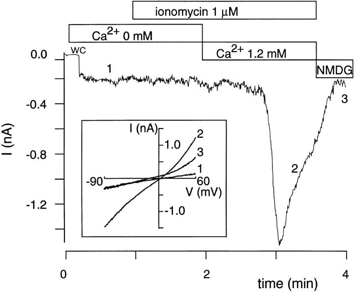
Since ionomycin might exert its effect by Ca2+ influx as well as by release of Ca2+ from intracellular stores and store depletion, we also tested ionomycin in the absence of extracellular Ca2+ (n = 4). No stimulation of the current was observed. Next, we performed experiments (Fig. 5) in which ionomycin was applied first in the absence of Ca2+ in the bath, followed by the combined application of ionomycin (1 μM) and Ca2+ (1.2 mM). Again, ionomycin had no effect in the absence of Ca2+ (n = 7), whereas large currents were evoked after readdition of extracellular Ca2+ (mean increase from baseline by 247 ± 193 pA; n = 7; no such currents occurred in controls, n = 5). Interestingly, there was a delay of 15–45 s before these currents started to develop. This delay was not completely explained by technical problems with the bath exchange. For example, the block of the currents by NMDG in Fig. 5 occurred much faster than the stimulation by Ca2+. Taken together, our findings demonstrate that ionomycin requires extracellular Ca2+ to stimulate TRPC3, consistent with the requirement for Ca2+ influx for the action of the ionophore.
Figure 5.
Requirement for extracellular Ca2+ for ionomycin effects on TRPC3 currents. A cell expressing TRPC3 was kept first in a Ca2+-free solution (5 mM EGTA) and then in a solution with 1.2 mM Ca2+, as indicated by the bars. Extracellular cations were changed to NMDG in the end. The cell was stimulated with ionomycin (1 μM) present in the bath over the indicated time. The insert shows current traces during voltage ramps from −90 to +60 mV which were obtained at the time points indicated (1, 2, and 3) in the main trace.
If Ca2+ influx activates TRPC3, a similar effect should be observed if the cells were dialyzed with a pipette solution containing an elevated Ca2+ concentration. Fig. 6 A shows such an experiment in which the pipette solution contained 10 μM Ca2+. Again, spontaneous cation currents were present right after obtaining the whole-cell configuration, but under these conditions, a considerable current increase developed over the next 20 s, followed by a much slower decline than that after stimulation with ionomycin. Similar results were obtained in three experiments. When the pipette solution contained 10 mM EGTA without CaCl2, currents were initially present but decreased rapidly (Fig. 6 B; n = 8). No Ca2+-induced currents were evoked in cells not expressing TRPC3 (n = 4).
Figure 6.
Stimulation of TRPC3 currents by intracellular Ca2+. (A) For whole-cell experiments, a pipette solution with a Ca2+ concentration of 10 μM was used. The bath was changed to an NMDG solution during the times indicated by the bars. (B) An experiment in which the pipette solution contained 10 mM EGTA but no CaCl2. The tracings appear noisier than in the other figures because they were obtained with a filtering of 5 kHz. The holding potential was −60 mV.
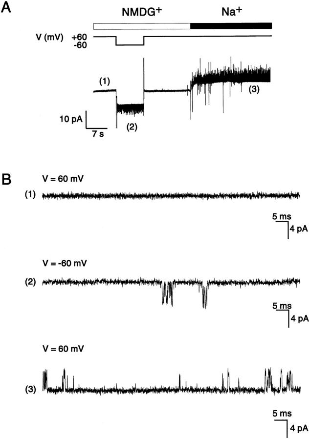
To estimate the single-channel conductance of TRPC3, we performed a simple noise analysis (Neher and Stevens, 1977) by correlating the mean current (I) within several 200-ms intervals with the current variance (s) during these intervals in experiments (n = 3) in which the initial TRPC3 current declined rapidly. From the linear regression analysis of the s/I plot, we calculated a single-channel conductance of 45, 49, and 52 pS (at −70 mV; data not shown). Channels with a comparable size were found in single-channel measurements in the cell-attached and inside-out mode on patches from cells coexpressing GFP and TRPC3 (n = 92 out of 110), but not in any patches (n = 34) from control cells (Fig. 7 A). The calculated slope conductance in inside-out patches was 66 pS (Fig. 7, B and C). The mean open time was shorter than 0.2 ms, as assessed from the dwell-time distribution of channel events recorded at 50 kHz and filtered at 5 kHz (Fig. 7 D). The channels were permeant to Na+ as well as to Cs+, but essentially not to NMDG or Cl−, as shown in Fig. 8. In this experiment, an inside-out patch was exposed first to a bath (facing the cytosolic side of the channel) containing NMDG as main cation and then to a bath with 140 mM Na-gluconate. The pipette contained the standard solution (140 mM CsCl). In NMDG, channel events were observed at a transmembrane potential of −60 mV, but not at +60 mV. After substitution of NMDG with Na+, channel events became visible at +60 mV, thus demonstrating that the currents were carried by Na+ and Cs+, and ruling out that they were carried by Cl−.
Figure 7.
Characteristics of TRPC3 in single-channel analysis of inside-out patches from cells expressing TRPC3 (and GFP). (A) Sample tracings at a membrane potential of −60 mV (corresponding to a holding potential of +60 mV). (B) Amplitude histogram of channel events at −60 mV, indicating a mean amplitude of −4.5 pA. (C) Amplitude–voltage relation of channel openings. Each point derives from amplitude histograms from 4–25 patches. The regression line yields a single-channel conductance of 66 pS. (D) Open-time distribution of TRPC3 channels. The dwell times were logarithmically binned, and the amplitudes of the bins were fitted to a monoexponential function (Sigworth and Sine, 1987). Note that the filtering (5 kHz) precludes recording of short events, such that the mean open time can be smaller than the calculated 0.13 ms.
Figure 8.
Cation selectivity of TRPC3 channels. (A) A continuous single-channel recording from an inside-out patch when the bath solution (facing the cytosolic side of the patch) initially contained NMDG as main cation (no Na+, no Ca2+) and was then changed to a solution with Na-gluconate (140 mM), as indicated by the bar. The pipette solution facing the extracellular side of the patch contained Cs+ (140 mM). The membrane potential was either +60 or −60 mV, as indicated. (B) Time-expanded recordings from the same patch, from times indicated (1, 2, and 3) in A. 1 was taken in NMDG solution at +60 mV, showing the absence of currents under these conditions. 2 was also taken in NMDG, but at −60 mV, showing inward currents carried by Cs+. 3 was taken in Na+ at +60 mV, showing outward currents carried by Na+.
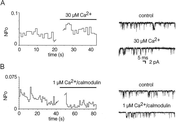
In cell-attached patches, channel activity (expressed as NPo) was constitutively present and considerably increased by ionomycin (1 μM) added to the bath solution (Fig. 9, n = 2 out of 4). As in the whole-cell measurements, the effect of ionomycin required extracellular Ca2+ because it was not observed in the absence of Ca2+ in the bath solution (n = 5, not shown). Thus, TRPC3 channels were activated in a Ca2+-dependent manner. Experiments in which the cytosolic side of inside-out patches was exposed to either 0, 1, 10, or 30 μM Ca2+, however, failed to reveal any obvious stimulation of the open probability by Ca2+ (Fig. 10 A). This situation was not changed when calmodulin (0.2 μM) was added along with Ca2+ (1 μM, Fig. 10 B). No obvious activation of channel activity was observed in 11 out of 14 patches. It should be noted that the remaining three patches showed moderate increases in activity in response to Ca2+ and calmodulin (increase in NPo by a factor of ∼3). Thus, the single-channel experiments confirmed the whole-cell data to the extent that TRPC3 is activated by Ca2+, but no evidence was obtained for a direct interaction of Ca2+ with the TRPC3 channel.
Figure 9.
Stimulation of TRPC3 channels by ionomycin in a cell-attached patch. The channel activity (expressed as NPo) of TRPC3 channels in a cell-attached patch was recorded over time. Ionomycin (0.5 μM) was added to the cell at the time indicated by the arrow. Note that the bath contained 1.8 mM Ca2+. The inserts show sample channel recordings at two different times. The holding potential was +60 mV.
Figure 10.
Absence of stimulation by Ca2+ and calmodulin of TRPC3 channels in inside-out patches. (A) Channel activity (expressed as NPo) when the Ca2+ concentration in the cytosolic solution was changed from 0 to 30 μM. (B) Channel activity in another patch when the cytosolic side was initially exposed to a solution with 0 Ca2+ and then to a solution with 1 μM Ca2+ plus 0.2 μM calmodulin. Representative tracings are shown on the right side. The membrane potential was −60 mV.
Discussion
This study was designed to functionally characterize the gene product of TRPC3 (or Htrp3), a member of the growing gene family of mammalian homologues of the trp gene from Drosophila. In particular, we tested whether TRPC3, previously shown to support receptor-mediated Ca2+ entry (Zhu et al., 1996), constitutes a Ca2+-permeable channel that enables currents similar to store-operated Ca2+ currents like ICRAC in mast cells (Hoth and Penner, 1992) and lymphocytes (Zweifach and Lewis, 1993). Indeed, expression of TRPC3 yielded whole-cell currents partly carried by Ca2+. In our hands, however, these currents were not activated by any measures known to deplete intracellular calcium stores. Therefore, TRPC3 currents should not be considered store-operated or responsible for capacitative calcium entry.
After expression of TRPC3 in CHO cells with intranuclear injection of the corresponding cDNA, currents were constitutively active, without requirement for previous depletion of calcium stores. No stimulation of TRPC3 was evoked with either InsP3 or thapsigargin, both the classical substances to induce store depletion and store-operated currents in the presence of high intracellular concentrations of the calcium chelator EGTA. Only the calcium ionophore ionomycin led to a profound increase in TRPC3 currents. Ionomycin induces Ca2+ flux through all kinds of biological membranes. Therefore, it can be used to mobilize calcium from intracellular stores, to an extent exceeding that of InsP3 (Morgan and Jacob, 1994). In the present study, however, the effects of ionomycin were strictly dependent on the presence of extracellular Ca2+ and must therefore be attributed to Ca2+ entry through the plasma membrane. Thus, we conclude that TRPC3 currents are activated by Ca2+. This conclusion is supported by our finding that dialysis of the cells with a pipette solution containing 10 μM Ca2+ led to an increase of the currents considerably above the level of spontaneous activity. On the other hand, release of Ca2+ from intracellular stores, evoked by application of ionomycin or dialysis of the cells with InsP3 or thapsigargin, was obviously not sufficient in the presence of intracellular EGTA. This might indicate the requirement for fairly high levels of Ca2+ in the vicinity of the channels, i.e., the submembraneous space. The steady decline of the currents in the course of the whole-cell experiments are consistent with the decline of [Ca2+]i by dialysis of the cell interior with the pipette solution, although other mechanisms of current inactivation probably play a role since the currents decreased also in the presence of ionomycin and elevated intracellular Ca2+ concentrations.
Our study extends that of Zhu et al. (1996), who showed that increases in [Ca2+]i induced by stimulation of membrane receptors were prolonged in cells expressing Htrp3 (TRPC3). Our measurements of [Ca2+]i revealed very similar results. We believe, however, that this kind of experiment does not unequivocally reveal the detailed mechanisms of how TRPC3 is activated. Led by the evidence from our electrophysiological measurements, we propose that any initial increase in [Ca2+]i by any mechanism will be enhanced in the presence of TRPC3 because the Ca2+-dependent activation of this Ca2+-permeable channel provides a positive feedback mechanism leading to Ca2+-induced Ca2+ entry. In general, it may be difficult to discriminate Ca2+-induced from store-operated Ca2+ entry with the fura-2 method as long as only changes in [Ca2+]i are measured. A more direct demonstration of cation entry may be required, such as quenching of fura-2 by entry of manganese, combined with probing the state of intracellular calcium stores. Using this technique, Jacob (1990) has shown that cation entry into endothelial cells takes place in the absence of an agonist when calcium stores are depleted, but not when the stores have been allowed to refill. Alternatively, cation influx may be quantified with the patch clamp technique that furthermore allows the intracellular application of InsP3 or other substances for store depletion. In this way, the store-operated regulation of ICRAC (Hoth and Penner, 1992), as well as of the human trp homologues TRPC1A (Zitt et al., 1996) and bCCE (Philipp et al., 1996), has been demonstrated. These currents develop slowly during exchange of the cytosol with the pipette solution, in sharp contrast to the TRPC3 currents.
The activation of TRPC3 by Ca2+ may represent either a direct interaction of Ca2+ with the channel or may be the result of a more complex process involving a Ca2+-dependent step. Since Ca2+-depleted cells showed no immediate but rather delayed responses to ionomycin applied along with extracellular Ca2+, and since these responses occurred only in a part of the cells, the hypothesis of a direct interaction of TRPC3 with Ca2+ already appears unlikely. To test the hypothesis more rigorously, we studied TRPC3 in inside-out patches and exposed the cytosolic side of the patches to various Ca2+ concentrations ranging from 0 to 30 μM. We did not find significant changes in channel activity. Thus, Ca2+ alone is not sufficient for a regulation of TRPC3. The fact that Ca2+-mediated activation was occasionally observed in the presence of calmodulin cannot be interpreted to indicate a calmodulin action on TRPC3, since the effect occurred inconsistently. Therefore, the exact cascade of events eventually leading to the activation of TRPC3 remains elusive; it may be speculated that not only Ca2+ but also calmodulin has a role. At any rate, the sustained channel activity in inside-out patches again argues against a role of store depletion for channel stimulation.
The expression of a Ca2+-activated, Ca2+-permeable channel may be considered hazardous for a cell because it may result in an uncontrolled Ca2+ influx leading to Ca2+ overload and cell death. Indeed, we noted that 2 d after injection of TRPC3 cDNA, the majority of cells were dead and that the surviving cells were those with little expression of TRPC3. We suggest that the spontaneous activity of TRPC3 channels leads to a lethal Ca2+ overload, enhanced by the Ca2+-triggered activation of the channels. After one day of TRPC3 expression, however, Ca2+ homeostasis appeared well compensated since basal [Ca2+]i levels were not higher than in control cells. The Na+ permeability of TRPC3 may help to keep Ca2+ influx at compensable levels since it is expected to lead to depolarization and therefore to a reduction of the driving force for Ca2+ influx. The finding that overexpression of TRPC3 is lethal is another striking difference to our experience with overexpression of TRPC1A, which resulted in fairly small currents routinely measured 2 d after microinjection. On the other hand, our results in an overexpression model do not exclude that TRPC3 may well have a role in normal cells expressing it in much lower numbers, with potent counterregulatory mechanisms available to keep net Ca2+ influx at bay. Ca2+-dependent Ca2+ entry may be used as convenient way to amplify an initial [Ca2+]i signal. Indeed, Ca2+-activated nonselective cation channels were among the first described ion channels demonstrated to carry receptor-mediated Ca2+ influx in nonexcitable cells (von Tscharner et al., 1986).
With a growing number of members of the trp family cloned and functionally characterized, it appears that its members obey very distinct regulatory principles. While some, like the parent member trp in Drosophila, can be activated by calcium store depletion (Vaca et al., 1994), it has been shown that this is not true for others. For example, trpl may be directly controlled by G proteins of the Gq/11 family (Obukhov et al., 1996) and also stimulated by Ca2+-calmodulin-dependent processes (Lan et al., 1996). Our present study demonstrates a Ca2+-dependent mechanism by which receptor stimulation can lead to the activation of a human trp homologue, with the same mechanism already demonstrated in mammalian calcium channels of unknown molecular structure (von Tscharner et al., 1986). Thus, the trp family may provide a broad spectrum of various Ca2+-permeable channels. Alternatively, proteins of the trp family may be subunits of heteromultimeric channels on which multiple regulatory pathways may act in concert, possibly in a cell-specific manner (Birnbaumer et al., 1996).
In summary, we have demonstrated that TRPC3 codes for a Ca2+-permeable nonselective cation channel. Since TRPC3 supports receptor-mediated Ca2+ influx, it may contribute to the refilling of intracellular calcium stores. However, depletion of calcium stores is not the mechanism by which activation of TRPC3 is initiated. In the opposite, TRPC3 is activated by a Ca2+-dependent process.
Acknowledgments
We thank Sabina Naranjo Kuchta and Evelyn Glaß for excellent technical assistance. This work was supported by the Bundesministerium für Forschung und Technologie, Deutsche Forschungsgemeinschaft and Fonds der Chemischen Industrie.
Abbreviations used in this paper
- GFP
green fluorescent protein
- InsP3
inositol-1,4,5-trisphosphate
- NMDG
N-methyl-d-glucamine
Footnotes
Address all correspondence to Günter Schultz, Institut für Pharmakologie, Thielallee 69-73, Freie Universität Berlin, D-14195 Berlin, Germany. Tel.: 49-30-8386360. Fax: 49-30-8315954.
References
- Bahnson TD, Pandol SJ, Dionne VE. Cyclic GMP modulates depletion-activated Ca2+entry in pancreatic acinar cells. J Biol Chem. 1993;268:10808–10812. [PubMed] [Google Scholar]
- Berridge MJ. Capacitative calcium entry. Biochem J. 1995;312:1–11. doi: 10.1042/bj3120001. [DOI] [PMC free article] [PubMed] [Google Scholar]
- Birnbaumer L, Zhu X, Jiang M, Boulay G, Peyton M, Vannier B, Brown D, Platano D, Sadeghi H, Stefani E, Birnbaumer M. On the molecular basis and regulation of cellular capacitative calcium entry: roles for Trp proteins. Proc Natl Acad Sci USA. 1996;93:15195–15202. doi: 10.1073/pnas.93.26.15195. [DOI] [PMC free article] [PubMed] [Google Scholar]
- Clapham DE. Intracellular calcium. Replenishing the stores. Nature (Lond) 1995;375:634–635. doi: 10.1038/375634a0. [DOI] [PubMed] [Google Scholar]
- Cubitt AB, Heim R, Adams SR, Boyd AE, Gross LA, Tsien RY. Understanding, improving and using green fluorescent proteins. Trends Biochem Sci. 1995;20:448–455. doi: 10.1016/s0968-0004(00)89099-4. [DOI] [PubMed] [Google Scholar]
- Dippel E, Kalkbrenner F, Wittig B, Schultz G. A heterotrimeric G protein complex couples the muscarinic m1 receptor to phospholipase C-β. Proc Natl Acad Sci USA. 1996;93:1391–1396. doi: 10.1073/pnas.93.4.1391. [DOI] [PMC free article] [PubMed] [Google Scholar]
- Hamill OP, Marty A, Neher E, Sakmann B, Sigworth FJ. Improved patch-clamp techniques for high-resolution current recording from cells and cell-free membrane patches. Pflügers Arch. 1981;391:85–100. doi: 10.1007/BF00656997. [DOI] [PubMed] [Google Scholar]
- Hoth M, Penner R. Depletion of intracellular calcium stores activates a calcium current in mast cells. Nature (Lond) 1992;355:353–356. doi: 10.1038/355353a0. [DOI] [PubMed] [Google Scholar]
- Jacob R. Agonist-stimulated divalent cation entry into single cultured human umbilical vein endothelial cells. J Physiol (Lond) 1990;421:55–77. doi: 10.1113/jphysiol.1990.sp017933. [DOI] [PMC free article] [PubMed] [Google Scholar]
- Kuno M, Gardner P. Ion channels activated by inositol 1,4,5-trisphosphate in plasma membrane of human T-lymphocytes. Nature (Lond) 1987;326:301–304. doi: 10.1038/326301a0. [DOI] [PubMed] [Google Scholar]
- Lan L, Bawden MJ, Auld AM, Barritt GJ. Expression of Drosophila trpl cRNA in Xenopus laevis oocytes leads to the appearance of a Ca2+ channel activated by Ca2+and calmodulin, and by guanosine 5′[γ-thio]triphosphate. Biochem J. 1996;316:793–803. doi: 10.1042/bj3160793. [DOI] [PMC free article] [PubMed] [Google Scholar]
- Lückhoff A, Clapham DE. Inositol-1,3,4,5-tetrakisphosphate activates an endothelial Ca2+-permeable channel. Nature (Lond) 1992;355:356–358. doi: 10.1038/355356a0. [DOI] [PubMed] [Google Scholar]
- Lückhoff A, Clapham DE. Ca2+channels activated by depletion of internal calcium stores in A431 cells. Biophys J. 1994;67:177–182. doi: 10.1016/S0006-3495(94)80467-9. [DOI] [PMC free article] [PubMed] [Google Scholar]
- Montell C, Rubin GM. Molecular characterization of the Drosophila trplocus: a putative integral membrane protein required for phototransduction. Neuron. 1989;2:1313–1323. doi: 10.1016/0896-6273(89)90069-x. [DOI] [PubMed] [Google Scholar]
- Morgan AJ, Jacob R. Ionomycin enhances Ca2+influx by stimulating store-regulated cation entry and not by a direct action at the plasma membrane. Biochem J. 1994;300:665–672. doi: 10.1042/bj3000665. [DOI] [PMC free article] [PubMed] [Google Scholar]
- Mozhayeva GN, Naumov AP, Kuryshev YA. Variety of Ca2+-permeable channels in human carcinoma A431 cells. J Membr Biol. 1991;124:113–126. doi: 10.1007/BF01870456. [DOI] [PubMed] [Google Scholar]
- Neher E, Stevens CF. Conductance fluctuations and ionic pores in membranes. Annu Rev Biophys Bioeng. 1977;6:345–381. doi: 10.1146/annurev.bb.06.060177.002021. [DOI] [PubMed] [Google Scholar]
- Obukhov AG, Harteneck C, Zobel A, Harhammer R, Kalkbrenner F, Leopoldt D, Lückhoff A, Nürnberg B, Schultz G. Activation of the Drosophila cation channel trpl by α-subunits of the Gq-protein subfamily. EMBO (Eur Mol Biol Organ) J. 1996;15:5833–5838. [PMC free article] [PubMed] [Google Scholar]
- Parekh AB, Terlau H, Stühmer W. Depletion of InsP3 stores activates a Ca2+ and K+current by means of a phosphatase and a diffusible messenger. Nature (Lond) 1993;364:814–818. doi: 10.1038/364814a0. [DOI] [PubMed] [Google Scholar]
- Petersen CCH, Berridge MJ, Borgese MF, Bennett DL. Putative capacitative calcium entry channels: expression of Drosophila trpand evidence for the existence of vertebrate homologues. Biochem J. 1995;311:41–44. doi: 10.1042/bj3110041. [DOI] [PMC free article] [PubMed] [Google Scholar]
- Philipp S, Cavalie A, Freichel M, Wissenbach U, Zimmer S, Trost C, Marquart A, Murakami M, Flockerzi V. A mammalian capacitative calcium entry channel homologous to DrosophilaTRP and TRPL. EMBO (Eur Mol Biol Organ) J. 1996;15:6166–6171. [PMC free article] [PubMed] [Google Scholar]
- Putney JW. Capacitative calcium entry revisited. Cell Calcium. 1990;11:611–624. doi: 10.1016/0143-4160(90)90016-n. [DOI] [PubMed] [Google Scholar]
- Restrepo D, Miyamoto T, Bryant BP, Teeter JH. Odor stimuli trigger influx of calcium into olfactory neurons of the channel catfish. Science (Wash DC) 1990;249:1166–1168. doi: 10.1126/science.2168580. [DOI] [PubMed] [Google Scholar]
- Schubert R. Multiple ligand-ion solutions: a guide for solution preparation and computer program understanding. J Vasc Res. 1996;33:86–98. doi: 10.1159/000159136. [DOI] [PubMed] [Google Scholar]
- Sigworth FJ, Sine SM. Data transformations for improved display and fitting of single-channel dwell time histograms. Biophys J. 1987;52:1047–1054. doi: 10.1016/S0006-3495(87)83298-8. [DOI] [PMC free article] [PubMed] [Google Scholar]
- Vaca L, Sinkins WG, Hu Y, Kunze DL, Schilling WP. Activation of recombinant trp by thapsigargin in Sf9insect cells. Am J Physiol (Cell Physiol 36) 1994;267:C1501–C1505. doi: 10.1152/ajpcell.1994.267.5.C1501. [DOI] [PubMed] [Google Scholar]
- von Tscharner V, Prod'hom B, Baggiolini M, Reuter H. Ion channels in human neutrophils activated by a rise in free cytosolic calcium concentration. Nature (Lond) 1986;324:369–372. doi: 10.1038/324369a0. [DOI] [PubMed] [Google Scholar]
- Wes PD, Chevesich J, Jeromin A, Rosenberg C, Stetten G, Montell C. TRPC1, a human homolog of a Drosophilastore-operated channel. Proc Natl Acad Sci USA. 1995;92:9652–9656. doi: 10.1073/pnas.92.21.9652. [DOI] [PMC free article] [PubMed] [Google Scholar]
- Zhu X, Chu PB, Peyton M, Birnbaumer L. Molecular cloning of a widely expressed human homologue for the Drosophila trpgene. FEBS Lett. 1995;373:193–198. doi: 10.1016/0014-5793(95)01038-g. [DOI] [PubMed] [Google Scholar]
- Zhu X, Meisheng J, Peyton M, Boulay G, Hurst R, Stefani E, Birnbaumer L. trp, a novel mammalian gene family essential for agonist-activated capacitative Ca2+entry. Cell. 1996;85:661–671. doi: 10.1016/s0092-8674(00)81233-7. [DOI] [PubMed] [Google Scholar]
- Zitt C, Zobel A, Obukhov AG, Harteneck C, Kalkbrenner F, Lückhoff A, Schultz G. Cloning and functional expression of a human Ca2+-permeable cation channel activated by calcium store depletion. Neuron. 1996;16:1189–1196. doi: 10.1016/s0896-6273(00)80145-2. [DOI] [PubMed] [Google Scholar]
- Zweifach A, Lewis RS. The mitogen-regulated calcium current of T lymphocytes is activated by depletion of intracellular calcium stores. Proc Natl Acad Sci USA. 1993;90:6295–6299. doi: 10.1073/pnas.90.13.6295. [DOI] [PMC free article] [PubMed] [Google Scholar]


