Abstract
Glycosylation has been implicated in the regulation of CD44-mediated cell binding of hyaluronan (HA). However, neither the relative contribution of N- and O-linked glycans nor the oligosaccharide structures that alter CD44 affinity for HA have been elucidated. To determine the effect of selective alteration of CD44 oligosaccharide composition on the affinity of CD44 for HA, we developed a novel strategy based on the use of affinity capillary electrophoresis (ACE). Soluble recombinant CD44–immunoglobulin fusion proteins were overproduced in the mutant CHO cell line ldl-D, which has reversible defects in both N- and O-linked oligosaccharide synthesis. Using this cell line, a panel of recombinant glycosidases, and metabolic glycosidase inhibitors, CD44 glycoforms with defined oligosaccharide structures were generated and tested for HA affinity by ACE. Because ldl-D cells express endogenous cell surface CD44, the effect of any given glycosylation change on the ability of cell surface and soluble CD44 to bind HA could be compared. Four distinct oligosaccharide structures were found to effect CD44-mediated HA binding: (a) the terminal α2,3-linked sialic acid on N-linked oligosaccharides inhibited binding; (b) the first N-linked N-acetylglucosamine residue enhanced binding; (c) O-linked glycans on N-deglycosylated CD44 enhanced binding; and (d) N-acetylgalactosamine incorporation into non–N-linked glycans augmented HA binding by cell surface CD44. The first three structures induced up to a 30-fold alteration in the intrinsic CD44 affinity for HA (K d = 5 to >150 μM). The fourth augmented CD44-mediated cellular HA avidity without changing the intrinsic HA affinity of soluble CD44.
Cd44 is currently thought to be the principal cell surface receptor for hyaluronan (HA)1 (Aruffo et al., 1990; Miyake et al., 1990), a ubiquitous glycosaminoglycan (GAG) component of extracellular and pericellular matrices (for review see Laurent and Fraser, 1992). Interaction between cell surface CD44 and HA is proposed to mediate, at least in part, a variety of cellular functions, including cell migration (Thomas et al., 1992), cell– matrix adhesion (Carter and Wayner, 1988; Aruffo et al., 1990), cell–cell adhesion (St. John et al., 1990), lymphocyte activation (Huet et al., 1989; Denning et al., 1990; Galandrini et al., 1994; Lesley et al., 1994), and hematopoiesis (Kincade, 1994; Legras et al., 1997). CD44 is highly polymorphic due to alternative splicing of at least 10 exons encoding a segment of the membrane proximal extracellular domain (Screaton et al., 1992, 1993; Tolg et al., 1993) and variable N- and O-linked glycosylation (Brown et al., 1991; Jackson et al., 1995). Multiple distinct CD44 isoforms are generated by incorporation of different combinations of the variable exons, which provide new oligosaccharide attachment sites resulting in potentially functionally significant glycosylation changes (Brown et al., 1991; Bennett et al., 1995a ,b; Jackson et al., 1995). The standard and most broadly expressed isoform, CD44H, contains none of the variable exons (Stamenkovic et al., 1989). However, CD44H can be modified by different oligosaccharide structures depending on the type and activation state of the cell in which it is expressed (Telen et al., 1986; Jalkanen et al., 1988; Camp et al., 1991; Jalkanen and Jalkanen, 1992; Hathcock et al., 1993; Levesque and Haynes, 1996; Takahashi et al., 1996). The specific glycosyltransferases which regulate CD44 glycosylation and the resulting oligosaccharide structures have yet to be determined.
Our working hypothesis is that the structural heterogeneity of CD44 isoforms is responsible not only for determining the ligand repertoire of CD44, which includes fibronectin (Jalkanen and Jalkanen, 1992), chondroitin sulfate (Naujokas et al., 1993), osteopontin (Weber et al., 1996), and at least two heparin-binding growth factors (Bennett et al., 1995a ,b), but also for modulating the HA-binding ability. Functional diversity among CD44 molecules unrelated to variant exon usage is demonstrated by observations that CD44H or any particular splice-variant can be active for HA binding when expressed in some cell types but inactive in others (Stamenkovic et al., 1991; He et al., 1992; Bartolazzi et al., 1995). In addition, differences in the ability of a single CD44 isoform to bind HA are apparent in different activation states of a single cell type. This is most clearly illustrated in lymphocytes, which bind HA poorly in the resting state despite expression of CD44H. Upon activation by cytokines (Murakami et al., 1990; Hathcock et al., 1993; Katoh et al., 1995), CD3 cross-linking (Galandrini et al., 1994), lipopolysaccharide (Murakami et al., 1990; Katoh et al., 1995), phorbol esters (Lesley et al., 1990; Hyman et al., 1991; Galandrini et al., 1994), or an in vivo allogeneic immune reaction (Murakami et al., 1991; Lesley et al., 1994), certain lymphocytes acquire a CD44-mediated HA-binding property. The observed change in ability to bind HA requires hours to days and is associated with a change in glycosylation (Hathcock et al., 1993), suggesting that CD44 glycosylation may provide one mechanism for regulating HA affinity. Differences in CD44 glycosylation may explain both the cell type and cell activation state dependence of CD44 function.
Differences in HA binding between CD44H and variant CD44 isoforms expressed in a single cell type have been attributed to glycosylation. Inclusion of variant exons 8–10, for example, results in additional O-linked glycosylation, which reduces HA binding when CD44v8–10 is expressed in COS cells and in a human melanoma (Bennett et al., 1995a ). Several recent studies have provided evidence that glycosylation also plays a role in regulating CD44H-mediated cell attachment to surface-bound and soluble HA (Brown et al., 1991; Hathcock et al., 1993; Katoh et al., 1995; Lesley et al., 1995; Bartolazzi et al., 1996). In addition to these cell-based studies, two studies showed that glycosylation can alter the ability of the purified CD44H protein to bind HA, indicating that the oligosaccharide structures on CD44 affect the molecule's intrinsic affinity for HA (Lokeshwar et al., 1991; Katoh et al., 1995). Biosynthetic studies showed that the HA-binding function was acquired with N-glycan structures, and in the presence of tunicamycin, with the O-glycan structures, suggesting a positive role for both N- and O-linked oligosaccharides. However, other studies found no effect or an inhibitory role for O-linked glycosylation (Bennett et al., 1995a ; Lesley et al., 1995). Furthermore, metabolic or enzymatic N-deglycosylation was found to either augment (Katoh et al., 1995; Lesley et al., 1995) or abrogate (Sleeman et al., 1996; Bartolazzi et al., 1996) HA binding. Mutagenesis of single or multiple N-glycan attachment sites abrogated HA binding (Bartolazzi et al., 1996), while an inhibitory role for complex-type N-glycans was suggested by the positive effects of the metabolic oligosaccharide processing inhibitor deoxymannojirimycin (dMM) observed in some, but not all, cell lines (Lesley et al., 1995; Bartolazzi et al., 1996). Enzymatic hydrolysis of sialic acid was found to either augment or have no effect on HA binding, depending at least in part on the cell type in which CD44 was expressed (Katoh et al., 1995; Lesley et al., 1995). Other than sialic acid, the specific saccharide residues and structural motifs responsible for these complex effects of glycosylation have not been characterized. It is also not known whether the inhibitory effect of sialic acid on the ability of CD44 to bind HA occurs when the sialic acid is on N- or O-linked oligosaccharides, nor is it known what impact the underlying oligosaccharide structure might have.
The seemingly contradictory results obtained by the different groups may be due to the different cell types and different experimental approaches used. In addition, interpretation of some of these results is complicated by the limitations of the approaches used. Reagents that completely inhibit N-linked glycosylation of all cell surface and secreted glycoproteins potentially alter the fate of a wide variety of cellular proteins that may directly or indirectly affect CD44 function. Mutagenesis of potential N-linked glycosylation sites, on the other hand, may alter protein folding, stability, or conformation, which may be critical for appropriate HA binding. Enzymatic or metabolic deglycosylation of purified glycoprotein can result in protein aggregation that may artifactually alter functional assays. Furthermore, most of the functional assays used have been qualitative in nature and were not designed to quantitatively measure modest differences in ligand affinity, which may more closely approximate a physiologically relevant range. Finally, the relative contribution of O- and N-linked glycosylation remains unresolved, and the identification of glycoconjugates that alter the ability of CD44 to interact with HA is lacking.
To overcome the limitations of conventional approaches, we have adapted affinity capillary electrophoresis (ACE) to quantitatively assess the effects of glycosylation changes on CD44 affinity for HA in free solution, and therefore in the absence of cell surface constraints. Capillary electrophoresis (CE) is a molecular analytic method that provides superior speed, sensitivity, and resolution compared with conventional electrophoretic and chromatographic methods. In addition, capillary electrophoresis provides a rapid and convenient means to assess the affinity of any protein for its ligand(s) in free solution under near physiological conditions. In several model systems, ACE has recently been shown to provide a method of choice to quantitatively determine protein–ligand dissociation constants in protein–drug, enzyme–inhibitor, and antibody–antigen interactions (Chu et al., 1992, 1994; Kraak et al., 1992; Gomez et al., 1994; Mammen et al., 1995; Gao et al., 1996). In this work, we describe the first application of ACE to analyze adhesion receptor–ligand interactions. Comparison of results from our novel ACE method to more traditional cell-based flow cytometry and adhesion assays allowed us to attribute each effect of glycosylation to either (a) an intramolecular mechanism, in which a change in CD44 glycosylation alters the intrinsic affinity of the CD44 extracellular domain, or (b) an intermolecular mechanism, in which a glycosylation-dependent change in CD44-mediated HA binding requires molecular interactions in a cellular context and therefore is only apparent in cell-based assays. We have identified four glycoconjugates that have distinct effects on CD44-mediated HA binding, and which together can provide an explanation for some of the apparently contradictory observations reported in previous studies.
Materials and Methods
Flow Cytometry
After nearing or reaching confluence (HAM's F-12, 10% FBS), cell monolayers were cultured in serum-free media (HAM's F-12) containing either no additions or supplemented with 20 μM galactose (Gal) alone, 200 μM N-acetylgalactosamine (GalNAc) alone, both Gal and GalNAc, and/or 2 μg/ml Swainsonine or the combination of 90 μg/ml deoxymannojirimycin (dMM; Toronto Research Chemicals Inc., North York, Ontario, Canada) and 5 μg/ml kifunensine (KIF; Toronto Research Chemicals Inc.) for 4 d with one culture medium replacement on day 2. EDTA- detached cells (6 × 10⁵), preincubated with 2 ml of hybridoma supernatant from KM-81 rat anti–mouse CD44 mAb (American Type Culture Collection, Rockville, MD) or from an isotype-matched unrelated antibody for 30 min at 4°C, were stained with fluorescein-conjugated high–molecular weight HA (Anika, Woburn, MA) (2 μg/ml) or FITC-conjugated goat anti–rat IgG (Cappel, Malvern, PA) in 500 μl wash buffer (DME, 0.15% polyvinylpyrrolidone, 0.02% sodium azide, supplemented with 1 M sodium Hepes, pH 7.0, to 15 mM final concentration) for 1 h in the dark at 4°C, washed twice with ice-cold wash buffer, and resuspended in PBS for FACS® analysis. Single cells were gated from doublets, debris, and damaged cells by light scatter.
Cell Adhesion
96-well microplates (flat well MaxiSorp; Nunc, Roskilde, Denmark) were coated with 100 μl/well 1 mg/ml high–molecular weight HA (human umbilical cord, Sigma, St. Louis, MO) or 1% BSA in PBS overnight at 4°C. After a 30-min wash with 1% BSA in PBS, plates were blocked with 1% denatured BSA in PBS for 30 min at room temperature. Ldl-D (American Type Culture Collection; Krieger et al., 1989) or CHO cells cultured in serum-free HAM's F-12 with or without 20 μM Gal and/or 200 μM GalNAc were EDTA-detached, preincubated with KM81 anti-CD44 hybridoma supernatant or an isotype-matched unrelated antibody, and allowed to adhere to the HA- or BSA-coated wells (105 cells in 100 μl serum-free media/well) for 30 min at 37°C. Nonadherent cells were removed by three washes with 1% BSA in PBS under agitation. Adherent cells were quantitated using CellTiter 96 (Promega Corp., Madison, WI) by measuring the enzymatic conversion of a tetrazolium dye during a 4-h incubation at 37°C. After overnight solubilization, the amount of dye formation was determined by absorbance at 595 nm on a microplate reader. The percent of adherent cells is calculated by dividing absorbance of the washed wells by that of identical unwashed wells. The mean of four wells was used for each reported value.
Production of CD44 Receptorglobulins Glycan Variants
A cDNA construct encoding a soluble fusion protein composed of the extracellular domain of CD44H fused to human IgGFc (CD44 receptorglobulin [CD44Rg]; Aruffo et al., 1990) was stably transfected into CHO and ldl-D cells. Confluent monolayers of the transfected cells were switched to culture medium lacking serum and supplemented with no sugar, 20 μM Gal, 200 μM GalNAc, or both Gal and GalNAc with or without 90 μg/ml dMM/5 μg/ml KIF or 2 μg/ml Swainsonine. After 2 d, the culture medium was discarded to allow depletion of cellular stores of Gal and/or GalNAc. Cell monolayers were then cultured for an additional 10 d with one medium replacement (or 5 d for dMM/KIF-containing cultures). CD44Rg from filtered culture supernatants, adjusted to pH 8.0 with NaOH, was bound to protein A–Sepharose, rinsed with H2O, eluted with 20 mM H3PO4, neutralized with 100 mM NaOH, quantitated by UV absorbance at 280 nm (BSA standard), aliquoted, lyophilized, and stored at −70°C. Aliquots were resuspended in H2O immediately before glycosidase digestion and/ or capillary electrophoresis analysis.
Affinity Capillary Electrophoresis
Affinity capillary electrophoresis (ACE) was performed on a capillary electropherograph (model 3850; Isco, Lincoln, NE) using an uncoated silica capillary (75 μM i.d., 64 cm length, 40.5-cm inlet to detector window, ISCO), a 5-s vacuum injection (10 nl), 15 kV (95 μA), and 210-nm UV absorbance detection in 50 mM NaPO4, pH 7.4. Buffer chambers and capillary containing hyaluronan 60-mers (HA60, Anika, Woburn, MA) were equilibrated at electrophoresis conditions for 13 minutes after each change in HA60 concentration. Samples (10 μl) contained 1–2 μM CD44Rg and 480 μg/ml mesityl oxide (MO). Mobility (μ) of a given molecule is defined as its anodal migration relative to MO, the neutral marker, and is determined from migration times by μ = l d(l c/V)(1/t eo − 1/t), where l d is the inlet to detector window length, l c is the capillary length, V is applied voltage, t eo is the migration time for MO, t is the migration time for the molecule of interest, and the units for μ are V−1min−1cm2. Migration times for HA60 are determined from the inverted peak resulting from the lack of HA60 in the sample plug. The mobility of CD44Rg in the presence of ligand is reported as a relative mobility, where a relative mobility of 0.0 is the mobility of CD44Rg in the absence of ligand, and a relative mobility of 1.0 is given as the mobility of the free ligand (HA60). Therefore, the relative mobility of CD44Rg at a given concentration of HA60 is given by relative mobility = (μ pl − μ p)/ (μ l − μ p), where μ pl is the mobility of CD44Rg in the presence of HA60, μ p is the mobility of CD44Rg in the absence of HA60, and μl is the mobility of HA60. μ pl and μ l are determined from the migration times of MO, CD44Rg, and HA60 for each ACE run containing HA60. μ p is determined from the migration times of MO and CD44Rg from replicate CE runs performed in the absence of HA60 and is used to determine the relative mobility for each ACE run performed in the presence of HA60 on the same day.
Radiolabeling and Immunoprecipitation
[35S]methionine labeling of CHO or ldl-D cells was performed and cell lysates were prepared as previously described (Camp et al., 1991). Cell lysates, precleared for 1 h at 4°C with goat anti–rat IgG coupled protein A–Sepharose CL4B (Zymed Labs, So. San Francisco, CA) in the presence of nonimmune rat IgG, were immunoprecipitated using goat anti–rat IgG coupled protein A–Sepharose CL4B in the presence of KM-81 rat anti– mouse CD44 IgG2a monoclonal antibody. Immunoprecipitates were subjected to SDS–10% PAGE, and the gels were dried and analyzed after exposure for autoradiography.
Glycosidase Digestion
Glycosidases were obtained from Glyko (Novato, CA) except endoglycosidase F (Endo F; N-glycosidase F–free, Boehringer Mannheim Corp., Indianapolis, IN). 2 μg CD44Rg adjusted to recommended pH with H3PO4 or NaOH was incubated with 4.8 μg MO and with or without (mock) 0.3–2 μl of each glycosidase in a final volume of 10 μl at 37°C until reaction completion as assessed by CD44Rg migration shift and peak width on CE analysis. Upon completion of the glycosidase reactions, samples were analyzed for HA affinity by ACE in the presence of HA60. Aggregation of CD44Rg after deglycosylation was assessed by changes in CE mobility; all ACE experiments were performed using monomer CD44Rg. Enzyme-only control CE runs lacking CD44Rg were performed to assure no interference of the CD44Rg peak by glycosidase protein.
Results
Selective Rescue of N- and O-linked Glycosylation in a Cell Line with a Reversible Glycosylation Defect Has Distinct Effects on CD44-mediated HA Binding
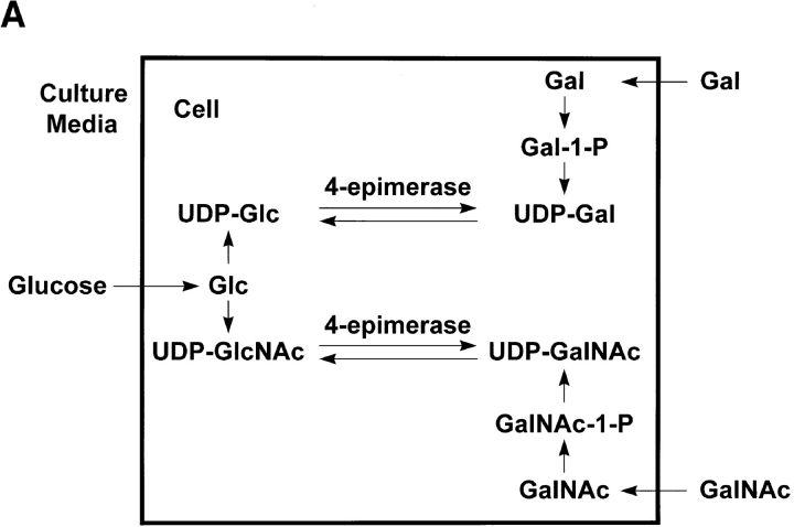
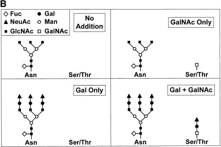
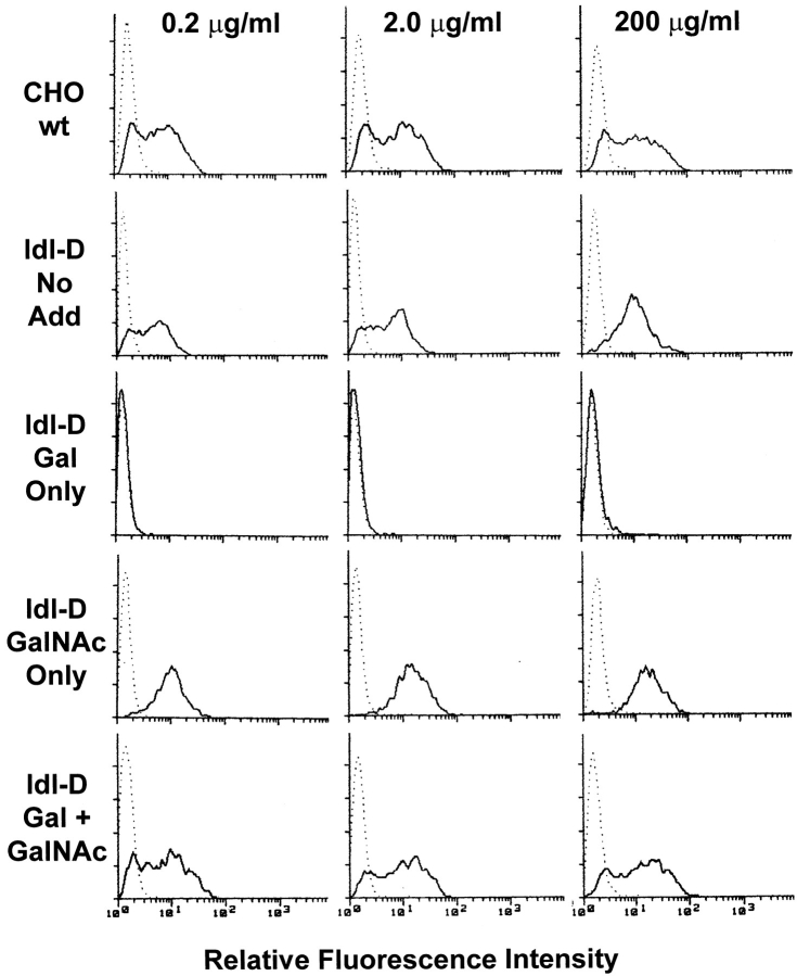
Wild-type (wt) CHO fibroblasts, which constitutively express CD44H, display heterogeneous soluble fluorescein (FITC)-labeled HA binding. The ability of the anti-CD44 antibody KM81 to block the binding of soluble HA to intact cells, as assessed by FACS® analysis, indicates that HA binding to CHO cells and their ldl-D derivative (see below) is CD44 mediated. Assessment of the effects of glycosylation changes on the affinity for HA of CD44-bearing cells was facilitated by the use of a mutant CHO clone, ldl-D, which lacks 4-epimerase activity (Fig. 1 A). Ldl-D cells are unable to use glucose from the culture media to generate UDP-Gal and UDP-GalNAc, the high-energy sugar donors for all synthetic Gal and GalNAc transfer reactions. Therefore, they generate glycoproteins with truncated or absent oligosaccharide side chains (Krieger et al., 1989). Reversal of the inability to synthesize N- and O-linked glycans can be achieved by supplementing the ldl-D cell culture media with the appropriate monosaccharide, i.e., Gal and GalNAc, respectively (Fig. 1 B). Selective glycosylation rescue of the mutant ldl-D cells resulted in marked differences in their ability to bind soluble HA (Fig. 2), while Gal and/or GalNAc addition to serum-free cultures of wild-type CHO cells had no effect on their binding of FITC- labeled HA (data not shown). Restoration of the ability to synthesize GalNAc-containing glycan structures resulted in increased HA binding, while selective incorporation of Gal-inhibited binding. These two effects appeared to be independent of each other. Supplementation of the cell culture medium with both Gal and GalNAc, which fully reverses the deficiency in oligosaccharide synthesis, resulted in ldl-D cell binding of HA comparable to that of wild-type CHO cells. These findings indicate that there are at least two glycosylation-dependent effects on CD44-mediated HA binding by whole cells: GalNAc-dependent enhancement and Gal-dependent inhibition.
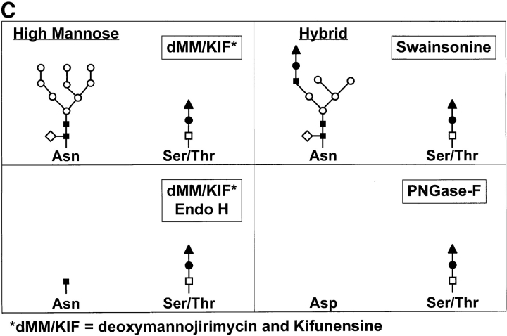
Figure 1.
Generation of CD44 glycovariants. (A) Ldl-D cells lack 4-epimerase. The reversible N- and O-linked glycosylation defect in ldl-D cells is due to the lack of a functional 4-epimerase. Culture medium supplementation with Gal and/or GalNAc reverse the N- and O-linked glycosylation defects, respectively. (B) Ldl-D oligosaccharide structures. GalNAc is required to initiate O-linked glycosylation, which is absent without GalNAc supplementation. Gal is required to complete the terminal modifications of complex-type N-linked oligosaccharides and many O-linked oligosaccharides. Truncated N- and O-linked oligosaccharide structures are generated in the absence of Gal supplementation. (C) Glycans after mannosidase inhibition and glycosidase treatment. High mannose–type N-linked oligosaccharides are generated in the presence of dMM and KIF, metabolic inhibitors of α mannosidase I. Hybrid type N-linked oligosaccharides are generated in the presence of swainsonine, an α mannosidase II inhibitor. The oligosaccharide structures remaining on CD44Rg after Endo H digestion of high mannose–type N-linked oligosaccharides or PNGase-F digestion are shown.
Figure 2.
Effect of glycosylation on the binding of soluble HA to CHO or ldl-D cells. HA binding to ldl-D cells was assessed by FACS® analysis performed using the indicated concentrations of FITC-HA, and CHO or ldl-D cells that were cultured for 4 d with the indicated saccharide additions. HA binding was performed after preincubation of cells with blocking anti-CD44 antibody KM-81 (dotted line) or an isotype-matched unrelated antibody (solid line). The same results were obtained from four other experiments.
Since glycosylation-rescued ldl-D cells display a similar level of HA-binding heterogeneity as the parental CHO cells, it appears unlikely that the HA-binding heterogeneity is the result of genetic diversity among the parental CHO population. Moreover, altered glycosylation can reduce this heterogeneity, as shown by the uniform high affinity binding of HA in the presence of GalNAc supplementation alone (Fig. 2). Loss of HA-binding heterogeneity is not due to the absence of Gal per se, since HA-FITC–binding heterogeneity is observed in the absence of sugar supplementation at low concentrations of HA-FITC.
High levels of CD44 cell surface protein expression by CHO and ldl-D cells were observed by FACS® analysis under each of the glycosylation conditions. Conditions allowing GalNAc incorporation resulted in slightly higher surface CD44 expression than conditions lacking GalNAc incorporation. The mean fluorescence intensities for CD44 expression were: CHO wt, 151; ldl-D Gal/GalNAc, 209; ldl-D GalNAc only, 164; ldl-D Gal only, 125; and ldl-D no addition, 109. These moderate variations in surface expression do not account for the marked differences in CD44-mediated binding of soluble HA.
Galactose-dependent Inhibition of HA Binding Is Related to the Number of Complex-Type N-linked Oligosaccharide Termini While GalNAc-dependent Enhancement of Binding Is Attributed to Non–N-linked Glycans
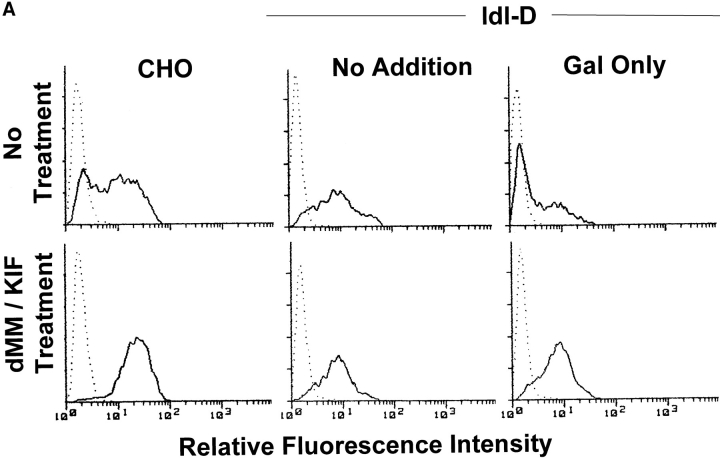
The galactose-dependent inhibitory and the GalNAc-dependent enhancing effects on cellular avidity for HA could be due to N- or O-linked glycosylation of the CD44 glycoprotein or glycoconjugates of other cellular proteins that indirectly effect CD44 function. To examine the role of the N-linked oligosaccharides, we treated CHO cells with three metabolic mannosidase inhibitors, dMM, KIF, and swainsonine, to block N-linked oligosaccharide processing, thus preventing complex-type oligosaccharide synthesis (Kaushal and Elbein, 1994) (Fig. 1 C). We found that treatment with a combination of dMM and KIF, both α-mannosidase I inhibitors, converted all cell surface CD44 N-linked glycans to high-mannose structures, while treatment with dMM alone only partially blocked N-linked glycoconjugate processing to complex-type oligosaccharides. Treatment with swainsonine, an α-mannosidase II inhibitor, resulted in cell surface expression of CD44 molecules containing hybrid type N-linked oligosaccharides in which only one branch terminates in sialic acidα2-3Galβ1-4GlcNAcβ1- (Fig. 1 C). Cells grown in the presence of dMM and KIF displayed increased HA binding compared with those with intact complex-type N-glycans (CHO, ldl-D Gal only, and ldl-D Gal/GalNAc; Fig. 3 A and data not shown). In contrast, dMM/KIF treatment had no effect on HA binding by cells lacking galactose-containing oligosaccharides (ldl-D no addition, ldl-D GalNAc only; Fig. 3 A and data not shown). In other words, cells expressing CD44 containing truncated N-linked glycans bound soluble HA as well as cells expressing CD44 containing high-mannose structures. The observation that ldl-D cells grown in the presence of Gal and dMM/KIF or in the absence of Gal display comparable avidity for HA indicates that the inhibitory effect of galactose can be attributed to its incorporation into N-linked structures. By contrast, the inability of dMM/KIF treatment to modify the observed GalNAc-dependent gain of function suggests that this effect may primarily be attributed to non–N-linked glycans, possibly O-linked structures.
Figure 3.
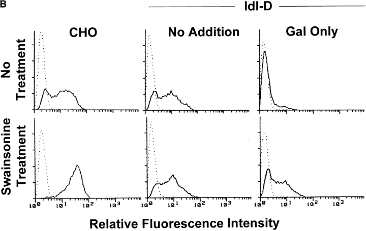
Effect of mannosidase inhibitors on the binding of soluble HA to CHO or ldl-D cells. HA binding to ldl-D cells was assessed by FACS® analysis performed using 2 μg/ml FITC-HA, and CHO or ldl-D cells that were cultured for 4 d with the indicated metabolic inhibitor and/or saccharide additions. HA binding was performed after preincubation of cells with blocking anti-CD44 antibody KM-81 (dotted line) or an isotype-matched unrelated antibody (solid line). A and B were from separate experiments. The results from A were repeated in two other experiments and from B in one other experiment with similar results.
Similar to dMM/KIF-treated cells containing high-mannose oligosaccharides, hybrid-type oligosaccharide-bearing cells, as a result of swainsonine treatment, displayed enhanced HA binding compared to cells bearing intact complex-type structures (CHO, ldl-D Gal only, ldl-D Gal/ GalNAc; Fig. 3 B and data not shown). However, unlike the effect of dMM/KIF treatment, cells treated with swainsonine had a slightly poorer avidity for HA when grown in the presence than when grown in the absence of Gal (ldl-D Gal only < ldl-D no addition and ldl-D Gal/GalNAc < ldl-D GalNAc only; Fig. 3 B and data not shown). These observations indicate that hybrid structures containing a single branch terminating with NeuAc-Gal provide a partial inhibitory effect, weaker than that of intact complex-type oligosaccharides containing multiple NeuAc-Gal terminating branches. As expected, swainsonine had no significant effect on HA binding by cells grown under conditions that resulted in expression of truncated complex-type oligosaccharides (ldl-D no addition, ldl-D GalNAc only; Fig. 3 B and data not shown). The level of CD44 surface expression as measured by FACS® analysis was modestly increased in the presence of GalNAc but unaltered by dMM/KIF or swainsonine treatment under all glycosylation conditions (data not shown).
Selective N- and O-linked Glycosylation Promotes Differential Adhesion of ldl-D Cells to a Hyaluronan-coated Surface
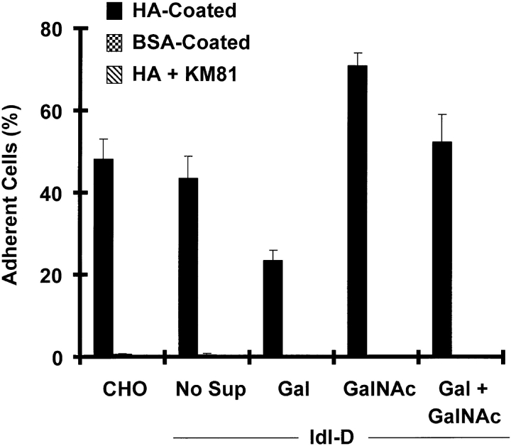
To determine whether these same glycosylation-dependent effects apply to CD44-mediated cell attachment to HA, we tested the ability of ldl-D cells to adhere to HA-coated plastic after selective or complete glycosylation reconstitution (Fig. 4). Similar to binding of soluble HA, we observed GalNAc-dependent augmentation and Gal-dependent reduction of ldl-D cell attachment to surface-bound HA. Cell adhesion to substrate coated with HA could be completely blocked by the anti-CD44 antibody KM-81 but not by unrelated antibodies, confirming that it was CD44 mediated. Treatment of the cells with dMM/ KIF resulted in increased adhesion under conditions of galactose incorporation (CHO, ldl-D Gal/GalNAc, and ldl-D Gal only) but had no significant effect on adhesion under conditions lacking galactose incorporation (ldl-D no addition, ldl-D GalNAc only, data not shown). These observations are consistent with inhibition of CD44-mediated cell attachment to HA by galactose-containing termini of complex-type N-linked oligosaccharides and enhancement of adhesion by GalNAc-containing non–N-linked glycans and are similar to those on the effects of the same glycosylation changes on soluble HA binding. It must be noted, however, that cell attachment and soluble HA-binding assays may preferentially measure different aspects of CD44 function. Nonetheless, our results suggest that glycosylation regulates these CD44 functions by similar mechanisms.
Figure 4.
Effect of glycosylation on CD44-mediated ldl-D cell adhesion to HA. CHO or ldl-D cells grown with the indicated saccharide supplementation for 4 d were seeded onto BSA- (BSA-Coated) or HA-coated plates after preincubation with blocking anti-CD44 monoclonal antibody, KM-81 (HA + KM81) or an isotype-matched unrelated antibody (HA-Coated). The percentage of adherent cells is reported as the mean of quadruplicate wells with the error bars representing the standard deviation. The same results were obtained in two other independent experiments.
Differences in Glycosylation Do Not Cause Redistribution of Cell Surface CD44
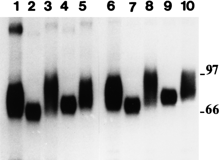
The glycosylation-dependent alterations of the ability of cell surface CD44 to bind HA could be due to changes in glycosylation of CD44 itself, resulting in an altered intrinsic HA affinity of the CD44 glycoprotein or altered CD44 distribution, oligomerization, or interaction with accessory molecules. To address the possibility of glycosylation-dependent redistribution of the CD44 molecules on the cell surface, cell surface CD44 expression of ldl-D monolayers cultured in the presence or absence of Gal and/or GalNAc was assessed by immunofluorescence using the anti-CD44 mAb KM-81. Diffuse CD44 cell surface staining was observed under all four saccharide supplementation conditions showing that the HA-avidity changes were not due to gross CD44 clustering or capping (data not shown). SDS-PAGE of endogenous cell surface CD44H immunoprecipitated from ldl-D cell lysates displayed glycosylation-dependent migration shifts, confirming the alteration of CD44 oligosaccharide structures as a result of selective or combined saccharide supplementation (Fig. 5). In addition to monosaccharides, serum can serve as an exogenous source of Gal and GalNAc via cellular uptake and degradation of serum glycoproteins (Krieger et al., 1989). Under nonreducing conditions, a small amount of CD44 multimer formation could be observed. However, the amount of CD44 multimerization apparent on nonreducing SDS-PAGE was unaltered by glycosylation changes in ldl-D cells (Fig. 5), rendering unlikely the possibility that changes in CD44H oligomerization provide a mechanism for the observed alterations in HA binding.
Figure 5.
Effect of glycosylation on SDS-PAGE mobility of endogenous CD44H from ldl-D cells. Immunoprecipitates of CD44 from [35S]methionine-labeled ldl-D cells cultured for 4 d with: 10% FBS (lanes 1 and 6), no supplementation (lanes 2 and 7), Gal only (lanes 3 and 8), GalNAc only (lanes 4 and 9), or Gal + GalNAc (lanes 5 and 10) were analyzed by SDS-PAGE under nonreducing (lanes 1–5) and reducing (lanes 6–10) conditions. Molecular mass (kD) is indicated.
Development of a Novel Strategy to Analyze Soluble CD44 Binding to HA
Ldl-D and wild-type CHO cells were stably transfected with a cDNA encoding a soluble fusion protein, termed CD44 receptorglobulin (CD44Rg), composed of the extracellular domain of CD44 and the Fc portion of human IgG1 (CD44Rg; Aruffo et al., 1990; Thomas et al., 1992). N- and O-linked glycan variants of CD44Rg were purified from supernatants of ldl-D and wt CHO cells and their affinity for HA assessed by ACE. ACE relates changes in electrophoretic mobility of a protein upon complexation with ligand present in the buffer to the affinity of the receptor–ligand pair. The dissociation constant can be calculated from the magnitude of the change in protein mobility as a function of ligand concentration. Purified HA 60-mers were used as the CD44 ligand to avoid potential problems associated with size heterogeneity of high–molecular weight HA polymer. Binding of high–molecular weight and 60-mer HA to CD44 on the cell surface was observed to be comparable, as assessed by flow cytometry (data not shown).
Because the dissociation rate for CD44Rg and HA60 was rapid compared with electrophoresis duration (data not shown), we included HA60 in the capillary and electrophoresis buffer and CD44Rg in the sample only. Binding of the negatively charged HA60 to CD44Rg in free solution imparts a greater negative charge-to-mass ratio on CD44Rg, thus delaying its migration, which is driven by electroosmosis and resisted by electrophoretic attraction to the anode inlet. During electrophoresis, the interaction between CD44Rg and HA60 is in rapid equilibrium. Thus, the shift in migration time of CD44Rg induced by the presence of ligand is related to the degree to which it is complexed with HA60. Specifically, during CE the molecules are in free solution and are resolved on the basis of their charge to mass ratio, where electrophoretic mobility is proportional to
 |
1 |
where μ is electrophoretic mobility, Z is the ionic charge, M is the mass, and C is a proportionality constant specific to a given group of molecules (Rickard et al., 1991; Chu et al., 1992). We found considerable variation in electroosmotic flow from one experiment to another, which can be corrected for by including an internal neutral marker, MO, in the sample. Mobilities determined relative to MO yield reproducible results. The coefficient of variation (CV) for electroosmotic flow was found to be 8.72%, while the CV for CD44Rg mobility was 2.16% (data not shown). The mobility of a given molecule is experimentally determined from its anodal migration relative to MO by:
 |
2 |
where l d is the length of capillary to the detector window (40.5 cm), l c is the length of the capillary (64 cm), V is applied voltage (15,000 V), t eo is the migration time for MO, and t is the migration time for the molecule of interest (Gomez et al., 1994). Determining the mobility shifts of CD44Rg as a function of varying concentrations of HA60 in the buffer and capillary allows determination of the affinity of the CD44Rg glycan variants for HA. The change in CD44Rg mobility induced by ligand is reported as a relative mobility (R M), defined as the mobility of CD44Rg in the presence of ligand relative to the mobility of CD44Rg without ligand (R M = 0) and the mobility of free ligand (HA60) (R M = 1). Specifically, the mobility shift is given by
 |
3 |
where μ pl is the mobility of CD44Rg in presence of ligand, μ p is the mobility of CD44Rg in the absence of ligand, and μ l is the mobility of HA60.
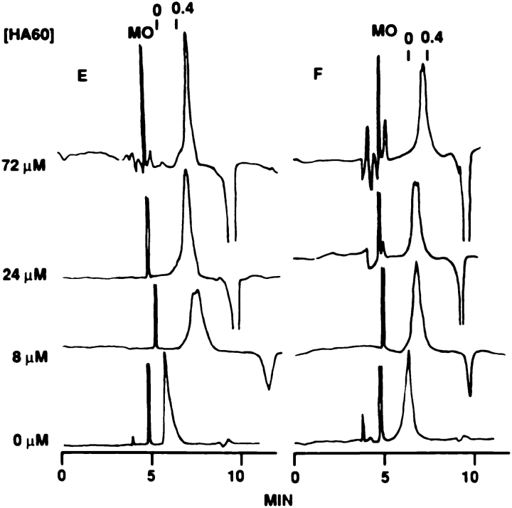
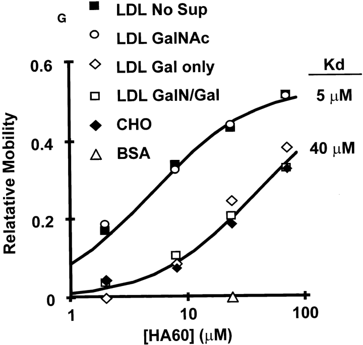
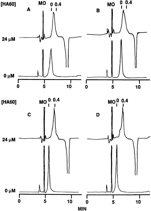
HA60 induced a concentration-dependent mobility shift of wt CHO-derived CD44Rg but failed to change the mobility of the noninteracting protein, BSA (Fig. 6, A and B). CD44Rg molecules synthesized by ldl-D cells cultured in the presence of Gal displayed the same relative mobilities as wt CHO cell–derived CD44Rg, but those synthesized in the absence of Gal displayed greater relative mobilities because of a higher affinity for HA at any given concentration of HA60 (Fig. 6, A and C–F). Dissociation constants (K d) were determined by fitting curves to the data using
 |
4 |
Figure 6.
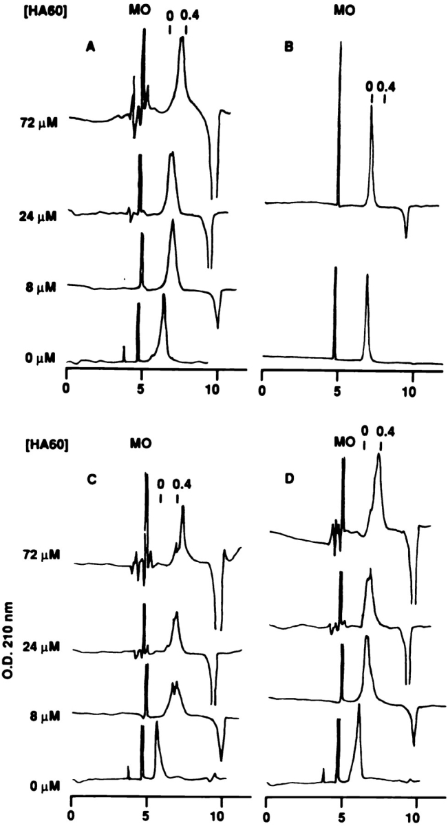
Effect of glycosylation on the intrinsic CD44Rg affinity for HA. Affinity capillary electrophoresis in the presence of the indicated concentrations of HA60 of (A) wt CHO–derived CD44Rg, (B) BSA, or (C–F) ldl-D–derived CD44Rg synthesized in the presence of (C) no saccharide addition, (D) Gal only, (E) GalNAc only, or (F) Gal + GalNAc. The narrow peak due to MO, a neutral marker included as an internal control, is labeled. The broader unlabeled peak corresponds to CD44Rg. The inverted peak at the migration time for HA60 is due to the absence of HA60 in the sample. Mobility values for CD44Rg and HA60 were calculated from migration times relative to MO. The mobility of CD44Rg in the presence of ligand is reported as a relative mobility (R M), where R M is 0.0 at the mobility of the uncomplexed CD44Rg and R M is 1.0 at the mobility of HA60. The migration times corresponding to R M of 0.0 and 0.4, indicated in each panel for the top electrophoretogram, were calculated using the mobility of CD44Rg from the CE runs at 0 μM HA60 and the migration times for MO and HA60 from the same electrophoretogram. The relative mobilities of CD44Rg from A–F are plotted in G. Displayed curves were fit to the equation R M = R M (max) * [HA60]/(K d * [HA60]) where R M is the relative mobility, R M (max) is the maximum relative mobility (mobility of CD44Rg at saturating concentrations of HA60), K d is the dissociation constant, and [HA60] is the concentration of HA 60-mer. The determined values were R M (max), 0.54 and K d, 5 and 40 μM.
where R M is the relative mobility, R M (max) is the maximum relative mobility (mobility of CD44Rg at saturating concentrations of HA60), K d is the dissociation constant, and [HA60] is the concentration of HA 60-mer. Affinities determined from Fig. 6 G revealed dissociation constants (K d) of 40 and 5 μM for HA60 binding of Gal-bearing and Gal-lacking CD44Rg glycoproteins, respectively. Thus, incorporation of Gal into the oligosaccharides of CD44 results in an eightfold lower affinity of soluble CD44 for HA60 and provides a plausible explanation for the reduced binding of soluble HA by ldl-D cells cultured in Gal-supplemented medium (Fig. 2). In contrast, the intrinsic affinity for HA of the CD44 molecule is independent of GalNAc incorporation into CD44 oligosaccharides, unlike whole cells where GalNAc supplementation increases CD44-mediated binding of HA. These observations indicate that GalNAc-dependent enhancement of CD44-mediated HA binding by cells is governed by a mechanism requiring the cellular context and is not caused by structure/function changes limited to the extracellular domain of the CD44 glycoprotein itself.
N-linked Oligosaccharide Structures Can Both Enhance and Reduce CD44 Affinity for HA: Reduction of Affinity is Due to α2,3-linked Sialylation
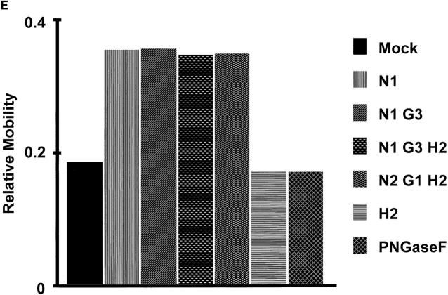
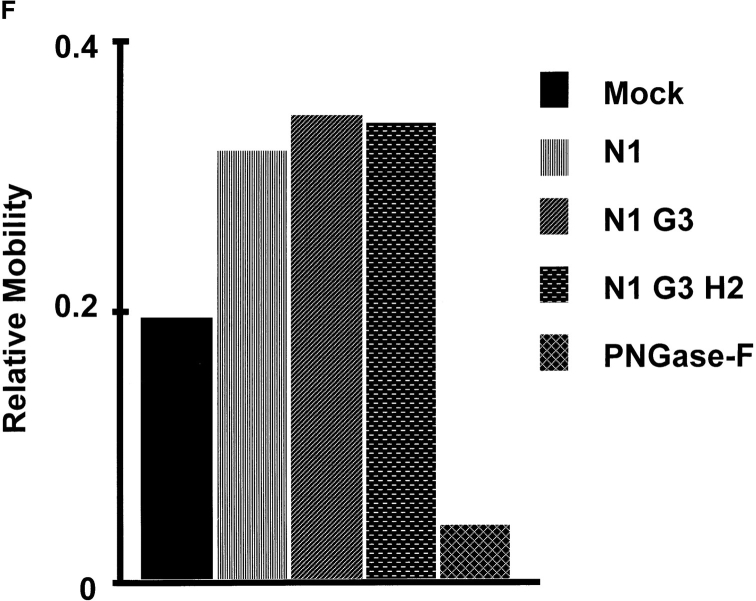
To further define the specific oligosaccharide structures involved in the regulation of CD44 affinity for HA, we analyzed CD44Rg-HA binding by ACE after glycosidase digestion of the CD44Rg glycan variants. A variety of CD44Rg glycovariants were found to give R M (max) values of about 0.54, despite having different CE mobilities in the absence of ligand (Fig. 6). We could therefore compare different CD44 glycovariants in an ACE experiment using a single concentration of HA60 and estimate the K d from the saturation equation (Eq. 4) by assuming a R M (max) of 0.54. Sequential trimming of CD44Rg N-linked oligosaccharides with specific glycosidases followed by determination of relative affinities for HA60 by ACE allowed determination of the functional contributions for each saccharide residue. Gal is required for the terminal modifications of complex N-linked and many O-linked oligosaccharides, while GalNAc is required for any O-linked structure (Fig. 1 B). Since the observed Gal-associated inhibition of HA binding by CD44Rg is GalNAc independent, Gal-dependent inhibition must be related to the terminal glycosylation of CD44 N-linked oligosaccharides. The N-linked oligosaccharides of CHO cell CD44Rg terminate with NeuAcα2,3-Galβ1,4-GlcNAcβ- (Skelton, T.P., and I. Stamenkovic, unpublished). Removal of sialic acid with an α2,3-specific neuraminidase (N1) was sufficient to convert GalNAc + Gal–supplemented ldl-D cell–derived CD44Rg from a moderate to a high-affinity binder of HA (Fig. 7), and additional glycosidase trimming of the terminal residues had no further enhancing effect. The mean dissociation constants from several such experiments are shown in Table I. For simplicity, relative mobilities corresponding to K d values of 5–19, 20–79, and 80–319 μM will hitherto be referred to as high-, intermediate-, and low-affinity binding, respectively. These observations demonstrate that the inhibitory effect of galactose incorporation is due to the ability of galactose to accept terminal sialic acid residues and not to the galactose residue per se. To confirm that terminal N-linked glycan modifications are responsible for the decreased CD44 affinity for HA, we performed the same functional analysis, after stepwise glycosidase removal of the oligosaccharide termini, on CD44Rg lacking O-linked oligosaccharides and observed comparable changes in HA binding affinity (ldl-D Gal only; Fig. 7 F). Inhibition of HA binding associated with sialylation is therefore due to N-linked but not to O-linked sialic acid.
Figure 7.
Effects of glycosidase digestion on the intrinsic CD44Rg affinity for HA. Glycosidase-digested CD44Rg glycovariants were analyzed for HA affinity by ACE at 24 μM HA60. The ACE electrophoretograms for ldl-D (Gal + GalNAc) CD44Rg treated with (A) mock, (B) PNGase-F, (C) NANase-I (N1), and (D) NANase-I + GALase-III glycosidase digestions are shown. ACE mobility shifts induced by 24 μM HA60 for CD44Rg glycovariants synthesized by ldl-D in the presence of (E) Gal + GalNAc and (F) Gal-only medium supplementation are shown. Specificities of glycosidases are: NANase-I (N1) and -II (N2), α2-3 and α2-3,6-linked sialic acid, respectively; GALase-I (G1) and -III (G3), β1-3,4,6 and β1-4 galactose, respectively; HEXase-II; (H2) βGlcNAc or βGalNAc. Relative mobilities (R M) of 0.19 (Mock), 0.36 (N1), and 0.17 (PNGase-F) from E correspond to dissociation constants (K d) of 44, 12, and 52 μM, respectively. Similar results were obtained from two other experiments.
Table I.
Micromolar Dissociation Constants (Kd) for HA of each CD44Rg Glycovariant
| Cell line | Media addition | N-Glycans | O-Glycans | K d | ± SD | Enzyme treatment | N-Glycans | O-Glycans | K d | |||||||||
|---|---|---|---|---|---|---|---|---|---|---|---|---|---|---|---|---|---|---|
| μM | μM | |||||||||||||||||
| CHO | Intact | Intact | 51 | ± 19 | PNG-F | Absent | Intact | 76 | ||||||||||
| ldl-D | Gal/GalNAc | Intact | Intact | 45 | ± 4 | PNG-F | Absent | Intact | 62 | |||||||||
| ldl-D | Gal | Intact | Absent | 47 | ± 13 | PNG-F | Absent | Absent | >150 | |||||||||
| ldl-D | No Addition | Truncated | Absent | 8 | ± 1 | PNG-F | Absent | Absent | >150 | |||||||||
| ldl-D | GalNAc | Truncated | Truncated | 8 | ± 2 | PNG-F | Absent | Truncated | 112 | |||||||||
| CHO | dMM/KIF | High | Intact | 12 | Endo H | GlcNAc | Intact | 6 | ||||||||||
| mannose | ||||||||||||||||||
| ldl-D | dMM/KIF | High | Absent | 10 | Endo H | GlcNAc | Absent | 5 | ||||||||||
| mannose | ||||||||||||||||||
| CHO | Swainsonine | Hybrid | Intact | 22 | ||||||||||||||
| CHO | NANase | Asialo- | Present | 12 | ||||||||||||||
| CHO | NANase/ | Asialo- | Present | 12 | ||||||||||||||
| GALase | Agalacto- | |||||||||||||||||
| CHO | NANase/ | Trimmed | Present | 9 | ||||||||||||||
| GALase/ | ||||||||||||||||||
| HEXase | ||||||||||||||||||
| ldl-D | Gal/GalNAc | NANase | Asialo- | Present | 13 | |||||||||||||
| ldl-D | Gal/GalNAc | NANase/ | Trimmed | Present | 13 | |||||||||||||
| GALase/ | ||||||||||||||||||
| HEXase | ||||||||||||||||||
| ldl-D | Gal | NANase | Asialo- | Absent | 18 | |||||||||||||
| ldl-D | Gal | NANase | Asialo- | Absent | 14 | |||||||||||||
| GALase | Agalacto- | |||||||||||||||||
| ldl-D | Gal | NANase/ | Trimmed | Absent | 14 | |||||||||||||
| GALase/ | ||||||||||||||||||
| HEXase |
Summary table of the micromolar dissociation constant (K d) for each CD44Rg glycovariant obtained from the mean of two to seven ACE experiments. The standard deviation of the mean K d is shown for values obtained from four or more experiments. PNG-F, peptide N-glycanase F; NANase, α-neuraminidase; GALase, β-galactosidase; HEXase, β-N-acetyl-hexosaminidase.
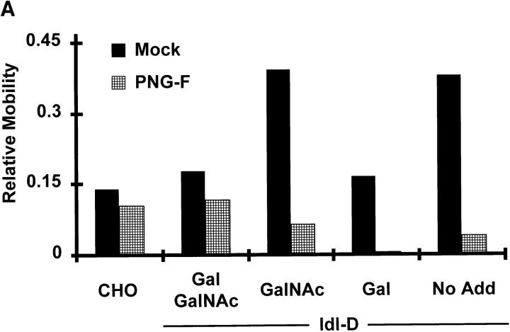
To determine whether the remaining core N-linked structure has an effect on HA affinity, the entire N-linked oligosaccharide was enzymatically removed by peptide N-glycosidase F (PNGase-F) before ACE analysis (Fig. 1 C). PNGase-F treatment of Gal/GalNAc-reconstituted ldl- D–derived CD44Rg resulted in moderate HA affinity, similar to or slightly lower than that of the untreated molecule (Fig. 7 E, Table I). Thus, in addition to removing the inhibitory effect of the terminal sialic acid, a positive effect of the N-glycan was also abrogated. Successful enzymatic hydrolysis was confirmed by a small shift in CD44Rg CE mobility (in the absence of ligand), as well as a marked loss in the neuraminidase-inducible CE mobility change. Since the results in Figs. 6 and 7 indicated only a negative effect of the N-linked termini, this enhancing effect on HA binding appeared to be due to some aspect of the N-glycan core. A more marked loss of affinity was observed after N-deglycosylation of a CD44Rg glycan variant having only N-linked and no O-linked oligosaccharides (derived from ldl-D cells supplemented with Gal alone) (Fig. 7 F, Table I). This finding suggests that O-glycosylation can partially compensate for the lack of N-glycosylation, providing a third glycosylation effect on the intrinsic HA affinity of CD44. To further address these N-glycan–dependent effects, CD44Rg synthesized by wt CHO cells and ldl-D cells under each of the glycosylation conditions were analyzed by ACE after PNGase-F or mock digestion (Fig. 8 A, Table I). The enhancing effect of N-glycans on CD44Rg affinity for HA was apparent in that the HA-binding ability of each CD44Rg glycovariant was reduced after N-glycan removal. The decrease in affinity was more pronounced for glycovariants lacking terminal sialic acid (ldl-D GalNAc and ldl-D no addition) because the enhancing effect of N-glycosylation was not partially offset by the inhibitory effect of sialic acid. In addition, the fully deglycosylated CD44Rg (PNGase-F-treated ldl-D Gal and ldl-D no addition) displayed poorer HA affinity than CD44Rg, which retained truncated (ldl-D GalNAc) or intact (ldl-D Gal/GalNAc and wt CHO) O-linked glycan structures, confirming that the reduction in affinity after N-deglycosylation was moderated by the presence of O-linked oligosaccharides. Thus, while the O-glycans appear to have no effect on affinity in the presence of N-linked glycans (Figs. 6; 7, E and F; and 8 A; Table I), the presence of O-glycosylation enhances affinity when N-glycosylation is absent (Figs. 7 and 8 A, Table I). Taken together, these experiments reveal an enhancing effect on HA binding of some aspect of the CD44 N-glycan core structure, loss of which can be partially compensated for by O-glycans, and an inhibitory effect of the terminal sialic acid of N-linked oligosaccharides.
Figure 8.
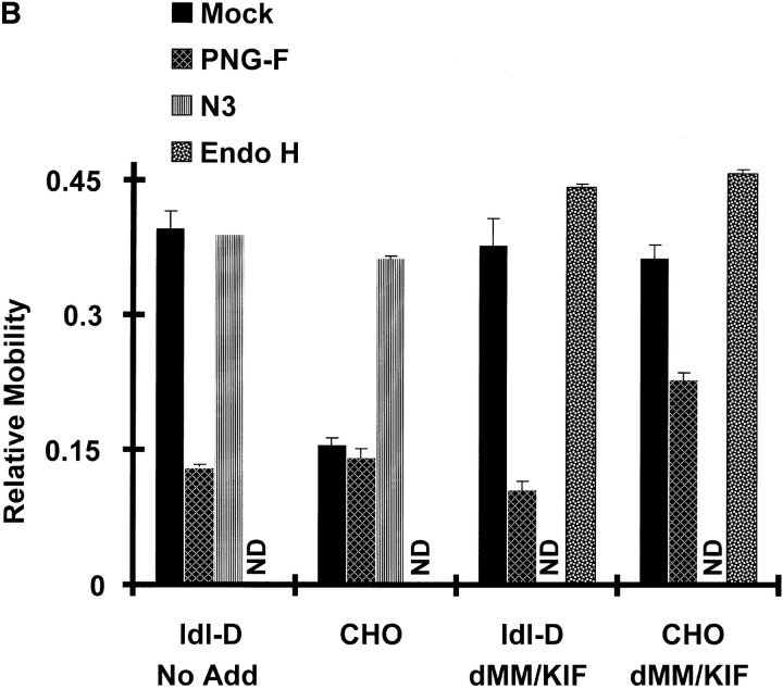
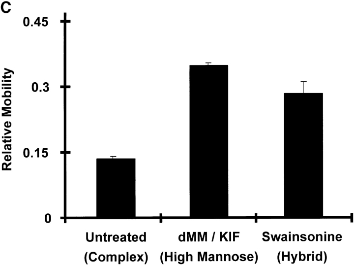
Characterization of three distinct effects of CD44 glycosylation on its intrinsic affinity for HA by ACE analysis of CD44Rg glycovariants. CD44Rg with defined oligosaccharide modifications were analyzed for HA affinity by ACE at 24 μM HA60. Error bars are 1 SD. (A) CD44Rg glycan variants were synthesized by ldl-D cells cultured in the presence of the indicated saccharide additions were treated with (PNGase-F) or without (Mock) enzymatic removal of N-linked oligosaccharides. (B) CD44Rg glycan variants generated in CHO cells bearing intact O-linked and complex-type (CHO) or high-mannose N-linked oligosaccharides (CHO dMM/KIF), or in ldl-D cells, grown without saccharide supplementation, lacking O-linked and bearing truncated complex-type (ldl-D No Add) or high-mannose N-linked oligosaccharides (ldl-D dMM/KIF) were treated with the indicated glycosidases before affinity analysis. Structures expected for peptide N-glycanase-F (PNGase-F) and Endo H digestions are shown in Fig. 1 C. Specificity for the neuraminidase (N3) is α2-3,6,8 sialic acid. (C) CD44Rg glycan variants were generated in CHO cells bearing intact O-linked and complex-type (Untreated), high-mannose (dMM/KIF), or hybrid-type (Swainsonine) N-linked oligosaccharides. The same results were obtained in at least one other experiment for each panel.
A Single N-linked GlcNAc Residue Is Sufficient to Promote CD44–HA Binding
To determine the minimal CD44 N-linked oligosaccharide structure required to promote high-affinity binding of HA, we used a combination of metabolic inhibition of N-linked glycan synthesis by dMM/KIF, which blocks N-linked glycosylation processing at a high-mannose structure (Kaushal and Elbein, 1994; Fig. 1 C), and hydrolysis by the endoglycosidase Endo H, which cleaves high-mannose oligosaccharides, leaving a single GlcNAc attached to the asparagine residue (Tai et al., 1975; Fig. 1 C). Endo H cleavage of high-mannose oligosaccharides from CD44Rg derived from wild-type CHO cells or ldl-D cells (without monosaccharide supplementation) cultured in the presence of dMM/KIF generated CD44Rg glycan variants that displayed high affinity for HA (Fig. 8 B, Table I). In comparison, PNGase-F treatment of CHO cell–derived and ldl-D cell–derived CD44Rg resulted in intermediate and low HA affinity, respectively (Fig. 8 B, Table I). Successful modification of the oligosaccharide structures by synthetic blockade or glycosidase digestion was confirmed by changes in CE mobility of the CD44Rg glycan variants (data not shown). These observations indicate that a single N-linked GlcNAc residue is sufficient to provide the enhancing effect on CD44 affinity for HA attributed to core N-linked oligosaccharide structures (Figs. 7 and 8 A).
Hybrid Structures Provide an Intermediate Level of Inhibition of Intrinsic CD44 Affinity for HA Compared with High-mannose and Complex-Type N-linked Oligosaccharides
In experiments that addressed the ability of whole cells to bind soluble HA, cells bearing hybrid oligosaccharide structures were found to display HA avidity that was intermediate compared with the high avidity of high mannose–bearing cells and the low avidity of cells synthesizing complex-type oligosaccharides (Fig. 3). To determine if these effects are caused by changes in the intrinsic ability of CD44 to bind HA, we examined the HA affinity of CD44Rg glycovariants bearing hybrid and high mannose–only N-linked structures by ACE. Similar to whole cells, CD44Rg containing hybrid structures bearing a single sialylated oligosaccharide branch displayed intermediate HA affinity (Fig. 8 C) compared with the high affinity of counterparts containing only high mannose and the lower affinity of CD44Rg containing complex-type oligosaccharides with multiple sialylated branches (Fig. 1 C).
Discussion
To identify CD44-associated oligosaccharides that are functionally relevant to CD44–HA interaction, we have taken advantage of a combination of selective synthetic blockade provided by ldl-D cells or metabolic mannosidase inhibitors and recombinant glycosidase digestion to generate glycoforms of CD44 with precisely defined changes in oligosaccharide structure for affinity analysis by ACE. This approach has helped identify four distinct oligosaccharide structure– and linkage-dependent effects of glycosylation on the ability of cell surface and soluble CD44 to bind HA. They are (a) a reduction in binding ability by α2,3sialylation of N-linked oligosaccharides, (b) an increase in binding ability by the asparagine-linked GlcNAc of the invariant N-linked glycan core structure, (c) an increase in binding ability by O-linked glycosylation of N-deglycosylated CD44; and (d) a GalNAc-dependent increase in HA binding by cell surface CD44. The first three effects were demonstrated using soluble CD44, indicating alterations in the affinity for HA of the CD44 glycoprotein itself, in the absence of cellular constraints, with a K d ranging from 5 to >150 μM. The fourth effect augmented CD44-mediated cell binding of HA without changing the affinity for HA of the soluble CD44 glycoprotein itself.
Three Types of Glycosylation Changes That Affect CD44 Affinity for HA Are Uncovered by ACE
A summary of the K d values obtained from several ACE experiments on each CD44Rg glycovariant is shown in Table I. The differences in the intrinsic affinity for HA among the purified CD44Rg glycovariants can be explained by three glycosylation-dependent effects. First, the higher affinity of CD44Rg glycovariants having truncated, high-mannose, residual single GlcNAc, or enzymatically desialylated N-glycans compared with CD44Rg molecules having intact N-glycans is explained by the absence of inhibitory terminal sialic acid residues on N-linked oligosaccharides. By extension, a K d of 22 μM for the CD44Rg glycovariant bearing hybrid-type N-glycans is consistent with partial sialic acid–mediated inhibition and suggests that the degree of inhibition is directly related to the number of oligosaccharide branches terminating in sialic acid. Second, the higher HA affinity of CD44Rg glycoproteins with an N-glycan present (intact, truncated, high-mannose, GlcNAc, exoglycosidase-treated, or hybrid) compared with CD44Rg molecules lacking an N-glycan (PNGase-F treated) is explained by the enhancing effect of the asparagine-linked GlcNAc. Finally, the higher HA affinity of N-deglycosylated CD44Rg glycovariants containing O-glycans (intact or truncated) compared with completely deglycosylated CD44Rg proteins (O-glycans absent) is attributed to an enhancing effect of the O-glycans on N-deglycosylated CD44.
Calculation of the standard deviation for the K d values obtained from several independent ACE experiments using CD44Rg derived from CHO or ldl-D cells under each of the tested glycosylation conditions illustrates the degree of variability in CD44Rg-HA affinity measured by this assay (Table I). Significantly higher interexperimental variation in receptor–ligand affinity was associated with CD44Rg produced in CHO cell or ldl-D cells/Gal–only supplementation than with the other CD44Rg glycovariants. The observed variation was found to be caused by differences in the degree of sialylation of CD44Rg derived from one tissue culture batch to the next and was demonstrated by batch-to-batch differences in neuraminidase-inducible changes in CD44Rg CE mobility (data not shown). After neuraminidase-mediated removal of sialic acid, CD44Rg molecules derived from different production batches were found to display the same CE mobility. Differences in CHO- and ldl-D/Gal cell–derived CD44Rg sialylation may be explained by variable release of soluble neuraminidase from cells cultured under suboptimal nutrient conditions (Warner et al., 1993; Ferrari et al., 1994). Neuraminidase released into the culture medium after CHO cell lysis can desialylate recombinant soluble glycoproteins (Warner et al., 1993). A similar mechanism may underlie the observed variability in CD44Rg sialylation and is supported by the much tighter K d values associated with nonsialylated CD44Rg glycovariants (ldl-D no addition and ldl-D GalNAc). Interestingly, the degree of variation in HA affinity of CD44Rg derived from ldl-D cells supplemented by Gal and GalNAc was not as high as that of CD44Rg synthesized under the other two sialylation-competent synthetic conditions (CHO and ldl-D Gal only). A possible explanation for this difference is that the ldl-D Gal/GalNAc cells (as well as ldl-D no addition and ldl-D GalNAc cells) were consistently found to be more effectively growth arrested by serum starvation than wt CHO cells or ldl-D Gal–only cells. It therefore appears plausible that during the long serum-free culture periods required for generation of adequate quantities of CD44Rg, wt CHO and ldl-D Gal–only cells may grow beyond the ability of the media to support viability, resulting in significant cell lysis and release of a soluble neuraminidase into the culture media.
The K d values obtained (Table I) allowed us to estimate the magnitude of each of the glycosylation-dependent effects on CD44Rg affinity for HA. Thus, we observed a four- to eightfold inhibition of HA binding attributable to N-linked sialic acid. However, considering the possible partial loss of CD44Rg-associated sialic acid in the cell culture medium in some of these experiments, the eightfold decrease in HA affinity displayed by the more highly sialylated CD44Rg molecules is more likely to reflect the actual degree of sialic acid–mediated inhibition of HA binding by endogenous CD44 in CHO cells. Asparagine-linked GlcNAc provides a greater than 30-fold enhancement of HA affinity demonstrated by the K d values of fully deglycosylated CD44Rg (>150 μM; ldl-D no addition PNGase-F) and CD44Rg containing N-linked GlcNAc as its only glycan (5 μM; ldl-D no addition dMM/KIF Endo H). The presence of intact O-glycans on N-deglycosylated CD44Rg provides about a 2.5-fold affinity enhancement (K d of 62 μM versus >150 μM).
Essential to the determination of the effect of different glycans on CD44 function was the use of affinity capillary electrophoresis. To our knowledge, this study represents the first described application of ACE to the analysis of a physiological cell surface receptor–ligand interaction. ACE revealed that the dissociation rate of HA from CD44Rg in free solution (<5 s in ACE experiments, data not shown) was much higher than the dissociation rate of soluble HA from the intact cell (>30 min in FACS® experiments). The greater HA avidity in the intact cell is presumably due to multivalent interactions between the high–molecular weight FITC-HA and multiple cell surface CD44 molecules. This interpretation is supported by observations that glycan modifications which alter the intrinsic CD44 affinity for HA yield a corresponding change in whole cell avidity, which is a function of intrinsic receptor–ligand affinity and valency. In light of the observation that GalNAc supplementation of ldl-D cell culture medium augments cell surface but not soluble CD44–HA binding, it appears likely that glycosylation can affect cell surface CD44-mediated cell adhesion to HA by mechanisms other than intrinsic receptor–ligand affinity. Such additional cell context-dependent mechanism(s) may allow CD44 to form higher valency interactions with HA molecules or may alter CD44 affinity for HA by promoting interactions between CD44 and other receptors on the cell surface.
ACE Analysis of CD44 Glycovariant Ability to Bind HA Provides Explanations for the Seemingly Contradictory Results of Earlier Studies
The multiple effects of glycosylation on CD44 affinity characterized in this study offer a means to reconcile the apparently conflicting results of previous reports. The inhibition of affinity by the terminal sialic acid of N-linked oligosaccharides is consistent with the studies by Katoh et al. (1995), who found activation of HA binding by neuraminidase treatment of some cell lines, a subset of splenic B cells, and CD44-coated beads. In addition, this mechanism of inhibition is consistent with studies that reported an augmentation of HA binding by some cell lines treated with dMM (Lesley et al., 1995; Bartolazzi et al., 1996; Sleeman et al., 1996) or dMM/KIF (this study). The inability of neuraminidase or dMM treatment to enhance HA binding by some CD44-expressing cell lines may be explained by the absence of sialic acid on the N-linked oligosaccharides of CD44 in these cells. In one such example, an SDS-PAGE mobility shift for CD44 was observed after neuraminidase treatment (Lesley et al., 1995) but may be explained by a loss of O-linked sialic acid, which would have no effect on intrinsic HA affinity. Alternatively, CD44 expressed in non–HA-binding cell lines may be maintained in an inactive state regardless of its N-linked glycosylation pattern by additional cell context-based mechanisms, one of which may be provided by glycosaminoglycans (GAG) (Lesley et al., 1995). Our results are also consistent with the high level of CD44-mediated HA binding by the Lec 8 cell line (Katoh et al., 1995), a CHO mutant with a nonreversible defect in UDP-galactose transport, which is the functional equivalent of ldl-D cells supplemented by GalNAc alone.
The results of the present study provide an explanation for the apparent discrepancy between the biosynthetic studies of Lokeshwar and Bourguignon (1991), where an enchancing effect on HA binding by CD44 O-glycosylation was reported, and those of Lesley et al. (1995), which found that inhibiting CD44 O-glycan extension by benzyl-α-GalNAc had no effect on HA binding. The biosynthetic experiments that detected a positive role for O-glycosylation were performed in the presence of tunicamycin, an inhibitor of N-glycosylation. As a result, the nonglycosylated ER CD44 precursor did not bind HA but acquired HA binding ability upon O-glycosylation in the Golgi (Lokeshwar and Bourguignon, 1991). In the absence of tunicamycin, the N-glycosylated ER precursor was able to bind HA. In contrast, the lack of an effect of metabolic inhibition of CD44 O-glycosylation (confirmed by SDS-PAGE mobility change) in a number of cell lines reported by Lesley et al. (1995) was observed under conditions where CD44 was N-glycosylated. Taken together, the observations of these two studies are consistent with the enhancing effect of any N-glycan core and the enhancing effect of O-glycans on CD44 molecules lacking N-glycans described in the present study. These results can therefore be explained by glycosylation-dependent changes in the intrinsic affinity of the CD44 molecule for ligand without invoking the involvement of other molecules or cell type–specific differences. Reports of inhibition of HA binding by O-glycosylation of CD44 itself appear to be limited to examples of variable exon usage that provides new attachment sites for inhibitory O-glycans (Bennett et al., 1995). The report that CD44-mediated HA binding could be augmented by para-nitrophenol-xyloside, an inhibitor of GAG synthesis, suggests an inhibitory role for these glycoconjugates (Lesley et al., 1995). However, GAGs were not found on the CD44 molecule in that study, suggesting that the observed inhibitory effect may be due to GAGs associated with other cell surface structures. Taken together, these reports are consistent with our finding that O-glycosylation of the standard form of CD44, CD44H, has no effect on the intrinsic affinity for HA in the (presumably) physiological situation of N-glycan–modified CD44.
Our finding that a single N-linked GlcNAc is sufficient to induce a 30-fold enhancement in CD44 affinity for HA offers a potential explanation for the apparent discrepancy between a loss of CD44–HA binding (Bartolazzi et al., 1996) and a 10-fold increase in CD44–HA binding (Katoh et al., 1995) after enzymatic N-deglycosylation. In the first study, PNGase-F was used as the deglycosylating enzyme, while the second study was performed using a combination of PNGase-F and Endo-F, an enzyme that cleaves between the first and second GlcNAc, leaving a single N-linked GlcNAc. Studies using tunicamycin, on the other hand, are more difficult to interpret, in part because of the broad effects of blocking N-glycosylation of all cellular glycoproteins. Activation or inhibition of CD44–HA binding can be obtained depending on the duration of tunicamycin treatment (Sleeman et al., 1996), and even after prolonged treatment, a significant fraction of CD44 molecules remain N-glycosylated (Katoh et al., 1995; Sleeman et al., 1996). In the extended serum-free culture conditions used in the present study, tunicamycin was toxic to the cells.
Potential Physiological and Nonphysiological Glycosylation-dependent Regulation of the Ability of CD44 to Bind HA
Clearly, it is important to distinguish between functional alterations associated with glycosylation changes that may be physiologically relevant and those that may arise only in an experimental system. Thus, the low HA affinity of the fully deglycosylated CD44Rg and the increased affinity contributed by the presence of O-linked glycosylation in the absence of N-linked glycans are unlikely to reflect physiological phenomena. In contrast, effects observed after more modest changes in specific saccharide residues of the terminal oligosaccharide modifications are more likely to approximate physiological regulatory mechanisms. The cell could therefore potentially regulate CD44 function by modulating specific glycosyltransferase activity that would result in synthesis of CD44 molecules with altered branching or sialylation of their N-linked oligosaccharides. However, it is also possible that the effects on CD44 function of the first GlcNAc of N-linked glycans and of O-linked GalNAc may have biological significance in a site-specific fashion. In other words, modulation of the efficiency of the N-oligosaccharide transfer to specific CD44 N-glycan attachment sites or regulation of one of the newly identified GalNAc-transferases that initiate site-specific O-linked glycosylation (Clausen, H., and E. Bennett. 1997. Glycoconj. J. 14: S8; Nehrke, K., F.K. Hagen, K.G. Ten Hagen, J. Zara, and T.A. Tabak. 1997. Glycoconj. J. 14:S8) are two possible mechanisms by which the cell could control CD44 function.
The finding that a single GlcNAc residue is sufficient to provide the enhancing effect of N-glycans on ligand binding, observed for CD44 in this study, was also previously observed for the cell adhesion molecule CD2 (Wyss et al., 1995). A single Asn-linked GlcNAc stabilizes human CD2 by counterbalancing an unfavorable cluster of five positive charges via hydrogen bonds and van der Waals contacts to the polypeptide. Complete deglycosylation results in unfolding of the CD2 protein and loss of binding to its natural ligand CD58 at a site distant from the glycosylation-charge cluster interaction. Interestingly, in contrast to human CD2, rat CD2 lacks an N-linked glycosylation site but maintains a stable protein conformation by replacing one of the cluster's positively charged lysines with a negatively charged glutamic acid. It thus seems that the structural stabilizing role of N-glycosylation of CD2 does not involve specific structures but rather is a product of the coevolution of glycosylation and amino acid changes within the physiochemical restraints for protein folding and stability. In the case of CD44, N-linked oligosaccharide structures may have evolved a specific regulatory function, while the coevolving amino acid changes may have developed the required interactions with the existing glycan core. In this way, CD44-associated N-linked glycans may have acquired a nonspecific protein stabilizing role, similar to the N-linked oligosaccharide of CD2. Our structure–function studies of the N-linked oligosaccharides on CD44 have identified candidates for both a nonspecific protein stabilizing role, shown by the positive effect of a single N-linked GlcNAc, and a specific regulatory role, shown by the negative effect of terminal sialylation.
Terminal α2,3-sialylation of complex-type oligosaccharides provides a plausible mechanism for the cellular regulation of HA binding by CD44. Since sialic acid is the terminal modification on N-linked oligosaccharides, alteration of the expression/function of the relevant sialyltransferase can provide a means for the cell to regulate CD44-mediated adhesiveness. Recent evidence indicates that N-linked oligosaccharide sialylation regulates the ability of at least three I-type lectins, CD22, CD33, and sialoadhesin, to interact with their ligands (Braesch-Andersen et al., 1994; Freeman et al., 1995). CD22, a B lymphocyte–specific receptor thought to be involved in B cell activation, loses its ability to bind ligands upon sialylation by a β-galactoside α2,6 sialyltransferase which is upregulated in activated B cells. Linkage-specific sialylation of CD22 may therefore provide a regulatory mechanism for its function (Sgroi and Stamenkovic, 1994). Our present data suggest an analogous mechanism as a candidate physiological regulator of CD44-mediated HA binding, where cell activation by various stimuli may alter the relevant α2,3-sialyltransferase activity, resulting in altered sialylation of CD44 and HA affinity.
The relatively high HA affinity of CD44Rg bearing hybrid-type oligosaccharides (Fig. 8 C) and the relatively high HA avidity of cells cultured in the presence of swainsonsine (Fig. 3 B) suggest that regulation of oligosaccharide branching offers an alternative mechanism for determining the amount of CD44 sialylation. Thus, regulation of the activity of glycosyltransferases that compete for common oligosaccharide intermediates to determine the final branching pattern of N-linked oligosaccharides may be implicated in controlling CD44 function. For example, increased activity of GlcNAc–transferase III or of an earlier acting β1-4galactosyltransferase would be expected to generate a greater number of hybrid-type oligosaccharides and produce CD44 molecules with relatively high affinity for HA. On the other hand, increased GlcNAc–transferase V activity would be expected to generate an increased number of complex-type branches and/or extended sialylated branches and produce a CD44 molecule with lower HA affinity. These possibilities are particularly interesting in view of the association of changes in GlcNAc-transferase III (Yoshimura et al., 1995) and GlcNAc–transferase V (Dennis and Laferte, 1989; Fernandes et al., 1991) activity with highly metastatic tumors and the implication of CD44 in the metastatic process.
How might the enhancing effect of GalNAc on cell surface CD44 binding of HA be explained? It is possible that the GalNAc-dependent activation of HA binding by whole cells, which is mediated by CD44 but not attributable to altered intrinsic CD44 affinity for HA, is related to a cell context–associated regulatory mechanism. Although GalNAc can occasionally be found on complex-type N-linked oligosaccharides, the observed GalNAc-associated augmentation of HA binding was unaltered by culturing the cells in the presence of dMM/KIF (Fig. 3 A and data not shown). Thus, activation of HA binding by GalNAc appears to be unrelated to the N-glycan structures. Furthermore, we were unable to show a GalNAc-related difference in CD44 cell surface distribution or in CD44 oligomerization. Because the absence of GalNAc prevents O-linked glycosylation on all cell surface glycoproteins, it would appear likely that the observed effect is nonphysiological. However, we cannot exclude the possibility that the O-linked oligosaccharides on CD44 may regulate its interaction with functionally relevant accessory molecules and thereby promote a physiological regulatory mechanism that would not be apparent in ACE analysis. Perhaps more likely, the loss of all surface O-linked glycosylation can be expected to have profound effects on the membrane mobility and interactions of surface glycoproteins (Wier and Edidin, 1988) that could account for the observed differences in cellular avidity.
Finally, glycosylation is probably not the only regulatory mechanism of CD44–HA interaction. The heterogeneity of HA binding among cells in a CHO cell population observed by FACS® analysis does not appear to be due to glycosylation-dependent variation in intrinsic CD44 affinity for HA since CD44Rg molecules synthesized by the same cell population display uniform affinity for HA as assessed by ACE. One possible explanation for this discrepancy is that CD44-mediated HA binding by the cell is regulated in part by a mechanism that is unrelated to glycosylation and that at any given time may be active in only a fraction of cells.
In summary, we have developed a novel approach for analyzing the effects of glycosylation on adhesion molecule function that may be generally applicable. Our results show that glycosylation can regulate the HA-binding ability of CD44 in at least four different ways, which may explain some of the apparently contradictory observations on the functional role of CD44 glycosylation. Of the four observed glycosylation-dependent effects, α2,3 sialylation of N-linked glycans on the CD44H polypeptide and possibly O-linked structures provide candidate participants in the physiological regulation of CD44–HA interaction.
Acknowledgments
This work was supported by National Institutes of Health (NIH) grant CA55735. I. Stamenkovic is a Scholar of the Leukemia Society of America. T. Skelton is supported by NIH training grant NCD2932-CA 09216.
Abbreviations used in this paper
- ACE
affinity capillary electrophoresis
- CE
capillary electrophoresis
- dMM
deoxymannojirimycin
- Endo H
endoglycosidase H
- GAG
glycosaminoglycan
- Gal
galactose
- GalNAc
N-acetylgalactosamine
- GlcNAc
N-acetylglucosamine
- HA
hyaluronan
- HA60
hyaluronan 60-mer
- Kd
dissociation constant
- KIF
kifunensine
- MO
mesityl oxide
- PNGase-F
peptide N-glycosidase-F
- Rg
receptorglobulin
- RM
relative mobility
- wt
wild type
Footnotes
Address all correspondence to Ivan Stamenkovic, Department of Pathology, Harvard Medical School and Pathology Research, Massachusetts General Hospital East, 149 13th Street, Charlestown Navy Yard, Boston, MA 02129. Tel: (617) 726-5634. Fax: (617) 726-5684.
References
- Aruffo A, Stamenkovic I, Melnick M, Underhill CB, Seed B. CD44 is the principal cell surface receptor for hyaluronate. Cell. 1990;61:1303–1313. doi: 10.1016/0092-8674(90)90694-a. [DOI] [PubMed] [Google Scholar]
- Bartolazzi A, Jackson DG, Bennett KL, Aruffo A, Dickson R, Stamenkovic I. Regulation of growth and dissemination of a human lymphoma by CD44 splice variants. J Cell Sci. 1995;108:1723–1733. doi: 10.1242/jcs.108.4.1723. [DOI] [PubMed] [Google Scholar]
- Bartolazzi A, Nocks A, Aruffo A, Spring F, Stamenkovic I. Glycosylation of CD44 is implicated in CD44-mediated cell adhesion to hyaluronan. J Cell Biol. 1996;132:1199–1208. doi: 10.1083/jcb.132.6.1199. [DOI] [PMC free article] [PubMed] [Google Scholar]
- Bennett KL, Modrell B, Greenfield B, Bartolazzi A, Stamenkovic I, Peach R, Jackson DG, Spring F, Aruffo A. Regulation of CD44 binding to hyaluronan by glycosylation of variably spliced exons. J Cell Biol. 1995a;131:1623–1633. doi: 10.1083/jcb.131.6.1623. [DOI] [PMC free article] [PubMed] [Google Scholar]
- Bennett KL, Jackson DG, Simon JC, Tamczps E, Peacj R, Modrell B, Stamenkovic I, Plowman G, Aruffo A. CD44 isoforms containing exon V3 are responsible for the presentation of heparin-binding growth factor. J Cell Biol. 1995b;128:687–698. doi: 10.1083/jcb.128.4.687. [DOI] [PMC free article] [PubMed] [Google Scholar]
- Braesch-Andersen S, Stamenkovic I. Sialylation of the B lymphocyte molecule CD22 by α2,6-sialyltransferase is implicated in the regulation of CD22-mediated adhesion. J Biol Chem. 1994;269:11783–11786. [PubMed] [Google Scholar]
- Brown TA, Bouchard T, St. John T, Wayner E, Carter WG. Human keratinocytes express a new CD44 core protein (CD44E) as a heparan-sulfate intrinsic membrane proteoglycan with additional exons. J Cell Biol. 1991;113:207–221. doi: 10.1083/jcb.113.1.207. [DOI] [PMC free article] [PubMed] [Google Scholar]
- Camp RL, Kraus TA, Pure E. Variations in the cytoskeletal interaction and posttranslational modification of the CD44 homing receptor in macrophages. J Cell Biol. 1991;115:1283–1292. doi: 10.1083/jcb.115.5.1283. [DOI] [PMC free article] [PubMed] [Google Scholar]
- Carter WG, Wayner EA. Characterization of the class III collagen receptor, a phosphorylated, transmembrane glycoprotein expressed in nucleated human cells. J Biol Chem. 1988;262:4193–4201. [PubMed] [Google Scholar]
- Chu YH, Avila LZ, Biebuyck HA, Whitesides GM. Use of affinity capillary electrophoresis to measure binding constants of ligands to proteins. J Med Chem. 1992;35:2915–2917. doi: 10.1021/jm00093a027. [DOI] [PubMed] [Google Scholar]
- Chu YH, Lees WJ, Stassinopoulos A, Walsh CT. Using affinity capillary electrophoresis to determine binding stoichiometries of protein-ligand interactions. Biochemistry. 1994;33:10616–10621. doi: 10.1021/bi00201a007. [DOI] [PubMed] [Google Scholar]
- Denning SM, Le PT, Singer KH, Haynes BF. Antibodies against the CD44 p80, lymphocyte homing receptor molecule augment human peripheral blood T cell activation. J Immunol. 1990;144:7–15. [PubMed] [Google Scholar]
- Dennis JW, Laferte S. Oncodevelopmental expression of GlcNAcβ1-6Manα1-6Manβ1 branched asparagine-linked oligosaccharides in murine tissues and human breast carcinomas. Cancer Res. 1989;49:945–950. [PubMed] [Google Scholar]
- Fernandes B, Sagman U, Auger M, Demetrio M, Dennis JW. β1-6 branched oligosaccharides as a marker of tumor progression in human breast and colon neoplasia. Cancer Res. 1991;51:718–723. [PubMed] [Google Scholar]
- Ferrari J, Harris R, Warner TG. Cloning and expression of a soluble sialidase from Chinese hamster ovary cells: sequence alignment similarities to bacterial sialidases. Glycobiology. 1994;4:367–373. doi: 10.1093/glycob/4.3.367. [DOI] [PubMed] [Google Scholar]
- Freeman S, Kelm S, Barber EK, Crocker PR. Characterization of CD33 as a new member of the sialoadhesin family of cellular interaction molecules. Blood. 1995;85:2005–2012. [PubMed] [Google Scholar]
- Galandrini R, Galluzzo E, Albi N, Grossi C, Velardi A. Hyaluronate is costimulatory for human T cell effector functions and binds to CD44 on activated T cells. J Immunol. 1994;153:21–31. [PubMed] [Google Scholar]
- Gao J, Mammen M, Whitesides GM. Evaluating electrostatic contributions to binding with the use of protein charge ladders. Science. 1996;272:535–537. doi: 10.1126/science.272.5261.535. [DOI] [PubMed] [Google Scholar]
- Gomez FA, Avila L, Chu YH, Whitesides GM. Determination of binding constants of ligands to proteins by affinity capillary electrophoresis: compensation for electroosmotic flow. Anal Chem. 1994;66:1785–1791. doi: 10.1021/ac00083a003. [DOI] [PubMed] [Google Scholar]
- Hathcock KS, Hirano H, Murakami S, Hodes RJ. CD44 expression on activated B cells. J Immunol. 1993;12:6712–6722. [PubMed] [Google Scholar]
- He Q, Lesley J, Hyman R, Ishihara K, Kincade PW. Molecular isoforms of murine CD44 and evidence that the membrane proximal domain is not critical for hyaluronate recognition. J Cell Biol. 1992;119:1711–1719. doi: 10.1083/jcb.119.6.1711. [DOI] [PMC free article] [PubMed] [Google Scholar]
- Huet S, Groux H, Caillou B, Valentin H, Prieur AM, Bernard A. CD44 contributes to T cell activation. J Immunol. 1989;143:798–801. [PubMed] [Google Scholar]
- Hyman R, Lesley J, Schulte R. Somatic cell mutants distinguish CD44 expression and hyaluronic acid binding. Immunogenetics. 1991;33:392–395. doi: 10.1007/BF00216699. [DOI] [PubMed] [Google Scholar]
- Jackson DG, Bell JI, Dickinson R, Timans J, Shields J, Whittle N. Proteoglycan forms of the lymphocyte homing receptor CD44 are alternatively spliced variants containing the v3 exon. J Cell Biol. 1995;128:673–685. doi: 10.1083/jcb.128.4.673. [DOI] [PMC free article] [PubMed] [Google Scholar]
- Jalkanen S, Jalkanen M. Lymphocyte CD44 binds the COOH-terminal heparin-binding domain of fibronectin. J Cell Biol. 1992;116:817–825. doi: 10.1083/jcb.116.3.817. [DOI] [PMC free article] [PubMed] [Google Scholar]
- Jalkanen S, Jalkanen M, Bargatze R, Tammi M, Butcher EC. Biochemical properties of glycoproteins involved in lymphocyte recognition of high endothelial venules in man. J Immunol. 1988;141:1615–1623. [PubMed] [Google Scholar]
- Katoh S, Zheng Z, Oritani K, Shimozato T, Kincade PW. Glycosylation of CD44 negatively regulates its recognition of hyaluronan. J Exp Med. 1995;182:419–429. doi: 10.1084/jem.182.2.419. [DOI] [PMC free article] [PubMed] [Google Scholar]
- Kaushal GP, Elbein AD. Glycosidase inhibitors in the study of glycoconjugates. Methods Enzymol. 1994;230:316–329. doi: 10.1016/0076-6879(94)30021-6. [DOI] [PubMed] [Google Scholar]
- Kincade PW. B lymphopoiesis: global factors, local control. Proc Natl Acad Sci USA. 1994;91:2888–2889. doi: 10.1073/pnas.91.8.2888. [DOI] [PMC free article] [PubMed] [Google Scholar]
- Kraak JC, Busch S, Poppe H. Study of protein-drug binding using capillary zone electrophoresis. J Chromatogr. 1992;608:257–264. doi: 10.1016/0021-9673(92)87132-r. [DOI] [PubMed] [Google Scholar]
- Krieger M, Reddyt P, Kozarsky K, Kingsley D, Hobbie L, Penman M. Analysis of the synthesis, intracellular sorting, and function of glycoproteins using a mammalian cell mutant with reversible glycosylation defects. Methods Cell Biol. 1989;32:57–84. doi: 10.1016/s0091-679x(08)61167-x. [DOI] [PubMed] [Google Scholar]
- Laurent TC, Fraser JR. Hyaluronan. FASEB (Fed Am Soc Exp Biol) J. 1992;6:2397–2404. [PubMed] [Google Scholar]
- Legras S, Levesque JP, Charrod R, Morimoto K, Le Bousse C, Clay D, Jasmin C, Smadja-Joffe F. CD44-mediated adhesiveness of human hematopoietic progenitors to hyaluronan is modulated by cytokines. Blood. 1997;89:1905–1914. [PubMed] [Google Scholar]
- Lesley J, Schulte R, Hyman R. Binding of hyaluronic acid to lymphoid cell lines is inhibited by monoclonal antibodies against Pgp-1. Exp Cell Res. 1990;187:224–233. doi: 10.1016/0014-4827(90)90085-o. [DOI] [PubMed] [Google Scholar]
- Lesley J, Howes N, Perschl A, Hyman R. Hyaluronan binding function of CD44 is transiently activated on T cells during an in vivo immune response. J Exp Med. 1994;180:383–387. doi: 10.1084/jem.180.1.383. [DOI] [PMC free article] [PubMed] [Google Scholar]
- Lesley J, English N, Perschl A, Gregoroff J, Hyman R. Variant cell lines selected for alterations in the function of the hyaluronan receptor CD44 show differences in glycosylation. J Exp Med. 1995;182:431–437. doi: 10.1084/jem.182.2.431. [DOI] [PMC free article] [PubMed] [Google Scholar]
- Levesque MC, Haynes BF. In vitro culture of human peripheral blood monocytes induces hyaluronan binding and up-regulates monocyte variant CD44 isoform expression. J Immunol. 1996;156:1557–1565. [PubMed] [Google Scholar]
- Lokeshwar VB, Bourguignon YW. Post-translational protein modification and expression of ankyrin-binding site(s) in GP85 (pgp-1/ CD44) and its biosynthetic precursors during T-lymphoma membrane biosynthesis. J Biol Chem. 1991;266:17983–17989. [PubMed] [Google Scholar]
- Mammen M, Gomez FA, Whitesides GM. Determination of the binding of ligands containing the N-2,4-dinitrophenyl group to bivalent monoclonal rat anti-DNP antibody using affinity capillary electrophoresis. Anal Chem. 1995;67:3526–3535. doi: 10.1021/ac00115a023. [DOI] [PubMed] [Google Scholar]
- Miyake K, Underhill CB, Lesley J, Kincaide PW. Hyaluronate can function as a cell adhesion molecule and CD44 participates in hyaluronate recognition. J Exp Med. 1990;172:69–75. doi: 10.1084/jem.172.1.69. [DOI] [PMC free article] [PubMed] [Google Scholar]
- Murakami S, Miyake K, June CH, Kincade PW, Hodes RJ. IL-5 induces a Pgp-1 (CD44) bright B cell subpopulation that is highly enriched in proliferative and Ig secretory activity and binds to hyaluronate. J Immunol. 1990;11:3618–3627. [PubMed] [Google Scholar]
- Murakami S, Miyake K, Abe R, Kincade PW, Hodes RJ. Characterization of autoantibody-secreting B cells in mice undergoing stimulatory (chronic) graft-versus-host reactions. J Immunol. 1991;146:1422–1427. [PubMed] [Google Scholar]
- Naujokas MF, Morin M, Anderson MS, Peterson M, Miller J. The chondroitin sulfate of invariant chain can enhance stimulation of T cell responses through interaction with CD44. Cell. 1993;74:257–268. doi: 10.1016/0092-8674(93)90417-o. [DOI] [PubMed] [Google Scholar]
- Rickard EC, Strohl MM, Nielsen RG. Correlation of electrophoretic mobilities from capillary electrophoresis with physicochemical properties of proteins and peptides. Anal Biochem. 1991;197:197–207. doi: 10.1016/0003-2697(91)90379-8. [DOI] [PubMed] [Google Scholar]
- St. John T, Meyer J, Idzerda R, Gallatin WM. Expression of CD44 confers a new adhesive phenotype on transfected cells. Cell. 1990;60:45–52. doi: 10.1016/0092-8674(90)90714-p. [DOI] [PubMed] [Google Scholar]
- Screaton GR, Bell MV, Jackson DG, Cornelis FB, Gerthe U, Bell JI. Genomic structure of DNA encoding the lymphocyte homing receptor CD44 reveals at least 12 alternatively spliced exons. Proc Natl Acad Sci USA. 1992;89:12160–12164. doi: 10.1073/pnas.89.24.12160. [DOI] [PMC free article] [PubMed] [Google Scholar]
- Screaton GR, Bell MV, Jackson DG. The identification of a new alternative exon with highly restricted tissue expression in transcripts encoding the mouse Pgp-1 (CD44) homing receptor. J Biol Chem. 1993;268:12235–12238. [PubMed] [Google Scholar]
- Sgroi D, Stamenkovic I. A B-cell Ig superfamily receptor with sialic acid-binding lectin activity. The Immunologist. 1994;2:161–166. [Google Scholar]
- Sleeman J, Rudy W, Hofmann M, Moll J, Herrlich P, Ponta H. Regulated clustering of variant CD44 proteins increases their hyaluronate binding capacity. J Cell Biol. 1996;135:1139–1150. doi: 10.1083/jcb.135.4.1139. [DOI] [PMC free article] [PubMed] [Google Scholar]
- Stamenkovic I, Amiot M, Pesando JM, Seed B. A lymphocyte molecule implicated in lymph node homing is a member of the cartilage link protein family. Cell. 1989;56:1057–1062. doi: 10.1016/0092-8674(89)90638-7. [DOI] [PubMed] [Google Scholar]
- Stamenkovic I, Aruffo A, Amiot M, Seed B. The hematopoietic and epithelial forms of CD44 are distinct polypeptides with different adhesion potentials for hyaluronate-bearing cells. EMBO (Eur Mol Biol Organ) J. 1991;10:343–348. doi: 10.1002/j.1460-2075.1991.tb07955.x. [DOI] [PMC free article] [PubMed] [Google Scholar]
- Tai T, Yamashita K, Ogata-Arakawa M, Koide N, Muramatsu T, Iwashita S, Inoue Y, Kobata A. Structural studies of two ovalbumin glycopeptides in relation to the endo-β-N-acetylglucosaminidase specificity. J Biol Chem. 1975;250:8569–8575. [PubMed] [Google Scholar]
- Takahashi K, Stamenkovic I, Cutler M, Dasgupta A, Tananbe KK. Keratan sulfate modification of CD44 modulates adhesion to hyaluronate. J Biol Chem. 1996;271:9490–9496. doi: 10.1074/jbc.271.16.9490. [DOI] [PubMed] [Google Scholar]
- Telen MJ, Shehata H, Haynes BF. Human medullary thymocyte p80 antigen and In (Lu)-related p80 antigen reside on the same protein. Hum Immunol. 1986;17:311–324. doi: 10.1016/0198-8859(86)90283-1. [DOI] [PubMed] [Google Scholar]
- Thomas L, Byers HR, Vink J, Stamenkovic I. CD44H regulates tumor cell migration on hyaluronate-coated substrate. J Cell Biol. 1992;118:971–977. doi: 10.1083/jcb.118.4.971. [DOI] [PMC free article] [PubMed] [Google Scholar]
- Tolg C, Hofmann M, Herrlich P, Ponta H. Splicing choice from ten variant exons establishes CD44 variability. Nucleic Acids Res. 1993;21:1225–1229. doi: 10.1093/nar/21.5.1225. [DOI] [PMC free article] [PubMed] [Google Scholar]
- Warner TG, Chang J, Ferrari J, Harris R, McNerney T, Bennett G, Burnier J, Sliwkowski MB. Isolation and properties of a soluble sialidase from the culture fluid of Chinese hamster ovary cells. Glycobiology. 1993;3:455–463. doi: 10.1093/glycob/3.5.455. [DOI] [PubMed] [Google Scholar]
- Weber GF, Ashkar S, Glimcher MJ, Cantor H. Receptor-ligand interaction between CD44 and osteopontin (Eta-1) Science. 1996;271:509–512. doi: 10.1126/science.271.5248.509. [DOI] [PubMed] [Google Scholar]
- Wier M, Edidin M. Constraint of the translational diffusion of a membrane glycoprotein by its external domains. Science. 1988;242:412–414. doi: 10.1126/science.3175663. [DOI] [PubMed] [Google Scholar]
- Wyss DF, Choi JS, Li J, Knoppers MH, Willis KJ, Arulanandam ARN, Smolyar A, Reinherz EL, Wagner G. Conformation and function of the N-linked glycan in the adhesion domain of human CD2. Science. 1995;269:1273–1278. doi: 10.1126/science.7544493. [DOI] [PubMed] [Google Scholar]
- Yoshimura M, Nishikawa A, Ihara Y, Nishiura T, Nakao H, Kanayama Y, Matuzawa Y, Taniguchi N. High expression of UDP-N-acetylglucosamine: β-D mannoside β-1,4-N-acetylglucosaminyltransferase III (GnT-III) in chronic myelogenous leukemia in blast crisis. Int J Cancer. 1995;60:443–449. doi: 10.1002/ijc.2910600404. [DOI] [PubMed] [Google Scholar]


