

Abstract
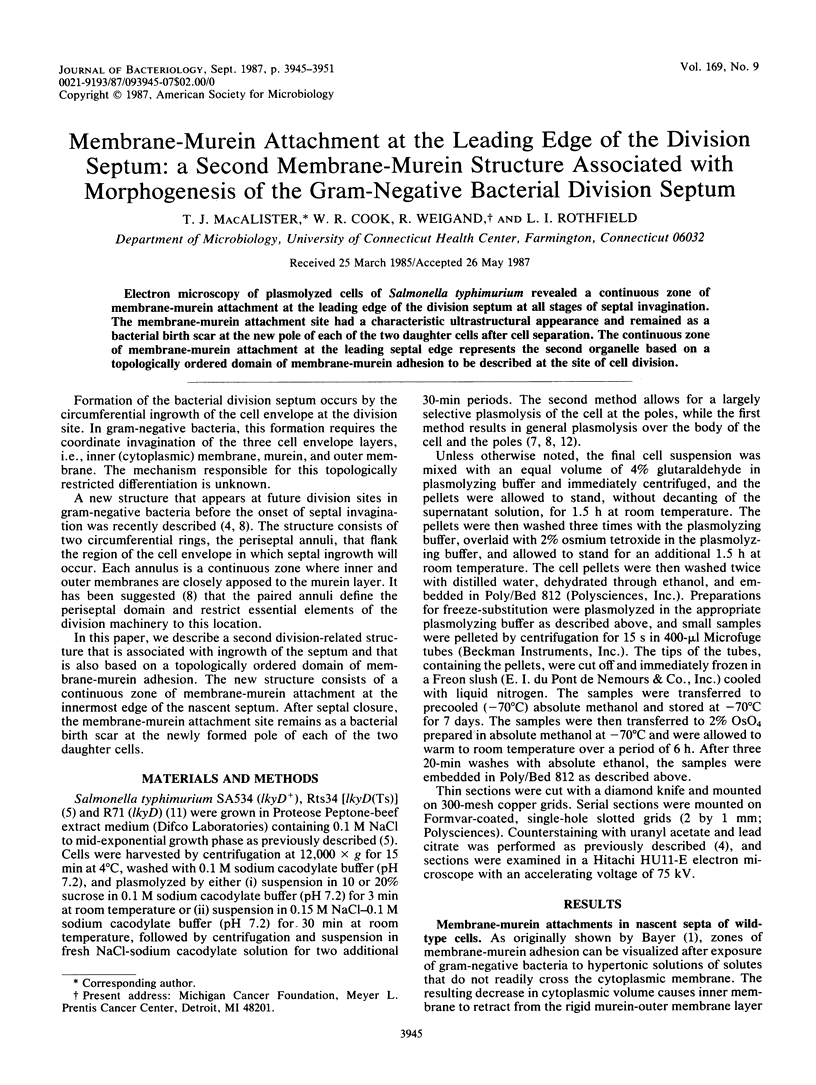
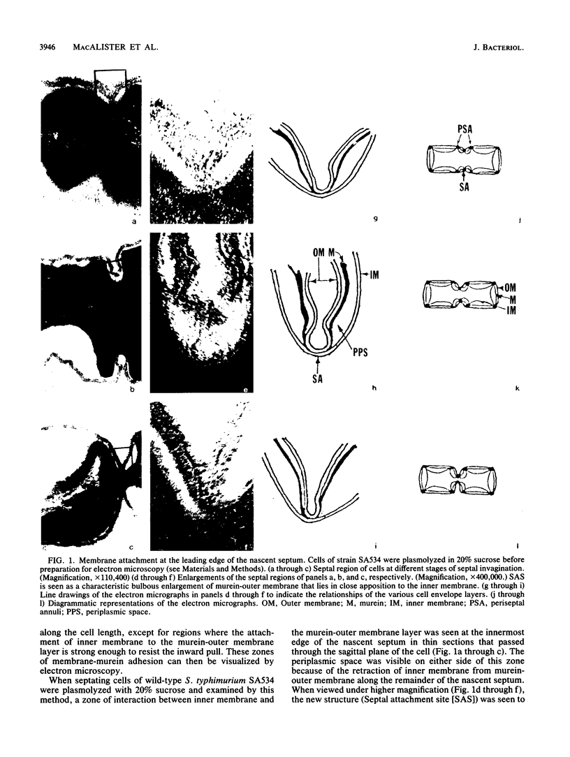
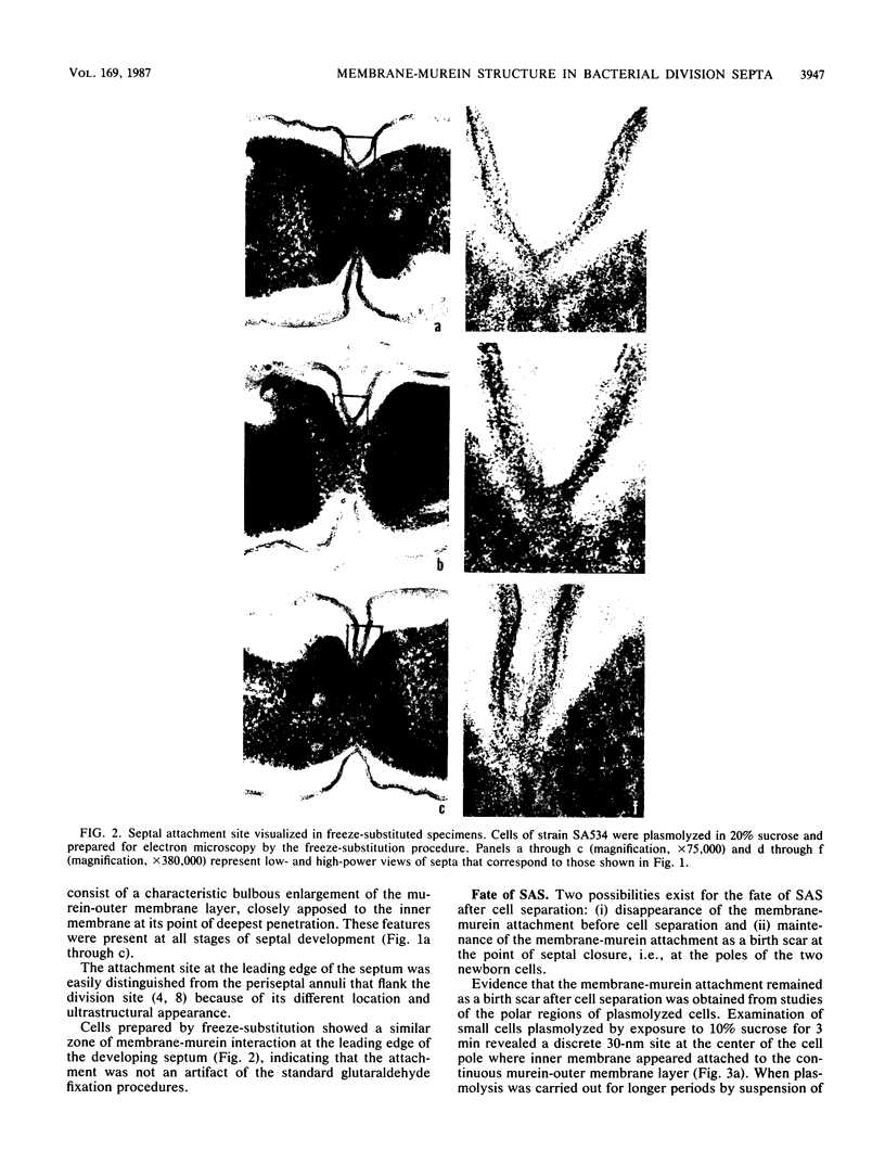
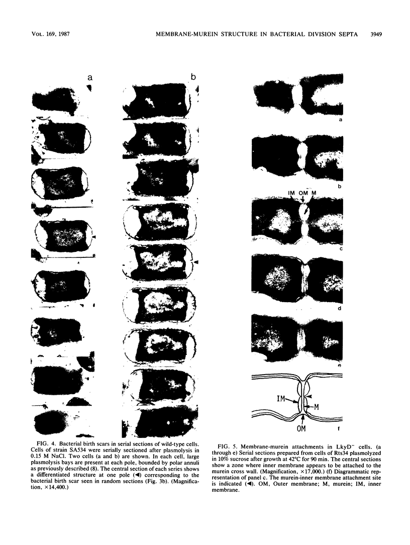
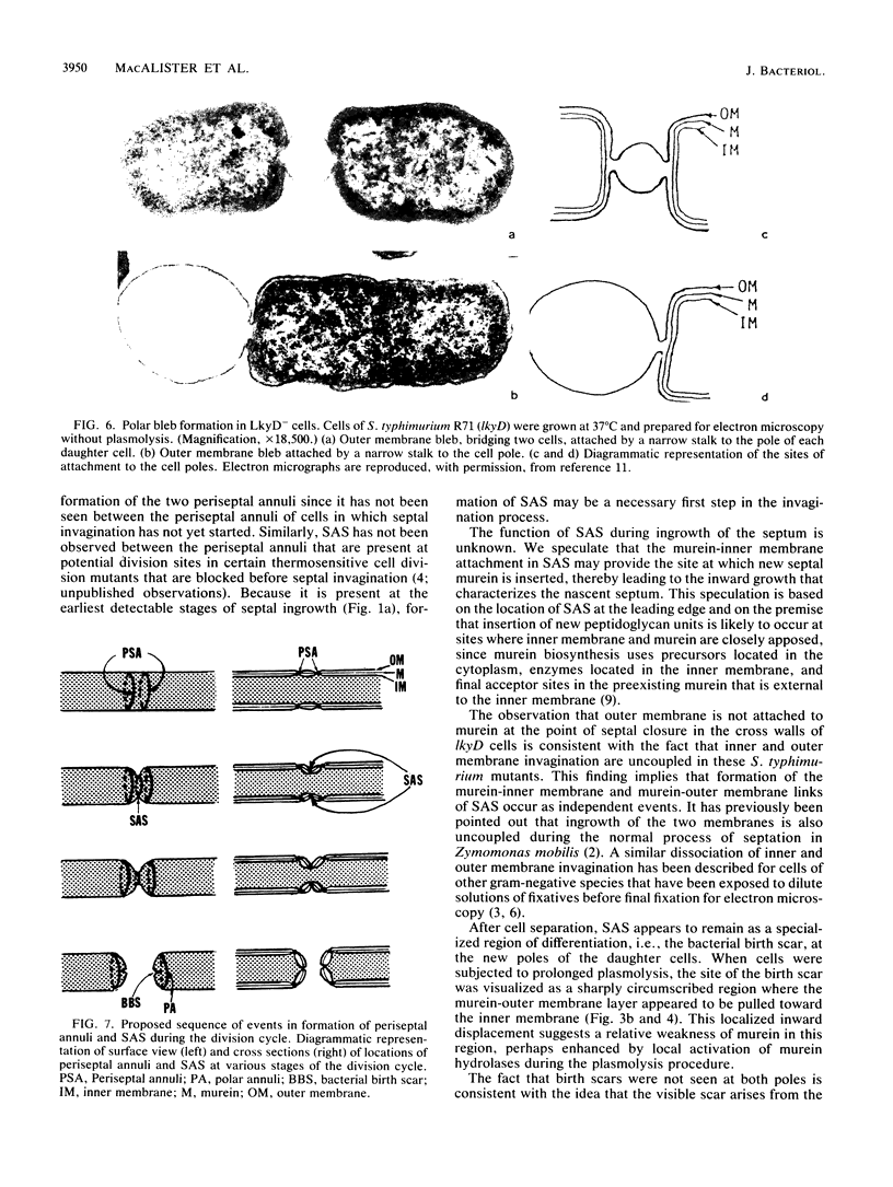
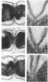
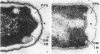
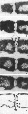
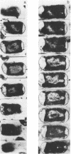
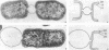
Electron microscopy of plasmolyzed cells of Salmonella typhimurium revealed a continuous zone of membrane-murein attachment at the leading edge of the division septum at all stages of septal invagination. The membrane-murein attachment site had a characteristic ultrastructural appearance and remained as a bacterial birth scar at the new pole of each of the two daughter cells after cell separation. The continuous zone of membrane-murein attachment at the leading septal edge represents the second organelle based on a topologically ordered domain of membrane-murein adhesion to be described at the site of cell division.
Full text
PDF
Images in this article
Selected References
These references are in PubMed. This may not be the complete list of references from this article.
- Bayer M. E. Areas of adhesion between wall and membrane of Escherichia coli. J Gen Microbiol. 1968 Oct;53(3):395–404. doi: 10.1099/00221287-53-3-395. [DOI] [PubMed] [Google Scholar]
- Burdett I. D., Murray R. G. Electron microscope study of septum formation in Escherichia coli strains B and B-r during synchronous growth. J Bacteriol. 1974 Sep;119(3):1039–1056. doi: 10.1128/jb.119.3.1039-1056.1974. [DOI] [PMC free article] [PubMed] [Google Scholar]
- Cook W. R., MacAlister T. J., Rothfield L. I. Compartmentalization of the periplasmic space at division sites in gram-negative bacteria. J Bacteriol. 1986 Dec;168(3):1430–1438. doi: 10.1128/jb.168.3.1430-1438.1986. [DOI] [PMC free article] [PubMed] [Google Scholar]
- Fung J. C., MacAlister T. J., Weigand R. A., Rothfield L. I. Morphogenesis of the bacterial division septum: identification of potential sites of division in lkyD mutants of Salmonella typhimurium. J Bacteriol. 1980 Aug;143(2):1019–1024. doi: 10.1128/jb.143.2.1019-1024.1980. [DOI] [PMC free article] [PubMed] [Google Scholar]
- Gilleland H. E., Jr, Murray R. G. Demonstration of cell division by septation in a variety of gram-negative rods. J Bacteriol. 1975 Feb;121(2):721–725. doi: 10.1128/jb.121.2.721-725.1975. [DOI] [PMC free article] [PubMed] [Google Scholar]
- MacAlister T. J., Costerton J. W., Thompson L., Thompson J., Ingram J. M. Distribution of alkaline phosphatase within the periplasmic space of gram-negative bacteria. J Bacteriol. 1972 Sep;111(3):827–832. doi: 10.1128/jb.111.3.827-832.1972. [DOI] [PMC free article] [PubMed] [Google Scholar]
- Macalister T. J., Macdonald B., Rothfield L. I. The periseptal annulus: An organelle associated with cell division in Gram-negative bacteria. Proc Natl Acad Sci U S A. 1983 Mar;80(5):1372–1376. doi: 10.1073/pnas.80.5.1372. [DOI] [PMC free article] [PubMed] [Google Scholar]
- Weigand R. A., Vinci K. D., Rothfield L. I. Morphogenesis of the bacterial division septum: a new class of septation-defective mutants. Proc Natl Acad Sci U S A. 1976 Jun;73(6):1882–1886. doi: 10.1073/pnas.73.6.1882. [DOI] [PMC free article] [PubMed] [Google Scholar]
- Wetzel B. K., Spicer S. S., Dvorak H. F., Heppel L. A. Cytochemical localization of certain phosphatases in Escherichia coli. J Bacteriol. 1970 Oct;104(1):529–542. doi: 10.1128/jb.104.1.529-542.1970. [DOI] [PMC free article] [PubMed] [Google Scholar]