

Abstract
The gapA gene encoding a novel RasGTPase-activating protein (RasGAP)–related protein was found to be disrupted in a cytokinesis mutant of Dictyostelium that grows as giant and multinucleate cells in a dish culture. The predicted sequence of the GAPA protein showed considerable homology to those of Gap1/Sar1 from fission yeast and the COOH-terminal half of mammalian IQGAPs, the similarity extending beyond the RasGAP-related domain. In suspension culture, gapA− cells showed normal growth in terms of the increase in cell mass, but cytokinesis inefficiently occurred to produce spherical giant cells. Time-lapse recording of the dynamics of cell division in a dish culture revealed that, in the case of gapA− cells, cytokinesis was very frequently reversed at the step in which the midbody connecting the daughter cells should be severed. Earlier steps of cytokinesis in the gapA− cells seemed to be normal, since myosin II was accumulated at the cleavage furrow. Upon starvation, gapA− cells developed and formed fruiting bodies with viable spores, like the wild-type cells. These results indicate that the GAPA protein is specifically involved in the completion of cytokinesis. Recently, it was reported that IQGAPs are putative effectors for Rac and CDC42, members of the Rho family of GTPases, and participate in reorganization of the actin cytoskeleton. Thus, it is possible that Dictyostelium GAPA participates in the severing of the midbody by regulating the actin cytoskeleton through an interaction with a member of small GTPases.
Cytokinesis is the final stage of the cell cycle, in which the cytoplasm of a cell is divided equally in the two daughter cells after the segregation of nuclei (Satterwhite and Pollard, 1992). In cytokinesis, an actin contractile ring first appears at the equator of a cell, which then constricts to generate the cleavage furrow. This constriction requires force generated by conventional myosin II. Thus, depletion of myosin II results in cytokinesis defects (Mabuchi and Okuno, 1977; De Lozanne and Spudich, 1987; Knecht and Loomis, 1987). The furrowing proceeds to form a narrow cytoplasmic bridge called the midbody that is eventually severed. These processes in cytokinesis should be spatially and temporally regulated, otherwise the components of the cell cannot be equally distributed between the daughter cells. In contrast with the detailed understanding of mitotic regulation, however, much less is known about the signal transduction pathways regulating cytokinesis.
Members of the Rho family of small GTPases, CDC42, Rac, and Rho proteins, regulate the formation of filopodia, lamellipodia, and stress fibers and focal adhesions, respectively, events that involve reorganization of the actin cytoskeleton (Ridley and Hall, 1992; Ridley et al., 1992; Nobes and Hall, 1995). Recently, it was found that these proteins are also involved in cytokinesis, an event in which the actin cytoskeleton plays a central role. In sand dollar (Mabuchi et al., 1993) and Xenopus (Kishi et al., 1993; Drechsel et al., 1996) eggs, microinjection of a Rho-specific inhibitor, C3 exoenzyme from Clostridium botulinum, prevents the progression of cytokinesis. Both a human cell line expressing a constitutively activated mutant of CDC42Hs (Dutartre et al., 1996) and a Dictyostelium strain lacking the racE gene encoding a Rac/CDC42-related protein (Larochelle et al., 1996) produced giant and multinucleate cells as a result of the impairment of cytokinesis. Rho-type GTPases appear to regulate these cytoskeletal events through cytoplasmic targets rather than nuclear ones (Vojtek and Cooper, 1995). Recently, putative cytoplasmic targets for CDC42/Rac (Hart et al., 1996; Brill et al., 1996; McCallum et al., 1996; Kuroda et al., 1996) and Rho (Watanabe et al., 1996; Amano et al., 1996a ,b; Matsui et al., 1996; Kimura et al., 1996) were identified. Some of them might be specifically involved in cytokinesis.
The cellular slime mold, Dictyostelium discoideum, is an ideal model organism for studying cytokinesis, since the basic mechanisms of Dictyostelium cytokinesis resemble those of higher eukaryotic cells. Furthermore, genetic and reverse genetic approaches are possible with the Dictyostelium system. Dictyostelium cells lacking myosin II generated by homologous recombination (De Lozanne and Spudich, 1987) or through expression of the corresponding antisense RNA (Knecht and Loomis, 1987) became multinucleate cells as a result of severe defects in cytokinesis. However, these cells were not lethal, since their growth was supported by traction-mediated cytofission, a process dependent on the attachment of cells to a solid surface (De Lozanne and Spudich, 1987; Fukui et al., 1990). This viable and multinucleate phenotype of Dictyostelium mutants enables us to identify genes involved in cytokinesis, either by disrupting genes encoding known proteins or by random tagging mutagenesis followed by cloning of the disrupted genes. Such screening will identify molecules regulating cytokinesis as well as those directly or indirectly associated with the contractile ring. Actually, genes encoding the subunits of myosin II (De Lozanne and Spudich, 1987; Manstein et al., 1989; Pollenz et al., 1992; Chen et al., 1994, 1995), actin-binding proteins (de Hostos et al., 1993; Haugwitz et al., 1994; Faix et al., 1996), calmodulin (Liu et al., 1992), and a Rac protein (Larochelle et al., 1996) have been identified as the cytokinesis genes in Dictyostelium.
To identify novel genes involved in cytokinesis, we have screened Dictyostelium cytokinesis mutants that grow as giant and multinucleate cells (Adachi et al., 1994) using an efficient tagging method called restriction enzyme–mediated integration (REMI)1 (Kuspa and Loomis, 1992). By analyzing one such mutant, we identified a Dictyostelium IQGAP-related protein, GAPA, which is required for cleavage of the midbody in the final stage of cytokinesis. Interacting with CDC42 and Rac, mammalian IQGAPs act as effectors for these GTPases to regulate reorganization of the actin cytoskeleton (Hart et al., 1996; Brill et al., 1996; McCallum et al., 1996; Kuroda et al., 1996). Like other IQGAPs, the GAPA protein in Dictyostelium cells could regulate late cytokinesis through the actomyosin system.
Materials and Methods
Dictyostelium Strains and Cell Culture
All the strains used in this study were derived from Dictyostelium discoideum AX2 (Watts and Ashworth, 1970). The cytokinesis mutant, D42-2, was described previously (Adachi et al., 1994). The wild-type AX2 and mutant strains were grown axenically in HL5 medium (Sussman, 1987) containing Proteose Peptone No. 2 or No. 3 (Difco Laboratories, Inc., Detroit, MI) at 22°C. For suspension culture, cells were shaken at 150 rpm. Penicillin G sodium and streptomycin sulfate (GIBCO BRL, Gaithersburg, MD) were always added to the medium at concentrations of 6 U/ml and 6 μg/ml, respectively. For marker selection, blasticidin S (Funakoshi Co., Tokyo, Japan) and/or G418 (Difco Laboratories, Inc.) were used at the final concentrations of 4 μg/ml and 10 μg/ml, respectively.
Southern Blot Analysis
Genomic DNA was isolated from Dictyostelium cells according to Bain and Tsang (1991). Gel electrophoresis and electroblotting of digested DNA were performed as previously described (Adachi et al., 1994). Southern hybridization and detection were carried out with a DIG DNA labeling and detection kit (Boehringer Mannheim Biochemicals, Indianapolis, IN) or a direct nucleic acid labeling and enhanced chemiluminescence detection system (Amersham Corp., Arlington Heights, IL) according to the manufacturer's instructions.
Reverse Transcription–PCR
Total RNA was isolated from 2 × 10⁷ vegetative cells using a QuickPrep® total RNA extraction kit (Pharmacia Biotech, Piscataway, NJ). 0.67 μg of total RNA was used for a single-tube reaction for first strand synthesis with a Ready-To-Go T-primed first strand kit (Pharmacia Biotech) according to the instruction manual. The reaction mixture was divided into two tubes. To each tube, distilled water, gapA primers (AGAAAAACTGTCATCTATAGCG and GTTGTTGTAATTTCATTGGGTG) or myosin II heavy chain primers (TGGTCTAGATTCACAAGCCACTATC and TTGTTCTCGAGCTTCTTCAATACGAGC), and AmpliTaq® Gold (Perkin-Elmer Corp., Norwalk, CT) were added, and then the PCR was performed as described in the manual. 10 μl of the reaction mixture was then subjected to 1.5% agarose gel electrophoresis, and the amplified DNA was detected according to Sambrook et al. (1989).
Molecular Cloning and Sequence Analysis of the gapA Gene
The general methods for recombinant DNA were described previously (Sambrook et al., 1989). Fragments of the gapA gene were cloned from the genomic DNA of the D42-2 strain by the plasmid rescue method as previously described (Adachi et al., 1994), except that Escherichia coli STBL2 (GIBCO BRL) was used as the host for the plasmids to prevent deletion of the Dictyostelium DNA in E. coli cells. First, EcoRI (12.6-kb) and HindIII (7.5-kb) fragments containing E. coli plasmid pUC118 (Vieira and Messing, 1987) with the 5′ or 3′ half of the gapA gene were rescued (see Fig. 1) and termed p42-2Eco and p42-2Hind, respectively. The DNA sequences of these fragments were determined with an SQ5500 DNA sequencer (Hitachi Co., Tokyo, Japan) and a Thermo Sequenase core sequencing kit with 7-deaza-dGTP (Amersham Corp.). The intact gapA gene was reconstructed as a 4.1-kb HindIII–ClaI fragment as follows. The gene fragment containing the insertion point of the D42-2 mutant was amplified by PCR from AX2 genomic DNA using an LA-PCR kit (Takara, Tokyo, Japan). The amplified DNA was cloned into pUC118 (Vieira and Messing, 1987), and the sequence from the BamHI site to the BsmI site of the insert was confirmed to be identical to the corresponding regions of the plasmid-rescued fragments. By ligation of this BamHI–BsmI fragment with the rescued HindIII–BamHI (p42-1Eco) and BsmI–ClaI (p42-2Hind) fragments, and cloning into pUC119 (Vieira and Messing, 1987), plasmid p42-2CPX carrying the intact gapA gene was obtained. The reconstructed HindIII–ClaI fragment was inserted into the Dictyostelium shuttle vector, pATANB43 (Dynes and Firtel, 1989), using the linker sequences to construct p43-L, which was used for the genetic complementation experiments.
Figure 1.

Restriction map around the Dictyostelium gapA gene and the tag integration site of the REMI mutant, D42-2. Genomic DNA from the D42-2 strain was digested with the restriction enzymes indicated, and then analyzed by Southern blotting using linearized pUC118, a portion of the tag, as a probe. Information obtained on restriction analysis of the rescued plasmids is also included. The four exons of the gapA gene are indicated by thick lines. bsr, blasticidin S-resistance marker.
Dictyostelium Transformation
The procedures for Dictyostelium transformation were previously described (Adachi et al., 1994). For regeneration of gapA− cells by homologous recombination, 10 μg of linearized p42-2Nde, the DNA fragment derived from D42-2 rescued with the NdeI restriction enzyme, was used for each electroporation with the AX2 strain. For complementation of the D42-2 strain with the p43-L plasmid, 2 μg of plasmid DNA was used for each electroporation, and the transformants were selected by G418.
Cell Staining
Dictyostelium cells were grown axenically on coverslips as described above. Fluorescence microscopy was performed according to Fukui et al. (1987). Briefly, cells on coverslips were fixed in methanol containing 1% formaldehyde for 5 min at −10°C, and then washed three times with PBS. For the observation of nuclei, the cells were stained with PBS containing 0.1 μg/ml of 4′,6-diamidino-2-phenylindole for 30 min at 37°C, washed with PBS, and then observed under an Axiovert 35 equipped with an LD Achroplan ×40 objective (Zeiss, Oberkochen, Germany). For the observation of myosin II, the cells were flattened by the agar-overlay technique (Fukui et al., 1987), and then fixed as described above. The fixed cells were first incubated with PBS containing anti–Dictyostelium myosin II mAbs DM2 (Yumura et al., 1984), for 30 min at 37°C, rinsed with PBS, and then stained with 25× diluted fluorescein-conjugated goat anti–mouse IgG antibodies (Biosource, Camarillo, CA) under the same conditions. After washing with PBS, the cells were observed with a Plan-Neofluar ×100 oil immersion objective.
Dictyostelium Development
108 axenically growing cells were washed twice with 17 mM potassium phosphate buffer (pH 6.5), and then spread on an 1.5% agarose plate (6 cm) containing the same buffer. Starved cells were developed at 22°C for 24 h.
Results
Molecular Cloning of the Dictyostelium Cytokinesis Gene from REMI Mutant D42-2
We have already reported three Dictyostelium cytokinesis mutants (Adachi et al., 1994) isolated by REMI (Kuspa and Loomis, 1992). On a substratum, these mutant strains grow and generate considerable numbers of giant and multinucleate cells that sometimes contain >100 nuclei as a result of their cytokinesis defects. Into the genomic DNA of these mutants, a single copy of the tag plasmid, pUCBsrΔBam, was inserted (Adachi et al., 1994).
One of the mutants, D42-2, was further analyzed. Fig. 1 shows the restriction map of genomic DNA around the tag integration site of D42-2 determined by Southern blotting. Involvement of the disrupted gene in cytokinesis was confirmed in two ways. First, mutant strains with a phenotype identical to that of D42-2 were regenerated by homologous recombination. The rescued NdeI fragment of D42-2 containing Dictyostelium DNA at both ends of the tag plasmid was introduced into the wild-type AX2 strain by electroporation. At high frequency (54%, n = 262), the transformants showed an identical phenotype to the original mutant (Fig. 2, C and G). Restriction maps of the genomic DNA of the regenerated mutants confirmed the location of insertion of the tag, as shown in Fig. 1 (data not shown). These results indicate that the integration of the tag is responsible for the mutant phenotype.
Figure 2.

Size and shape of axenically growing Dictyostelium cells. (A–D) Cells were grown on coverslips, fixed, and then stained with 4′,6-diamidino-2-phenylindole. A phase-contrast image was laid over a fluorescent one. (E–H) Cells were grown in suspension (150 rpm), and living cells were observed by phase-contrast microscopy. (A and E) The parental strain, AX2; (B and F) the cytokinesis mutant, D42-2; (C and G) one of the mutants regenerated through homologous recombination, D42-2HR11; (D and H) the D42-2 strain harboring the p43-L plasmid carrying an intact gapA gene. Bar, 50 μm.
Second, genetic complementation of the mutant phenotype with the intact gene was examined. To clone the entire gene, EcoRI and HindIII fragments of the D42-2 DNA including the 5′ and 3′ halves of the disrupted gene (Fig. 1) were rescued. Through ligation of the rescued fragments, the intact HindIII–ClaI fragment (4.1 kb) was reconstructed and inserted into a Dictyostelium shuttle vector, and the resultant plasmid, p43-L, was used to transform the D42-2 strain. All the transformants showed the wild-type phenotype (Fig. 2, D and H), indicating that this 4.1-kb fragment carries the gene involved in cytokinesis. Southern blot analysis showed that the gene exists as a single copy (Fig. 3 A). The absence of a transcript of the gene in vegetative mutant cells was confirmed by the reverse transcription– PCR method, using the myosin II heavy chain transcript as a control (Fig. 3 B).
Figure 3.

Southern blot (A) and reverse transcription–PCR (B) analyses of wild-type AX2 (W) and mutant D42-2 (M) cells. (A) 2.5 μg of genomic DNA digested with either ClaI or HindIII was loaded into each lane. The transferred DNA was probed with the full-length gapA cDNA. (B) DNA amplified with the same amount of total RNA and specific primers was loaded onto each lane. For details, see Materials and Methods.
A Gene Encoding a Novel IQGAP-related Protein Is Disrupted in the D42-2 Strain
DNA sequencing of the HindIII–ClaI fragment revealed an open reading frame interrupted by three putative introns with typical features of Dictyostelium introns (Figs. 1 and 4). The intron–exon boundaries were confirmed by sequence analysis of cDNA clones prepared from the vegetative mRNA (data not shown). It was found that the tag was inserted into the fourth exon of the gene in the D42-2 DNA (Figs. 1 and 4), and that the open reading frame is 2,580 bp in length and codes for a protein of 860 amino acids (molecular mass = 98.8 kD).
Figure 4.
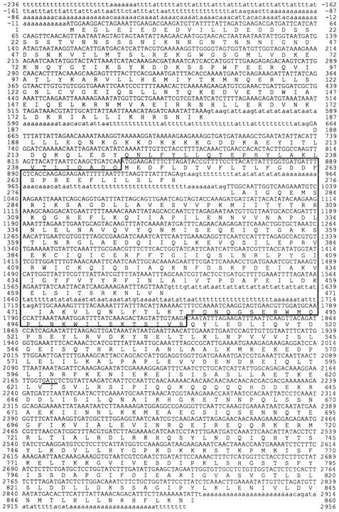
DNA sequence of the Dictyostelium gapA gene and the deduced amino acid sequence of the GAPA protein. The coding DNA sequences (exons) are indicated by capital letters, and the 5′ and 3′ flanking sequences as well as introns are shown in lowercase letters. (Boxed region) RasGAP-related domain (GRD). In cytokinesis mutant D42-2, the tag was inserted into the underlined GATC sequence in the fourth exon. These sequence data are available from EMBL/GenBank/DDBJ under accession number D88027.
The deduced amino acid sequence of this protein was compared with the NBRF-PIR and SWISS PLOT databases, which revealed that it is very closely related to Gap1/ Sar1 from fission yeast (Imai et al., 1991; Wang et al., 1991) and the COOH-terminal half of IQGAP1 from human (Weissbach et al., 1994) (Fig. 5). In the middle of the homologous regions of these three proteins lies a conserved sequence for RasGTPase-activating proteins (RasGAPs) called the GAP-related domain (GRD; Figs. 4 and 5). Thus, we named this protein GAPA, and the gene gapA, from its sequence similarity to RasGAPs. The homology of the Dictyostelium GAPA to the members of the RasGAP family other than IQGAP1, IQGAP2 (recently identified homologue of IQGAP1 [Brill et al., 1996]), and Gap1/Sar1 is restricted to the GAP-related domain. Fig. 6 shows amino acid sequence alignment of the most conserved region of the GRDs of typical RasGAP-related proteins. The most prominent feature is that the Phe-Leu residues in the invariant FLRXXXPAXXXP (X: any amino acid) motif are replaced by Tyr-Tyr residues only in GAPA and IQGAP1 (in IQGAP2 also Tyr-Tyr). It was reported that substitution of the invariant Leu with an Ile residue resulted in the loss of the activity of p120GAP (Brownbridge et al., 1993), and that RasGAP activity was not detected for IQGAPs (Hart et al., 1996; Brill et al., 1996; McCallum et al., 1996). In contrast with IQGAPs, both biochemical (Hart et al., 1996) and genetic (Imai et al., 1991; Wang et al., 1991) analyses indicated that yeast Gap1/Sar1 activates the GTPase of yeast RAS2, and the invariant Phe-Leu residues are conserved in this protein. These suggest that GAPA might be much more related functionally to IQGAPs than to Gap1/Sar1. On the basis of this finding, we looked for the sequence motifs that were found in the NH2-terminal half of IQGAPs (Weissbach et al., 1994; Hart et al., 1996) (Fig. 6) in the NH2-terminal region of GAPA. One putative IQ motif (I145AEIQELKRNMVAE158; the conserved residues are underlined) was found in the region where the homology between IQGAP1 and GAPA starts, although the conserved arginine was replaced by glutamic acid158. We found neither a calponin homology domain, an IQGAP repeat, nor a WW domain (Sudol et al., 1995).
Figure 5.

Schematic drawing of the similarity among the members of the IQGAP family. Identical residues (%) were calculated. CH, calponin homology domain; IR, IQGAP repeats; WW, WW domain; IQ, four repeats of the calmodulin-binding motif; GRD, RasGAP-related domain; Hs, Homo sapiens; Dd, Dictyostelium discoideum; Sp, Schizosaccharomyces pombe.
Figure 6.

Amino acid sequence alignment of the most conserved region of RasGAP-related proteins. Identical residues are shaded. The invariant FLRXXXPAXXXP motifs are boxed. Dm, Drosophila melanogaster; Sc, Saccharomyces cerevisiae. GenBank accession numbers: Dd GAPA, D88027; Hs IQGAP1, D63875; Sp Sar1/Gap1, D10457; Hs p120GAP, M23379; Dm GAP1, M86655; Sc Bud2, L19162; Hs NF1, M89914; Sc Ira1, M24378. The sequence of GAP1IP4BP was taken from the original paper (Cullen et al., 1995).
Reversion of Cytokinesis Observed in gapA− Cells
To determine the step(s) at which GAPA acts during cytokinesis, the phenotype of gapA− cells was further characterized. First, the growth of gapA− cells in suspension culture was examined. It is known that Dictyostelium cells lacking myosin II grow as multinucleate cells on a substratum (De Lozanne and Spudich, 1987; Knecht and Loomis, 1987) like gapA− cells. In this case, the growth is not supported by “real” cytokinesis but by traction-mediated cytofission, which is dependent on the solid surface (Fukui et al., 1990), and the cells cannot survive in suspension culture. Unlike myosin II–deficient cells, gapA− cells grew in suspension culture (Fig. 7). The rate of increase in cell number was, however, much lower than that of the wildtype cells (Fig. 7 A). In contrast, the rates of increase in cell mass, monitored as the turbidity of the culture, were similar to each other (Fig. 7 B). Thus, the mutant strain also produced spherical giant cells in suspension culture (Fig. 2, B and F). These results suggest that, in the gapA− cells, cytokinesis was not completed at a considerable frequency.
Figure 7.

Growth of Dictyostelium cells in axenic suspension. Wild-type AX2 and gapA− D42-2 cells were diluted in fresh medium at 105 cells per ml, and then shaken at 22°C and 150 rpm. Their growth was monitored as the increase in cell number (A) or turbidity (B; absorbance at 660 nm) with the same culture.
Second, the dynamics of cell division on a glass surface were monitored by means of time-lapse video recording. gapA− cells (Fig. 8, lower frames) stopped protruding pseudopodia, became rounded, and then became constricted to produce daughter cells linked by a thin cytoplasmic bridge (midbody; Fig. 8, arrowheads). By this stage, cytokinesis of the gapA− cells was similar to that of the wild-type cells (Fig. 8, upper frames), although it sometimes took longer for constriction in the mutant. This bridge, however, frequently remained intact for a long time, and finally cytokinesis was reversed to yield a single cell (Fig. 8, filled triangle). This reversion occurred for ∼50% (n = 44) of the smallest mutant cells that attempted to divide into two. The failed cytokinesis accumulated to produce giant and multinucleate cells. These results suggest that, in gapA− cells, earlier stages of cytokinesis are rarely affected, whereas the fidelity of the latest severing of the midbody is greatly reduced. Consistent with this notion, myosin II was localized at the cleavage furrow in the dividing mutant cells as in the dividing wild-type cells (Fig. 9). In interphase mutant cells, myosin II was also predominantly localized at the periphery of the cells, as in the wild-type cells. The localization of actin filaments in interphase mutant cells was also indistinguishable from that in the wildtype cells (data not shown).
Figure 8.

Reversion of cytokinesis observed in gapA− cells. Wildtype AX2 (upper frames) and mutant D42-2 (lower frames) cells were axenically grown on coverslips. Cell division was observed under a microscope connected to a time-lapse video recorder. The number in each frame indicates the time in min. In the lower frames (D42-2), the mutant cells (open and filled triangles) attempted to divide into two daughter cells. For the cell with the filled triangle, cytokinesis was reversed at the step where the two daughter cells were connected by a thin cytoplasmic bridge (midbody; arrowhead). In contrast, the cell with the open triangle completed cytokinesis like wild-type cells. Bar, 10 μm.
Figure 9.

Localization of myosin II at the cleavage furrow. (A and B) AX2; (C–F) D42-2. Axenically growing cells were fixed by the agar-overlay method (Fukui et al., 1987). Myosin II was visualized by means of indirect immunofluorescence using DM2 mAbs against Dictyostelium myosin II (B, D, and F). (A, C, and E) Phase contrast. Bar, 10 μm.
Development of gapA− Cells Is Normal
When starved on phosphate agar, gapA− cells developed and formed normal fruiting bodies with viable spores at the same time as the wild-type cells (Fig. 10). The increase in size of the plaque on the bacterial lawn and the terminal phenotype were also unaffected by the mutation (data not shown). These results indicate that the GAPA protein is not required for Dictyostelium development and phagocytosis.
Figure 10.

Development of Dictyostelium cells upon starvation. (Left) AX2; (Right) D42-2. Vegetative cells were washed and then plated onto phosphate agar. The developed phenotype was observed after 24 h. Bar, 100 μm.
The phenotypes of the REMI mutant shown above were identical to those of the null strain lacking most of the coding sequence of the gapA gene (data not shown).
Discussion
Although it has been established that the astral microtubules determine the position of the contractile ring (Rappaport, 1990), the molecular nature of this signal remains to be determined. The timing of the initiation and progression of cytokinesis should be strictly regulated, possibly by molecules controlling the cell cycle, such as Cdc2 kinase. Myosin II is required for furrowing, and activation and/or inactivation of the myosin ATPase by such molecules could control constriction of the contractile ring. Actually, it was demonstrated that phosphorylation of myosin II regulatory light chain by Cdc2/Cyclin could regulate the timing of cytokinesis (Satterwhite et al., 1992). In addition, myosin II could be regulated through the signal from a small GTPase Rho during constriction, considering the findings that Rho is involved in cytokinesis (Mabuchi et al., 1993; Kishi et al., 1993), and that the phosphorylation of myosin II regulatory light chain, which activates the ATPase of myosin II, is regulated by effectors of Rho, Rho kinase, and myosin phosphatase (Kimura et al., 1996; Amano et al., 1996a ). As for the termination of cytokinesis, however, the existence of a signal for severing of the midbody remains to be confirmed.
Dictyostelium cells lacking the IQGAP-related protein, GAPA, grow as giant and multinucleate cells both on a substratum and in suspension culture. Observation of cell division of gapA− cells indicated that multinucleate cells are produced through the frequent reversion of cytokinesis at the step in which the cytoplasmic bridge (midbody) connecting the daughter cells should be severed. Earlier stages of cytokinesis characterized by accumulation of myosin II at the furrow were not affected by the gapA− mutation. Development upon starvation and vegetative growth in terms of the increase in cell mass were also not affected. Thus, we propose that the GAPA protein is specifically involved in the signal required for accurate cleavage of the midbody.
Similar reversed cytokinesis has been reported for a cell line in which the expression level of calmodulin is reduced about twofold through expression of the corresponding antisense RNA (Liu et al., 1992). It might be possible that, in the signaling pathway required for cleavage of the midbody, interaction between GAPA and calmodulin is essential, and the resultant complex transduces the signal of elevation of the calcium level observed during cytokinesis (Chang and Meng, 1995). A putative IQ motif, a calmodulin-binding sequence, is present in the GAPA protein, although the binding to calmodulin has to be confirmed biochemically. In mammalian IQGAPs, four successive calmodulin-binding IQ-motifs (Hart et al., 1996; Brill et al., 1996) are present at a position corresponding to the putative IQ motif of GAPA (Fig. 4).
Since a RasGAP-related domain lies in the middle of the GAPA protein, GAPA could activate the GTPase activity of Ras. Genetic (Imai et al., 1991; Wang et al., 1991) and biochemical (Hart et al., 1996) analyses showed that Sar1/Gap1 from fission yeast, one of the GAPA-related proteins (Fig. 4), activates yeast Ras. However, the other GAPA relatives, mammalian IQGAPs, do not interact with Ras, but bind to and inhibit the GTPases of CDC42 and Rac, members of the Rho family of GTPases (Hart et al., 1996; Bain and Tsang, 1991; McCallum et al., 1996). Invariant Phe-Leu residues in the most conserved region of the RasGAP-related domain are replaced by Tyr-Tyr residues in both IQGAPs and GAPA, but not in Sar1/Gap1 (Fig. 6). Substitution of this conserved leucine of p120GAP resulted in loss of the GAP activity toward Ras (Brownbridge et al., 1993). Like mammalian IQGAPs, GAPA might interact with a CDC42/Rac-related GTPase but not with a Ras-related protein (five Ras-related proteins were found in Dictyostelium [Daniel et al., 1993]). Actually, a crude extract of gapA− cells retained the GAP activity toward H-ras and Dictyostelium RasG, a counterpart of Ras in vegetative Dictyostelium cells (data not shown).
In Dictyostelium, eight Cdc42/Rac-related proteins have been identified (Bush et al., 1993; Larochelle et al., 1996). RacE, a novel Rac/Cdc42-related protein, is specifically required for cytokinesis (Larochelle et al., 1996). In suspension culture, racE− cells become multinucleate cells, as well as do the gapA− cells. However, in contrast with gapA− cells, racE− cells never divide like myosin II–deficient cells. The phenotypical difference between gapA− and racE− cells suggests that these proteins participate in distinct pathways in cytokinesis. The fact that racE− cells resemble myosin II–deficient cells and that they only grow on dishes through traction-mediated cytofission might suggest that RacE could control the myosin function during earlier stages in cytokinesis, unlike GAPA.
Very recently, two other IQGAP-related proteins were found in Dictyostelium (Faix and Dittrich, 1996; Lee et al., 1997). Disruption of the genes encoding other IQGAPrelated proteins caused developmental defects in contrast with the gapA− cells, suggesting that their functions are distinct from that of GAPA. For one of them, DGAP1, disruption of the corresponding gene had no effect on cytokinesis, while overexpression of this protein caused cytokinesis defects (Faix and Dittrich, 1996). The cells overproducing DGAP1 by threefold became multinucleate and divided through traction-mediated cytofission when placed on a substratum. These phenotypes resemble those of racE− and myosin II–deficient cells. DGAP1 might interact with RacE and be involved in earlier stages of cytokinesis. Gene disruption of DdRasGAP1, another Dictyostelium IQGAP-related protein, caused cytokinesis defects only in suspension culture (Lee et al., 1997) unlike the gapA− cells. The presence of closely related but different IQGAP-related proteins in Dictyostelium cells implies distinct pathways of cytokinesis regulation that may involve different members of small GTPases.
Acknowledgments
We thank Drs. Takahisa Ohta and Masayoshi Kawaguchi for helpful discussions. We also thank Dr. Shigehiko Yumura for the anti–myosin II antibodies.
This work was supported by grants-in-aid for Scientific Research from the Ministry of Education, Science, and Culture of Japan to H. Adachi and K. Sutoh.
Footnotes
1. Abbreviations used in this paper: GRD, GTPase-activating protein– related domain; RasGAP, RasGTPase-activating protein; REMI, restriction enzyme–mediated integration.
Please address all correspondence to Hiroyuki Adachi, Department of Life Sciences, Graduate School of Arts and Sciences, University of Tokyo, 3-8-1 Komaba, Meguro-ku, Tokyo 153, Japan. Tel. and Fax: (81) 3-54546769. e-mail: f37771@m-unix.cc.u-tokyo.ac.jp
References
- Adachi H, Hasebe T, Yoshinaga K, Ohta T, Sutoh K. Isolation of Dictyostelium discoideumcytokinesis mutants by restriction enzyme-mediated integration of the blasticidin S resistance marker. Biochem Biophys Res Commun. 1994;205:1808–1814. doi: 10.1006/bbrc.1994.2880. [DOI] [PubMed] [Google Scholar]
- Amano M, Ito M, Kimura K, Fukata Y, Chihara K, Nakano T, Matsuura Y, Kaibuchi K. Phosphorylation and activation of myosin by rho-associated kinase (Rho-kinase) J Biol Chem. 1996a;271:20246–20249. doi: 10.1074/jbc.271.34.20246. [DOI] [PubMed] [Google Scholar]
- Amano M, Mukai H, Ono Y, Chihara K, Matsui T, Hamajima Y, Okawa K, Iwamatsu A, Kaibuchi K. Identification of a putative target for Rho as the serine-threonine kinase protein kinase N. Science (Wash DC) 1996b;271:648–650. doi: 10.1126/science.271.5249.648. [DOI] [PubMed] [Google Scholar]
- Bain G, Tsang A. Disruption of the gene encoding the p34/31 polypeptides affects growth and development of Dictyostelium discoideum. . Mol Gen Genet. 1991;226:59–64. doi: 10.1007/BF00273587. [DOI] [PubMed] [Google Scholar]
- Brill S, Li S, Lyman CW, Church DM, Wasmuth JJ, Weissbach L, Bernards A, Snijders AJ. The ras GTPase-activating-protein-related human protein IQGAP2 harbors a potential actin binding domain and interacts with calmodulin and rho family GTPases. Mol Cell Biol. 1996;16:4869–4878. doi: 10.1128/mcb.16.9.4869. [DOI] [PMC free article] [PubMed] [Google Scholar]
- Brownbridge GG, Lowe PN, Moore KJM, Skinner RH, Webb MR. Interaction of GTPase activating proteins (GAPs) with p21rasmeasured by a novel fluorescence anisotropy method. J Biol Chem. 1993;268:10914–10919. [PubMed] [Google Scholar]
- Bush J, Franek K, Cardelli J. Cloning and characterization of seven novel Dictyostelium discoideum rac-related genes belonging to the rho family of GTPases. Gene (Amst) 1993;136:61–68. doi: 10.1016/0378-1119(93)90448-c. [DOI] [PubMed] [Google Scholar]
- Chang DC, Meng C. A localized elevation of cytosolic free calcium is associated with cytokinesis in the Zebrafish embryo. J Cell Biol. 1995;131:1539–1545. doi: 10.1083/jcb.131.6.1539. [DOI] [PMC free article] [PubMed] [Google Scholar]
- Chen P, Ostrow BD, Tafuri SR, Chisholm RL. Targeted disruption of the Dictyostelium RMLCgene produces cells defective in cytokinesis and development. J Cell Biol. 1994;127:1933–1944. doi: 10.1083/jcb.127.6.1933. [DOI] [PMC free article] [PubMed] [Google Scholar]
- Chen T-LL, Kowalczyk PA, Ho G, Chisholm RL. Targeted disruption of the Dictyosteliummyosin essential light chain gene produces cells defective in cytokinesis and morphogenesis. J Cell Sci. 1995;108:3207–3218. doi: 10.1242/jcs.108.10.3207. [DOI] [PubMed] [Google Scholar]
- Cullen PJ, Hsuan JJ, Truong O, Letcher AJ, Jackson TR, Dawson AP, Irvine RF. Identification of a specific Ins(1,3,4,5)P4-binding protein as a member of the GAP1 family. Nature (Lond) 1995;376:527–530. doi: 10.1038/376527a0. [DOI] [PubMed] [Google Scholar]
- Daniel J, Bush J, Cardelli G, Spiegelman GB, Weeks G. Isolation of two novel ras genes in Dictyostelium discoideum: evidence for a complex, developmentally regulated ras-gene subfamily. Oncogene. 1993;9:501–508. [PubMed] [Google Scholar]
- de Hostos EL, Rehfueß C, Bradtke B, Waddell DR, Albrecht R, Murphy J, Gerisch G. Dictyosteliummutants lacking the cytoskeletal protein coronin are defective in cytokinesis and cell motility. J Cell Biol. 1993;120:163–173. doi: 10.1083/jcb.120.1.163. [DOI] [PMC free article] [PubMed] [Google Scholar]
- De Lozanne A, Spudich JA. Disruption of the Dictyosteliummyosin heavy chain gene by homologous recombination. Science (Wash DC) 1987;236:1086–1091. doi: 10.1126/science.3576222. [DOI] [PubMed] [Google Scholar]
- Drechsel DN, Hyman AA, Hall A, Glotzer M. A requirement for Rho and Cdc42 during cytokinesis in Xenopusembryos. Curr Biol. 1996;7:12–23. doi: 10.1016/s0960-9822(06)00023-6. [DOI] [PubMed] [Google Scholar]
- Dutartre H, Davoust J, Gorvel J-P, Chavrier P. Cytokinesis arrest and redistribution of actin-cytoskeleton regulatory components in cells expressing the Rho GTPase CDC42Hs. J Cell Sci. 1996;109:367–377. doi: 10.1242/jcs.109.2.367. [DOI] [PubMed] [Google Scholar]
- Dynes JL, Firtel RA. Molecular complementation of a genetic marker in Dictyosteliumusing a genomic DNA library. Proc Natl Acad Sci USA. 1989;86:7966–7970. doi: 10.1073/pnas.86.20.7966. [DOI] [PMC free article] [PubMed] [Google Scholar]
- Faix J, Dittrich W. DGAP1, a homologue of rasGTPase activating proteins that controls growth, cytokinesis, and development in Dictyostelium discoideum. . FEBS Lett. 1996;394:251–257. doi: 10.1016/0014-5793(96)00963-5. [DOI] [PubMed] [Google Scholar]
- Faix J, Steinmetz M, Boves H, Kammerer RA, Lottspeich F, Mintert U, Murphy J, Stock A, Aebi U, Gerisch G. Cortexillins, major determinants of cell shape and size, are actin-bundling proteins with a parallel coiled-coil tail. Cell. 1996;86:631–642. doi: 10.1016/s0092-8674(00)80136-1. [DOI] [PubMed] [Google Scholar]
- Fukui Y, Yumura S, Yumura TK. Agar-overlay immunofluorescence: high resolution studies of cytoskeletal components and their changes during chemotaxis. Methods Cell Biol. 1987;28:347–356. doi: 10.1016/s0091-679x(08)61655-6. [DOI] [PubMed] [Google Scholar]
- Fukui Y, De Lozanne A, Spudich JA. Structure and function of the cytoskeleton of a Dictyosteliummyosin-defective mutant. J Cell Biol. 1990;110:367–378. doi: 10.1083/jcb.110.2.367. [DOI] [PMC free article] [PubMed] [Google Scholar]
- Hart MJ, Callow MG, Souza B, Polakis P. IQGAP1, a calmodulin-binding protein with a rasGAP-related domain, is a potential effector for cdc42 Hs. EMBO (Eur Mol Biol Organ) J. 1996;15:2997–3005. [PMC free article] [PubMed] [Google Scholar]
- Haugwitz M, Noegel AA, Karakesisoglou J, Schleicher M. Dictyostelium amoebae that lack G-actin-sequestering profilins show defects in F-actin content, cytokinesis, and development. Cell. 1994;79:303–314. doi: 10.1016/0092-8674(94)90199-6. [DOI] [PubMed] [Google Scholar]
- Imai Y, Miyake S, Hughes DA, Yamamoto M. Identification of a GTPase-activating protein homolog in Schizosaccharomyces pombe. . Mol Cell Biol. 1991;11:3088–3094. doi: 10.1128/mcb.11.6.3088. [DOI] [PMC free article] [PubMed] [Google Scholar]
- Kimura K, Ito M, Amano M, Chihara K, Fukata Y, Nakafuku M, Yamamori B, Feng J, Nakano T, Okawa K, et al. Regulation of myosin phosphatase by Rho and Rho-associated kinase (Rho-kinase) Science (Wash DC) 1996;273:245–248. doi: 10.1126/science.273.5272.245. [DOI] [PubMed] [Google Scholar]
- Kishi K, Sasaki T, Kuroda S, Itoh T, Takai Y. Regulation of cytoplasmic division of Xenopus embryo by rho p21 and its inhibitory GDP/GTP exchange protein (rhoGDI) J Cell Biol. 1993;120:1187–1195. doi: 10.1083/jcb.120.5.1187. [DOI] [PMC free article] [PubMed] [Google Scholar]
- Knecht DA, Loomis WF. Antisense RNA inactivation of myosin heavy chain gene expression in Dictyostelium discoideum. . Science (Wash DC) 1987;236:1081–1086. doi: 10.1126/science.3576221. [DOI] [PubMed] [Google Scholar]
- Kuroda S, Fukata M, Kobayashi K, Nakafuku M, Nomura N, Iwamatsu A, Kaibuchi K. Identification of IQGAP as a putative target for the small GTPases, Cdc42 and Rac1. J Biol Chem. 1996;271:23363–23367. doi: 10.1074/jbc.271.38.23363. [DOI] [PubMed] [Google Scholar]
- Kuspa A, Loomis WF. Tagging developmental genes in Dictyosteliumby restriction enzyme-mediated integration of plasmid DNA. Proc Natl Acad Sci USA. 1992;89:8803–8807. doi: 10.1073/pnas.89.18.8803. [DOI] [PMC free article] [PubMed] [Google Scholar]
- Larochelle DA, Vithalani KK, De Lozanne A. A novel member of the rhofamily of small GTP-binding proteins is specifically required for cytokinesis. J Cell Biol. 1996;133:1321–1329. doi: 10.1083/jcb.133.6.1321. [DOI] [PMC free article] [PubMed] [Google Scholar]
- Lee S, Escalante R, Firtel RA. A Ras GAP is essential for cytokinesis and spatial patterning in Dictyostelium. . Development (Camb) 1997;124:983–996. doi: 10.1242/dev.124.5.983. [DOI] [PubMed] [Google Scholar]
- Liu T, Williams JG, Clarke M. Inducible expression of calmodulin antisense RNA in Dictyosteliumcells inhibits the completion of cytokinesis. Mol Biol Cell. 1992;3:1403–1413. doi: 10.1091/mbc.3.12.1403. [DOI] [PMC free article] [PubMed] [Google Scholar]
- Mabuchi I, Okuno M. The effect of myosin antibody on the division of starfish blastomeres. J Cell Biol. 1977;74:251–263. doi: 10.1083/jcb.74.1.251. [DOI] [PMC free article] [PubMed] [Google Scholar]
- Mabuchi I, Hamaguchi Y, Fujimoto H, Morii N, Mishima M, Narumiya S. A rho-like protein is involved in the organisation of the contractile ring in dividing sand dollar eggs. Zygote. 1993;1:325–331. doi: 10.1017/s0967199400001659. [DOI] [PubMed] [Google Scholar]
- Manstein DJ, Titus M A, De Lozanne A, Spudich JA. Gene replacement in Dictyostelium: generation of myosin null mutants. EMBO (Eur Mol Biol Organ) J. 1989;8:923–932. doi: 10.1002/j.1460-2075.1989.tb03453.x. [DOI] [PMC free article] [PubMed] [Google Scholar]
- Matsui T, Amano M, Yamamoto T, Chihara K, Nakahuku M, Ito M, Nakano T, Okawa K, Iwamatsu A, Kaibuchi K. Rho-associated kinase, a novel serine/threonine kinase, as a putative target for the small GTP binding protein Rho. EMBO (Eur Mol Biol Organ) J. 1996;15:2208–2216. [PMC free article] [PubMed] [Google Scholar]
- McCallum SJ, Wu WJ, Cerione RA. Identification of a putative effector for Cdc42Hs with high sequence similarity to the RasGAP-related protein IQGAP1 and a Cdc42Hs binding partner with similarity to IQGAP2. J Biol Chem. 1996;271:21732–21737. doi: 10.1074/jbc.271.36.21732. [DOI] [PubMed] [Google Scholar]
- Nobes CD, Hall A. Rho, rac, and cdc42 GTPases regulate the assembly of multimolecular focal complexes associated with actin stress fibers, lamellipodia, and filopodia. Cell. 1995;81:53–62. doi: 10.1016/0092-8674(95)90370-4. [DOI] [PubMed] [Google Scholar]
- Pollenz RS, Chen T-LL, Trivinos-Lagos L, Chisholm RL. The Dictyostelium essential light chain is required for myosin function. Cell. 1992;69:951–962. doi: 10.1016/0092-8674(92)90614-i. [DOI] [PubMed] [Google Scholar]
- Rappaport R. Role of mitotic apparatus in furrow initiation. Ann NY Acad Sci. 1990;582:15–21. doi: 10.1111/j.1749-6632.1990.tb21663.x. [DOI] [PubMed] [Google Scholar]
- Ridley AJ, Hall A. The small GTP-binding protein rho regulates the assembly of focal adhesions and actin stress fibers in response to growth factors. Cell. 1992;70:389–399. doi: 10.1016/0092-8674(92)90163-7. [DOI] [PubMed] [Google Scholar]
- Ridley AJ, Paterson HF, Johnston CL, Diekmann D, Hall A. The small GTP-binding protein rac regulates growth factor-induced membrane ruffling. Cell. 1992;70:401–410. doi: 10.1016/0092-8674(92)90164-8. [DOI] [PubMed] [Google Scholar]
- Sambrook, J., E.F. Fritsch, and T. Maniatis. 1989. Molecular Cloning: A Laboratory Manual. Second edition. Cold Spring Harbor Laboratory, Cold Spring Harbor, NY. 545 pp.
- Satterwhite LL, Pollard TD. Cytokinesis. Curr Opin Cell Biol. 1992;4:43–52. doi: 10.1016/0955-0674(92)90057-j. [DOI] [PubMed] [Google Scholar]
- Satterwhite LL, Lohka MJ, Wilson KL, Scherson TY, Cisek LJ, Corden JL, Pollard TD. Phosphorylation of myosin-II regulatory light chain by cyclin-p34cdc2: a mechanism for the timing of cytokinesis. J Cell Biol. 1992;118:595–605. doi: 10.1083/jcb.118.3.595. [DOI] [PMC free article] [PubMed] [Google Scholar]
- Sudol M, Bork P, Einbond A, Kastury K, Druck T, Negrini M, Huebner K, Lehman D. Characterization of the mammalian YAP (Yes-associated protein) gene and its role in defining a novel protein module, the WW domain. J Biol Chem. 1995;270:14733–14741. doi: 10.1074/jbc.270.24.14733. [DOI] [PubMed] [Google Scholar]
- Sussman M. Cultivation and synchronous morphogenesis of Dictyosteliumunder controlled experimental conditions. Methods Cell Biol. 1987;28:9–29. doi: 10.1016/s0091-679x(08)61635-0. [DOI] [PubMed] [Google Scholar]
- Vieira J, Messing J. Methods Enzymol. 1987;153:3–11. doi: 10.1016/0076-6879(87)53044-0. [DOI] [PubMed] [Google Scholar]
- Vojtek AB, Cooper JA. Rho family members: activators of MAP kinase cascades. Cell. 1995;82:527–529. doi: 10.1016/0092-8674(95)90023-3. [DOI] [PubMed] [Google Scholar]
- Wang Y, Boguski M, Riggs M, Rodgers L, Wigler M. Sar1, a gene from Schizosaccharomyces pombeencoding a protein that regulates ras1. Cell Regul. 1991;2:453–465. doi: 10.1091/mbc.2.6.453. [DOI] [PMC free article] [PubMed] [Google Scholar]
- Watanabe G, Saito Y, Madaule P, Ishizaki T, Fujisawa K, Morii N, Mukai H, Ono Y, Kakizuka A, Narumiya S. Protein kinase N (PKN) and PKN-related protein rhophilin as targets of small GTPase Rho. Science (Wash DC) 1996;271:645–648. doi: 10.1126/science.271.5249.645. [DOI] [PubMed] [Google Scholar]
- Watts DJ, Ashworth JM. Growth of myxamoebae of cellular slime mould Dictyostelium discoideumin axenic culture. Biochem J. 1970;119:171–174. doi: 10.1042/bj1190171. [DOI] [PMC free article] [PubMed] [Google Scholar]
- Weissbach L, Settleman J, Kalady MF, Snijders AJ, Murthy AE, Yan Y-X, Bernards A. Identification of a human RasGAP-related protein containing calmodulin-binding motifs. J Biol Chem. 1994;269:20517–20521. [PubMed] [Google Scholar]
- Yumura S, Mori H, Fukui Y. Localization of actin and myosin for the study of ameboid movement in Dictyosteliumusing improved immunofluorescence. J Cell Biol. 1994;99:894–899. doi: 10.1083/jcb.99.3.894. [DOI] [PMC free article] [PubMed] [Google Scholar]