Abstract
Context: Evaluating moment balance around the knee helps athletic trainers set appropriate targets for injury prevention and rehabilitation programs.
Objective: To examine the knee flexor (KF) to knee extensor (KE) moment ratios using the moments when each muscle group acts as an agonist and using the moments when the KE acts as an agonist and the KF acts as an antagonist.
Design: Cross-sectional.
Setting: University research laboratory.
Patients or Other Participants: Seventeen pubertal males (age = 13.7 ± 0.2 years, height = 1.61 ± 0.04 m, mass = 51.3 ± 2.7 kg).
Intervention(s): The subjects performed maximal isokinetic concentric KE (KECON) and eccentric KF (KFECC) efforts and performed eccentric KE (KEECC) and concentric KF efforts at 60°/s and 180°/s while we recorded the bipolar surface electromyographic (EMG) signal of the involved muscles. The KF antagonist moment was estimated from EMG-moment relationships determined during calibration KF efforts. Maximal moments were used to estimate the KF:KE ratios, and EMG-based moments were used to estimate the antagonist to agonist ratios.
Main Outcome Measure(s): We calculated KF:KE ratios for various angular positions, velocities, and movement directions.
Results: The KFECC:KECON ratio significantly increased as the knee extended (P < .05) at increased angular velocity (P < .05), reaching a value of 3.14 ± 1.95 at full extension. The estimated knee flexor antagonist to knee extensor agonist ratio also increased near full extension (0.32 ± 0.21).
Conclusions: Although the KFs have a higher capacity to produce maximal moment near knee extension and at increased angular velocities, knee joint movement is achieved through a balanced coactivation of the 2 antagonistic muscle groups to maintain joint stability and movement efficiency. The combined use of moment and EMG data can provide additional useful information regarding muscle balance around the knee.
Keywords: muscle balance, strength, rehabilitation, injury index, coactivation
Key Points
The information obtained when using the knee flexor to extensor moment ratio does not reflect the true balance of forces around the knee joint.
When combined, the electromyographic measures and moment data provide better information about the balance of forces between the hamstrings and quadriceps muscles at the knee than when these measures are used alone.
Alterations of muscle coactivation patterns during and after a training program could provide an index of the neuromuscular adaptations to exercise.
In clinical and scientific research, knee joint and thigh muscle function conventionally have been described using a variety of techniques, including isokinetic strength evaluation1,2 and electromyography (EMG).3,4 The knee flexor (KF) to knee extensor (KE) peak moment ratio is the most frequent isokinetic value used to estimate muscle balance1,5 because it expresses the function of 2 opposing (agonist and antagonist) muscle groups, providing a comprehensive description of reciprocal muscle function.1 Researchers currently propose evaluating functional or dynamic control ratios.2,5,6 This evaluation involves calculating the eccentric KF (KFECC) to concentric KE (KECON) ratio that represents knee extension or calculating the concentric KF (KFCON) to eccentric KE (KEECC ) ratio that represents knee flexion.1 The peak moment KF:KE ratio ranges from 0.5 to 0.6 and increases near full knee extension, exceeding 1.0.6–8 attributed this increase to the KFs demonstrating relative dominance near full extension as the stabilizer of the knee joint when the strain on the anterior cruciate ligament (ACL) is the greatest.9 They also attributed the shift of the KFECC:KECON ratio at angles of knee extension to a limitation in recruitment of the KE motor unit at joint angles of greatest ACL strain.8 However, they did not record the muscle activation patterns during the movement.
Isokinetic moment ratios are limited because they provide the balance of moments that the antagonist extensors and flexors exert and that are obtained using a maximal test of each muscle group separately. However, joint loading does not depend only on the maximal capacity of each muscle to produce force independently; it also depends on the magnitude of force that both muscles produce at the same time.10,11 Quantification of agonist and antagonist moments of force exerted during a task is difficult. Examining the EMG activity of the involved musculature provides an indirect index of coactivation. Increased hamstrings antagonist EMG levels have been linked to large amounts of loading of the knee joint and the ACL.3,10,11 Using advanced algorithms, authors of some studies12–14 have reported that the antagonist moment can account for almost 20% of the net joint moment, and this percentage increases up to 80% near full knee extension. Although these investigators have examined the role of the hamstrings in maintaining joint stability, they have not examined the balance of moments between the quadriceps and hamstrings.
Researchers assert that evaluation of the KF:KE ratio and of antagonist coactivation helps identify normal knee function, with which pathologic states can be compared. It may help explain the causes of hamstrings and knee injuries and may help athletic trainers develop a preventive approach through correct training and rehabilitation.1,2 However, Hoskins and Pollard15 concluded that insufficient evidence exists to conclude that hamstring weakness or hamstring-quadriceps imbalance is a risk factor for hamstring injury. Evaluation of the KF:KE ratio at specific joint angles may improve the usefulness of this value. Simultaneous examination of the muscle activation levels may provide additional important baseline information regarding the state of the neuromuscular activation around the knee, with which pathologic conditions could be compared. Furthermore, restoring knee muscle function is one of the main targets of rehabilitation programs,16 and early identification of muscle imbalances may help prevent knee joint injuries.6
The purpose of our study was to examine the KF:KE ratio by using the moment values when each muscle group acts as an agonist and when one muscle (KE) acts as an agonist and one muscle (KF) acts as an antagonist.
METHODS
Subjects
Seventeen pubertal males (age = 13.7 ± 0.2 years, height = 1.61 ± 0.04 m, mass = 51.3 ± 2.7 kg) volunteered to participate in our study. Parents and subjects gave informed consent before the subjects participated in the experiment, which was approved by the university's ethical committee. All subjects passed medical examinations within 10 days before the tests and had no injury to their lower limbs. Assessment of maturation status was performed using self-assessed indices of Tanner for pubic hair.17,18 Thirteen subjects rated themselves as Tanner stage 3, and 4 subjects rated themselves as Tanner stage 4.
Design
The subjects performed 2 series of contractions: maximal isokinetic efforts and calibration knee flexion efforts. We recorded moment and EMG data throughout testing. Maximal KE and KF moments were used to calculate KF:KE ratios. Calibration knee flexion efforts were performed to establish the relationship between biceps femoris (BF) EMG and moment. Establishing this relationship allowed the estimation of the moment that the KFs exert, based on the recorded antagonist EMG. Once we estimated the antagonist moment, we calculated the antagonist to agonist moment ratio.
Instrumentation
We performed isokinetic tests using a Cybex Norm dynamometer (Lumex Corp, Ronkonkoma, NY). The dynamometer signal was amplified using a DA100 B amplifier (Biopac Systems Inc, Goleta, CA; common mode rejection ratio, >90 dB; bandwidth = 0.05–500 Hz; noise voltage = 1 μV rms). The EMG measurements were taken using a TEL100D (Biopac Systems Inc) remote system. All signals were fed to a 12-bit analog-to-digital converter sampling at 1000 Hz.
Experimental Procedure
Bipolar surface electrodes (model SS2; Biopac Systems Inc; interelectrode distance = 15 mm) interfaced to a portable amplifier/transmitter (model TEL100M; Biopac Systems Inc; common mode rejection ratio, >110 dB at 50/60 Hz; bandwidth = 10–500 Hz) were placed on the vastus medialis (VM), vastus lateralis (VL), and BF muscles. We identified the locations for EMG electrode placement as the subject accomplished a maximal voluntary isometric contraction effort from the seated (VM and VL) and prone (BF) positions on the isokinetic dynamometer. We prepared the electrode locations by shaving the skin at each site and cleaning it with alcohol wipes. A customized attachment to the chair was placed underneath the subject, leaving a space between the back surface of the thigh and the chair. This position enabled the subject to perform knee flexion contractions and prevented the chair from applying pressure on the surface electrodes.
All tests were performed from the seated position (hip flexion angle = 75°) with the subject's trunk, waist, and thigh of the right leg stabilized with hook-and-loop straps. The axis of rotation of the dynamometer was aligned carefully with the approximate center of rotation of the knee on the posterior aspect of the lateral femoral condyle.
The subjects performed 3 submaximal and 3 maximal concentric and eccentric efforts with the KEs and KFs at 60°/s and 180°/s in randomized order. Range of motion (ROM) was from 100° to 0° of knee flexion (0° = full extension).
Calibration Efforts of the Knee Flexors
Each subject performed 5 isokinetic concentric and 5 eccentric efforts of the KFs at each angular velocity tested. The purpose of these tests was to obtain moment and EMG data at various levels of intensity when the flexors act as agonists and to use these data to predict the moment that the same muscles exerted when acting as antagonists. In particular, we asked the subjects to sequentially increase the level of intensity from 1 contraction to another until the moment reached approximately 60% of their maximum.14 The subjects were instructed to try to overcome the moment-angle curve from the previous repetition and exert moment throughout ROM.
Data Analysis
Knee Flexor to Extensor Ratio
We analyzed the repetition demonstrating the maximal KE moment. All moment values were corrected for gravity. The angular acceleration was calculated after smoothing and differentiation of the angular position data.19 The moment of inertia of the shank-foot system was estimated from anthropometric data, whereas the moment of inertia of the dynamometer lever arm was determined by modeling the moment as a system of 2 rectangular blocks.20 Subsequently, the moment that the dynamometer recorded was corrected for the inertial effects.21
The ROM was divided into 9 equal intervals from 0° to 90° of knee flexion. We did not analyze the part of the movement from 90° to 100°. The KFECC:KECON and KFCON:KEECC ratios were estimated for each angular position interval.
Quantification of Antagonist Moment
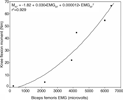
The EMG signals were full-wave rectified and averaged with a step of 10 milliseconds, yielding the averaged EMG (aEMG). The antagonist moment of the KFs was predicted using a method that the primary investigator (E.K.) and colleagues described in other studies.14,22 Briefly, for each angular position and muscle action (eccentric and concentric) in each subject, we used second-degree polynomials to fit the agonist hamstrings aEMG-moment curves formed at 6 levels of effort (Figure 1). The polynomials were used to predict the antagonist moment from the recorded antagonist aEMG. This method demonstrates a standard error of estimate that is less than 10%,14,23 and it provides reliable results in an adult population.22 Based on the results from the previous study of the primary investigator and colleagues,22 we assumed that the maximal KF moment is solely due to BF agonist activity. The coefficients of determination (r2) resulting from the fitting procedure ranged from .63 to .95.
Figure 1. Biceps femoris electromyography (EMGBF) to knee flexor moment (MKF) data (presented as points) from 1 subject at an angular position interval fitted using a second-degree polynomial (presented as a line).
Quantification of Antagonist to Agonist Moment Ratio
During isokinetic KE movement, the moment the dynamometer recorded represented the difference between the net moment exerted by the quadriceps (agonist KE [KEAGON]) and the hamstrings (antagonist KF [KFANT]):
 |
We estimated KFANT throughout ROM using the described procedure. After KFANT was estimated, the KEAGON moment was computed by rearranging the terms of Equation 1:
 |
After we determined KFANT and KEAGON moments, we calculated the KFANT:KEAGON ratio for each angular position interval throughout ROM.
Statistical Analysis
We considered the KFECC:KECON ratio to represent muscle balance during knee extension because it corresponds with the isokinetic test in which the quadriceps work concentrically as agonists. The KFCON:KEECC ratio was considered to represent knee flexion because it corresponds with the isokinetic test in which the quadriceps work eccentrically as agonists.
We used 2-way analysis of variance (ANOVA) to examine the effects of angular position and velocity on the KFECC: KECON and KFCON:KEECC ratios. The effects of movement direction (extension or flexion), angular position, and velocity on KFANT:KEAGON ratios were examined using a 3-way ANOVA. All statistical analyses were performed with SPSS (version 11.5; SPSS Inc, Chicago, IL). To further analyze the KFANT:KEAGON, KFECC:KECON, and KFCON:KEECC ratios, we calculated post hoc Tukey tests. The level of significance was set at P = .05.
RESULTS
Eccentric Knee Flexor to Concentric Knee Extensor Ratio
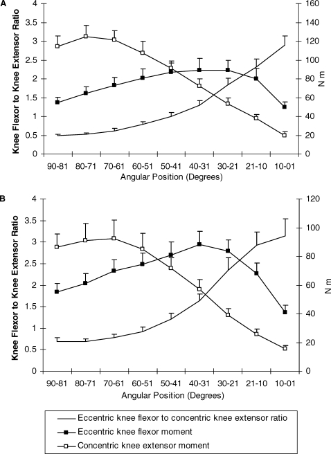
The KFECC:KECON ratios ranged from 0.48 ± 0.13 to 3.14 ± 1.95 (Figure 2). The main effect of angular velocity (F1,16 = 6.81, P = .019) and angular position (F1,8 = 41.49, P = .005) on KFECC:KECON ratios was significant. Post hoc Tukey tests indicated that the ratio (collapsed across angular positions) was significantly higher at 60°/s than at 180°/s (P = .02). Further, the ratios (collapsed across angular velocities) recorded at each angular position from 60° to 0° was significantly higher than the ratios recorded at angles from 90° to 71° (P = .002).
Figure 2. Eccentric knee flexor (KFECC) to concentric knee extensor (KECON) moment ratio, KFECC moment, and KECON moment during isokinetic knee extension at 60°/s (A) and 180°/s (B). Error bars indicate SD.
Concentric Knee Flexor to Eccentric Knee Extensor Ratio
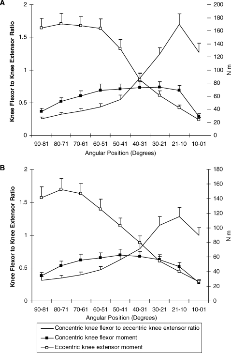
The KFCON:KEECC ratio throughout ROM ranged from 0.25 ± 0.12 to 1.69 ± 0.44 (Figure 3). The ANOVA results indicated a significant main effect of angular position (F1,8 = 78.03, P = .002) on KFCON:KEECC ratios. Post hoc Tukey tests indicated that the ratios (collapsed across angular velocities) recorded at angles from 50° to 0° were significantly higher than the ratios recorded at angles from 90° to 71° (P = .01).
Figure 3. Concentric knee flexor (KFCON) to eccentric knee extensor (KEECC) moment ratio, KFCON moment, and KEECC moment during isokinetic flexion at 60°/s (A) and 180°/s (B). Error bars indicate SD.
Antagonist Knee Flexor to Agonist Knee Extensor Ratios
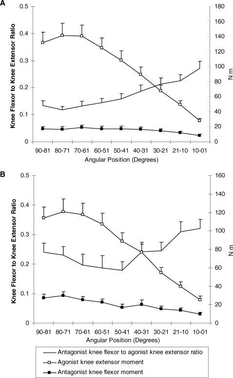
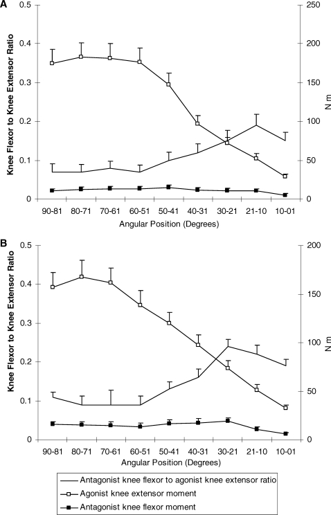
For knee extension, the KFANT:KEAGON ratio ranged from 0.18 ± 0.09 to 0.32 ± 0.21 (Figure 4). For knee flexion, the KFANT:KEAGON ranged from 0.06 ± 0.03 to 0.24 ± 0.17 (Figure 5). The ANOVA indicated a significant main effect of movement direction (F1,16 = 18.35, P = .001), angular velocity (F1,16 = 9.55, P = .007), and angular position (F8,128 = 12.55, P = .0007) on KFANT:KFAGON ratios. Post hoc Tukey tests revealed a higher ratio during knee extension than during flexion (P = .01). Ratios (collapsed across movement direction and angular positions) were higher at 60°/s than ratios recorded at 180°/s (P = .02). Further, the ratios recorded at angular positions from 0° to 30° were significantly higher than the ratios recorded at angles from 31° to 90° (P = .01). Finally, the ratios recorded at angular positions from 31° to 40° were higher than those recorded at angles from 41° to 80° (P = .03).
Figure 4. Antagonist knee flexor (KFANT) to agonist knee extensor (KEAGON) moment ratio, KFANT moment, and KEAGON moment during isokinetic knee extension at 60°/s (A) and 180°/s (B). Error bars indicate SD.
Figure 5. Antagonist knee flexor (KFANT) to agonist knee extensor (KEAGON) moment ratio, KFANT moment, and KEAGON moment during isokinetic knee flexion at 60°/s (A) and 180°/s (B). Error bars indicate SD.
DISCUSSION
We found that the KF:KE ratio increased significantly toward knee extension, with values exceeding 1.0, especially at 180°/s (Figures 2 and 3). The estimated KFANT:KEAGON ratios were lower (Figures 4 and 5), indicating that only part of the higher maximal moment capacity of the KFs relative to KEs is used when the KEs act as agonists.
Knee Flexor to Extensor Ratios
The KFECC:KECON ratio (Figure 2) and the KFCON:KEECC ratio (Figure 3) increased significantly as the knee approached extension. This finding corresponds with the findings of other researchers1,6,8 and could be explained by examining the moments that each muscle group exerted throughout the ROM. In particular, the KF moment was higher at angles from 40° to 30° of knee flexion, whereas the KE moment was higher at angles from 61° to 80° (Figures 2 and 3). Therefore, any conclusions regarding the balance of moments between the 2 antagonist muscle groups are highly dependent on the angular position examined. If the ratio is used as a predictor of knee joint or ACL injury, then the KF:KE ratio should be evaluated at angles ranging from 40° to 0°, where ACL strain is higher because of quadriceps forces.4,16
Antagonist Knee Flexor to Agonist Knee Extensor Ratio
The KFANT:KEAGON ratio increased as the knee approached full extension, reaching a value close to 0.30 (Figures 4 and 5). This increase was mainly due to a reduction in the KE moment as a result of the force-length relationship and the mechanical disadvantage of this muscle group near full knee extension.10 This indicates that, near full extension, the hamstrings produce approximately 25% to 30% of the moment that the quadriceps exert, and this supports previous findings that the hamstring muscles play a significant role in providing dynamic joint stabilization during active knee extension.4,6
Movement Direction Effects
Our finding that the KFECC:KECON ratio (Figure 2) was higher than the KFCON:KEECC ratio (Figure 3) confirms the findings of other researchers.1,6 Similarly, we noted that the KFANT:KEAGON ratio was lower during flexion (Figure 5) than during extension (Figure 4). This finding indicates that the hamstring muscles have a reduced capacity for dynamic knee joint stabilization during forceful knee flexion movements with simultaneous eccentric quadriceps muscle contraction.6 This reduced capacity could be attributed to the basic physiologic principles of the length-force relationship of skeletal muscle; the force-velocity relationship is characterized by eccentric contractions that generate higher forces than corresponding concentric contractions generate. Therefore, because of higher muscular forces exerted during eccentric actions, the ability of the hamstrings to produce antagonist muscle force might be critically important for maintaining joint stability during various movements that involve the knee actively flexing at high joint velocities.
Angular Velocity Effects
The results of our study indicate a decline in both KFECC: KECON (Figure 2) and KFANT:KEAGON (Figures 4 and 5) ratios from slow to fast angular velocities. This decline indicates that as angular velocity increases, the relative eccentric maximal moment capacity is higher for the KFs than for the KEs and can be attributed to the moment-velocity properties of human muscle. In our study, as the angular velocity increased, the concentric moment that the KEs produced declined, and the eccentric moment that the KFs produced remained unaltered. In contrast, the effect of angular velocity on the KFCON: KEECC ratio was not significant (Figure 3). Careful examination of the KF and KE moment data indicated that KFCON and KEECC moments showed a similar decline from 180°/s to 60°/s, which resulted in a similar KF:KE ratio at slow and fast speeds. Because the KFECC:KECON ratio and the KFCON: KEECC ratio are considered as indices of muscle balance during knee extension and flexion, respectively, our results, which correspond with the findings of Aagaard et al,6 indicate that angular velocity plays a more important role in the functional moment ratio during knee extension than during knee flexion.
Differences Between Knee Flexor to Extensor and Antagonist Knee Flexor to Agonist Knee Extensor Ratios
Our study shows that the information obtained using the KF:KE ratio was not the same as that obtained using EMG-based moments. The high KF:KE ratios indicate that the “braking” action of the hamstring muscles is equal in magnitude to the maximal quadriceps moment.1,6 This finding indicates that the maximal capacity of the hamstrings equals that of the quadriceps, so the hamstrings have the potential to stabilize the joint, when necessary. Furthermore, this capacity for muscular knee joint stabilization was augmented progressively at gradually more extended knee joint positions, as indicated by the very high KFECC:KECON ratios observed near full extension (Figures 2 and 3). However, if the quadriceps and the hamstrings produced the same amount of opposing moments around the knee, then the knee joint would be difficult to extend. In other words, as muscle coactivation increases, movement becomes less efficient.
In a typical movement, the agonist muscles produce the main force for the movement, and antagonist activity is higher at the initial and final phases of the movement to decelerate the limb and control the joint.24 Given the principle of reciprocal inhibition and the need for an optimal neuromuscular system, we suggest that the hamstrings would only produce the absolute necessary force to stabilize the knee and decelerate the limb.19,24,25 This explains the difference in values between the KF:KE and the KFANT:KEAGON ratios that we observed.
Implications for Training and Rehabilitation
One of the main targets of injury prevention or rehabilitation programs for the ACL is improving the strength of the hamstrings because of their role in counteracting anterior-directed ACL shear.10,12,16 Despite their limitations, isokinetics enable safe and reliable assessment of muscle strength, so using them to monitor training programs is recommended.2 However, research findings on the use of KF:KE ratios are conflicting. First, investigators1,26 generally disagree about the range of KF:KE ratio values in healthy, uninjured subjects. Second, they disagree about previous studies of the ability of the KF:KE ratio to discriminate between controls and subjects with knee ligament problems. For example, some researchers27,28 have found a higher KF:KE ratio in subjects with ACL reconstruction, and others29,30 have asserted that the role of moment improvement after ACL reconstruction is overestimated. Croisier et al31 showed that the KFECC:KECON ratio plays a significant role in the occurrence of hamstring muscle injury, but Hoskins and Pollard15 suggested that the evidence is inconclusive. Aagaard et al6 observed alterations in the KF: KE ratio after high-resistance exercise, but other investigators1,4,8,28,32,33 have stated the importance of identifying specific neural muscle activation patterns for dynamic joint stabilization. For example, St Clair Gibson et al34 attributed the lack of significant differences in the KFECC:KECON ratio between ACL-deficient and normal knees to altered muscular coordination strategies to maintain normal limb activity despite the strength losses. Because the KFANT:KEAGON ratio combines EMG activity of the antagonists with the moment capacity of each muscle group, it could be used to examine function of the knee or to monitor rehabilitation programs.10 Alterations of muscle coactivation patterns during and after a training program would provide an index of the neuromuscular adaptations to exercise. This information is not obtained when using the KF:KE ratios.
CONCLUSIONS
In conclusion, the KF:KE ratios that we found exceeded 1.0 near full knee extension, indicating an enhanced maximal force capacity of the hamstrings relative to the quadriceps. The EMG moment calibration technique that we applied indicated that, during active knee extension, the KFANT:KEAGON ratio increased toward knee flexion and reached a value close to 0.30, especially at the higher concentric angular velocity tested. Maximal moment ratios provide an index of balance ratios around the knee. However, examination of simultaneous balance of forces between the quadriceps and hamstrings can be better achieved when surface EMG measures are combined with isokinetic data.
REFERENCES
- Coombs R, Garbutt G. Developments in the use of the hamstring/quadriceps ratio for the assessment of muscle balance. J Sport Sci Med. 2002;1:56–62. [PMC free article] [PubMed] [Google Scholar]
- Kellis E, Baltzopoulos V. Isokinetic eccentric exercise. Sports Med. 1995;19:202–222. doi: 10.2165/00007256-199519030-00005. [DOI] [PubMed] [Google Scholar]
- Osternig LR, Caster BL, James CR. Contralateral hamstring (biceps femoris) coactivation patterns and anterior cruciate ligament dysfunction. Med Sci Sports Exerc. 1995;27:805–808. [PubMed] [Google Scholar]
- Solomonow M, Baratta R, D'Ambrosia R. The role of hamstrings in the rehabilitation of the anterior cruciate ligament-deficient knee in athletes. Sports Med. 1989;7:42–48. doi: 10.2165/00007256-198907010-00004. [DOI] [PubMed] [Google Scholar]
- Dvir Z, Eger G, Halperin N, Shklar A. Thigh muscle activity and anterior cruciate ligament insufficiency. Clin Biomech (Bristol, Avon) 1989;4:87–91. doi: 10.1016/0268-0033(89)90044-2. [DOI] [PubMed] [Google Scholar]
- Aagaard P, Simonsen EB, Magnusson SP, Larsson B, Dyhre-Poulsen P. A new concept for isokinetic hamstring:quadriceps muscle strength ratio. Am J Sports Med. 1998;26:231–237. doi: 10.1177/03635465980260021201. [DOI] [PubMed] [Google Scholar]
- Aagaard P, Simonsen EB, Trolle M, Bangsbo J, Klausen K. Isokinetic hamstring:quadriceps strength ratio: influence from joint angular velocity, gravity correction and contraction mode. Acta Physiol Scand. 1995;154:421–427. doi: 10.1111/j.1748-1716.1995.tb09927.x. [DOI] [PubMed] [Google Scholar]
- Hiemstra LA, Webber S, MacDonald PB, Kriellaars DJ. Hamstring and quadriceps strength balance in normal and hamstring anterior cruciate ligament-reconstructed subjects. Clin J Sport Med. 2004;14:274–280. doi: 10.1097/00042752-200409000-00005. [DOI] [PubMed] [Google Scholar]
- Beynnon BD, Fleming BC, Johnson RJ, Nichols CE, Renstrom PA, Pope MH. Anterior cruciate ligament strain behavior during rehabilitation exercises in vivo. Am J Sports Med. 1995;23:24–34. doi: 10.1177/036354659502300105. [DOI] [PubMed] [Google Scholar]
- Kellis E. Quantification of quadriceps and hamstring antagonist activity. Sports Med. 1998;25:37–62. doi: 10.2165/00007256-199825010-00004. [DOI] [PubMed] [Google Scholar]
- Solomonow M, Baratta B, Zhou BH. The synergistic action of the anterior cruciate ligament and thigh muscles in maintaining joint stability. Am J Sports Med. 1987;15:207–213. doi: 10.1177/036354658701500302. [DOI] [PubMed] [Google Scholar]
- Aagaard P, Simonsen EB, Andersen JL, Magnusson SP, Bojsen-Moller F, Dyhre-Poulsen P. Antagonist muscle coactivation during isokinetic knee extension. Scand J Med Sci Sports. 2000;10:58–67. doi: 10.1034/j.1600-0838.2000.010002058.x. [DOI] [PubMed] [Google Scholar]
- Doorenbosch CA, Harlaar J. A clinically applicable EMG-force model to quantify active stabilization of the knee after a lesion of the anterior cruciate ligament. Clin Biomech (Bristol, Avon) 2003;18:142–149. doi: 10.1016/s0268-0033(02)00183-3. [DOI] [PubMed] [Google Scholar]
- Kellis E, Baltzopoulos V. The effect of antagonist moment on the resultant knee joint moment during isokinetic testing of the knee extensors. Eur J Appl Physiol Occup Physiol. 1997;76:253–259. doi: 10.1007/s004210050244. [DOI] [PubMed] [Google Scholar]
- Hoskins W, Pollard H. The management of hamstring injury, part 1: issues in diagnosis. Man Ther. 2005;10:96–107. doi: 10.1016/j.math.2005.03.006. [DOI] [PubMed] [Google Scholar]
- Beynnon BD, Johnson RJ, Abate JA, Fleming BC, Nichols CE. Treatment of anterior cruciate ligament injuries, part 1. Am J Sports Med. 2005;33:1579–1602. doi: 10.1177/0363546505279913. [DOI] [PubMed] [Google Scholar]
- Faulkner RA. Maturation. In: Docherty D, ed. Measurements in Pediatric Exercise Science. Champaign, IL: Human Kinetics; 1996:129–158.
- Tanner JM. Growth at Adolescence. 2nd ed. Oxford, UK: Blackwell Scientific Publications; 1962.
- Winter DA. Biomechanics and Motor Control of Human Movement. 2nd ed. New York, NY: John Wiley & Sons; 1990:27–50.
- Robertson DGE. Body segment parameters. In: Robertson DGE, Caldwell GE, Hamill J, Kamen G, Whittlesey SN, eds. Research Methods in Biomechanics. Champaign, IL: Human Kinetics; 2004:55–71.
- Iossifidou A, Baltzopoulos V, Giakas G. Isokinetic knee extension and vertical jumping: are they related. J Sports Sci. 2005;23:1121–1127. doi: 10.1080/02640410500128189. [DOI] [PubMed] [Google Scholar]
- Kellis E, Kouvelioti V, Ioakimidis P. Reliability of a practicable EMG-moment model for antagonist moment prediction. Neurosci Lett. 2005;383:266–271. doi: 10.1016/j.neulet.2005.04.038. [DOI] [PubMed] [Google Scholar]
- Kellis E. Antagonist moment of force during maximal knee extension in pubertal boys: effects of quadriceps fatigue. Eur J Appl Physiol. 2003;89:271–280. doi: 10.1007/s00421-003-0795-5. [DOI] [PubMed] [Google Scholar]
- Enoka RM. Neuromechanics of Human Movement. 3rd ed. Champaign, IL: Human Kinetics; 2002.
- Smith AM. The coactivation of antagonist muscles. Can J Physiol Pharmacol. 1981;59:733–747. doi: 10.1139/y81-110. [DOI] [PubMed] [Google Scholar]
- Perrin DH. Isokinetic Exercise and Assessment. Champaign, IL: Human Kinetics; 1993.
- Patel RR, Hurwitz DE, Bush-Joshep CA, Bach BR, Jr, Andriacchi TP. Comparison of clinical and dynamic knee function in patients with anterior cruciate ligament deficiency. Am J Sports Med. 2003;31:68–74. doi: 10.1177/03635465030310012301. [DOI] [PubMed] [Google Scholar]
- Zhang LQ, Nuber GW, Bowen MK, Koh JL, Butler JP. Multiaxis muscle strength in ACL deficient and reconstructed knees: compensatory mechanism. Med Sci Sports Exerc. 2002;34:2–8. doi: 10.1097/00005768-200201000-00002. [DOI] [PubMed] [Google Scholar]
- Ciccotti M, Kerlan R, Perry J, Pink M. An electromyographic analysis of the knee during functional activities, II: the anterior cruciate ligament-deficient and -reconstructed profiles. Am J Sports Med. 1994;22:651–658. doi: 10.1177/036354659402200513. [DOI] [PubMed] [Google Scholar]
- Gokeler A, Schmalz T, Knopf E, Freiwald J, Blumentritt S. The relationship between isokinetic quadriceps strength and laxity on gait analysis parameters in anterior cruciate ligament reconstructed knees. Knee Surg Sports Traumatol Arthrosc. 2003;11:372–378. doi: 10.1007/s00167-003-0432-1. [DOI] [PubMed] [Google Scholar]
- Croisier JL, Forthomme B, Namurois MH, Vanderhommen M, Crielaard JM. Hamstring muscle strain recurrence and strength performance disorders. Am J Sports Med. 2002;30:199–203. doi: 10.1177/03635465020300020901. [DOI] [PubMed] [Google Scholar]
- Aagaard P. Training-induced changes in neural function. Exerc Sports Sci Rev. 2003;31:61–67. doi: 10.1097/00003677-200304000-00002. [DOI] [PubMed] [Google Scholar]
- Wojtys EM, Huston LJ, Taylor PD, Bastian SD. Neuromuscular adaptations in isokinetic, isotonic, and agility training programs. Am J Sports Med. 1996;24:187–192. doi: 10.1177/036354659602400212. [DOI] [PubMed] [Google Scholar]
- St Clair Gibson A, Lambert MI, Durandt JJ, Scales N, Noakes TD. Quadriceps and hamstrings peak torque ratio changes in persons with chronic anterior cruciate ligament deficiency. J Orthop Sports Phys Ther. 2000;30:418–427. doi: 10.2519/jospt.2000.30.7.418. [DOI] [PubMed] [Google Scholar]


