

Abstract
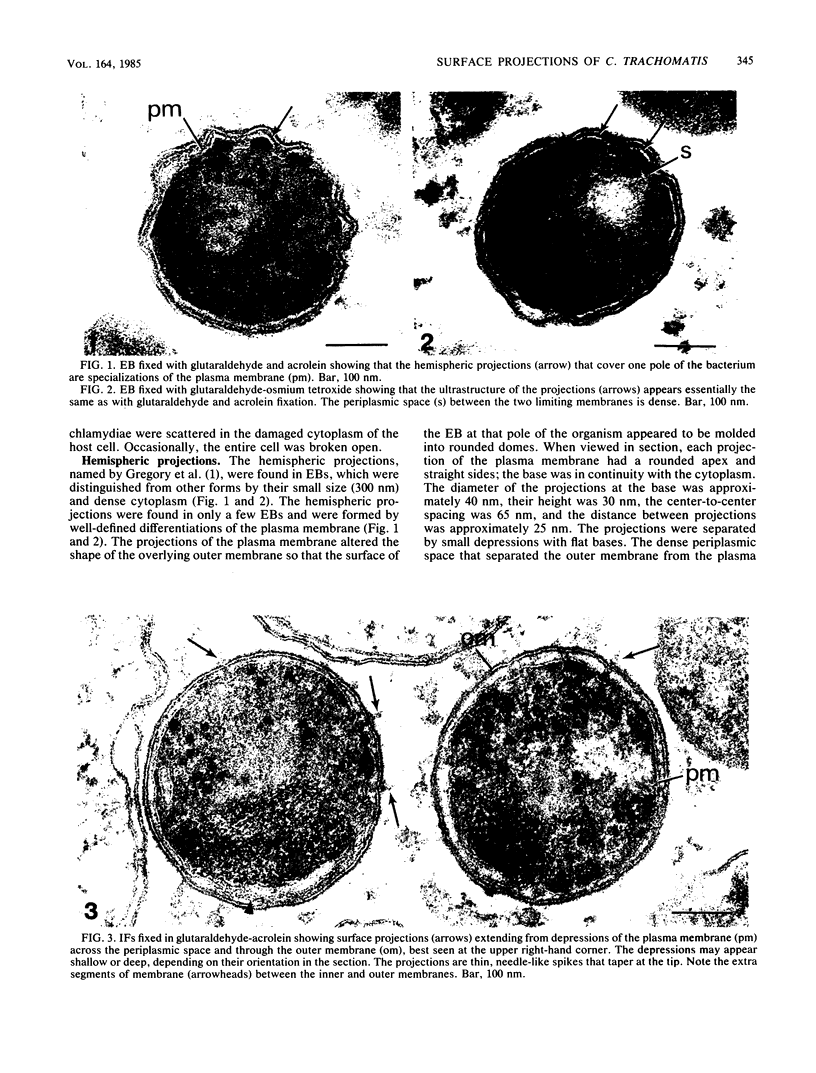
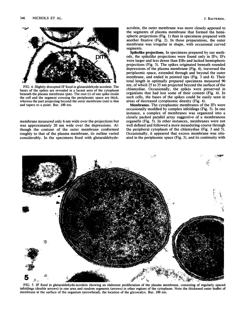
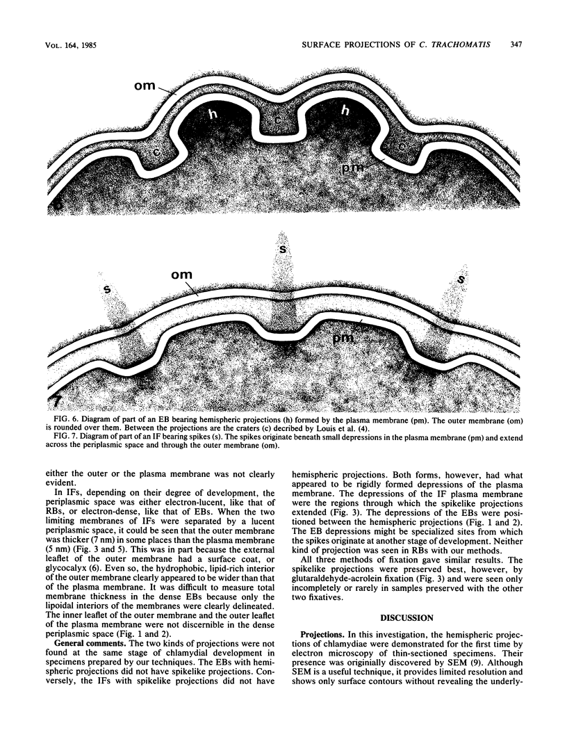
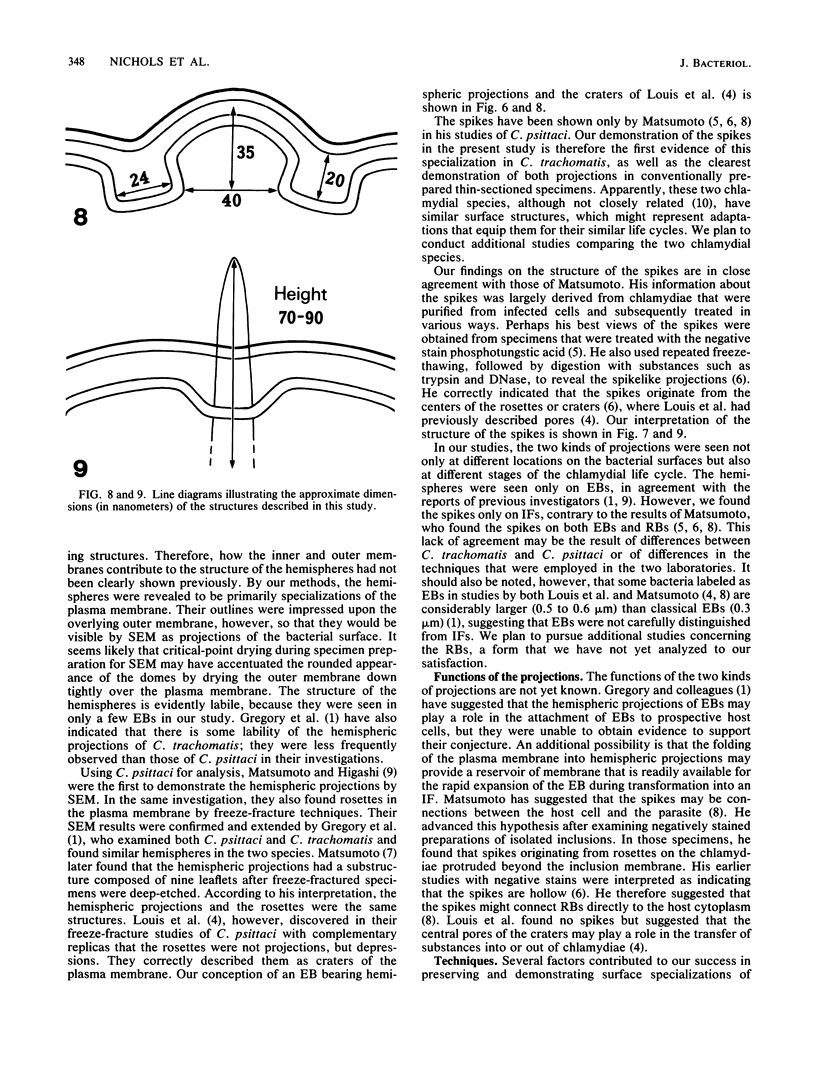
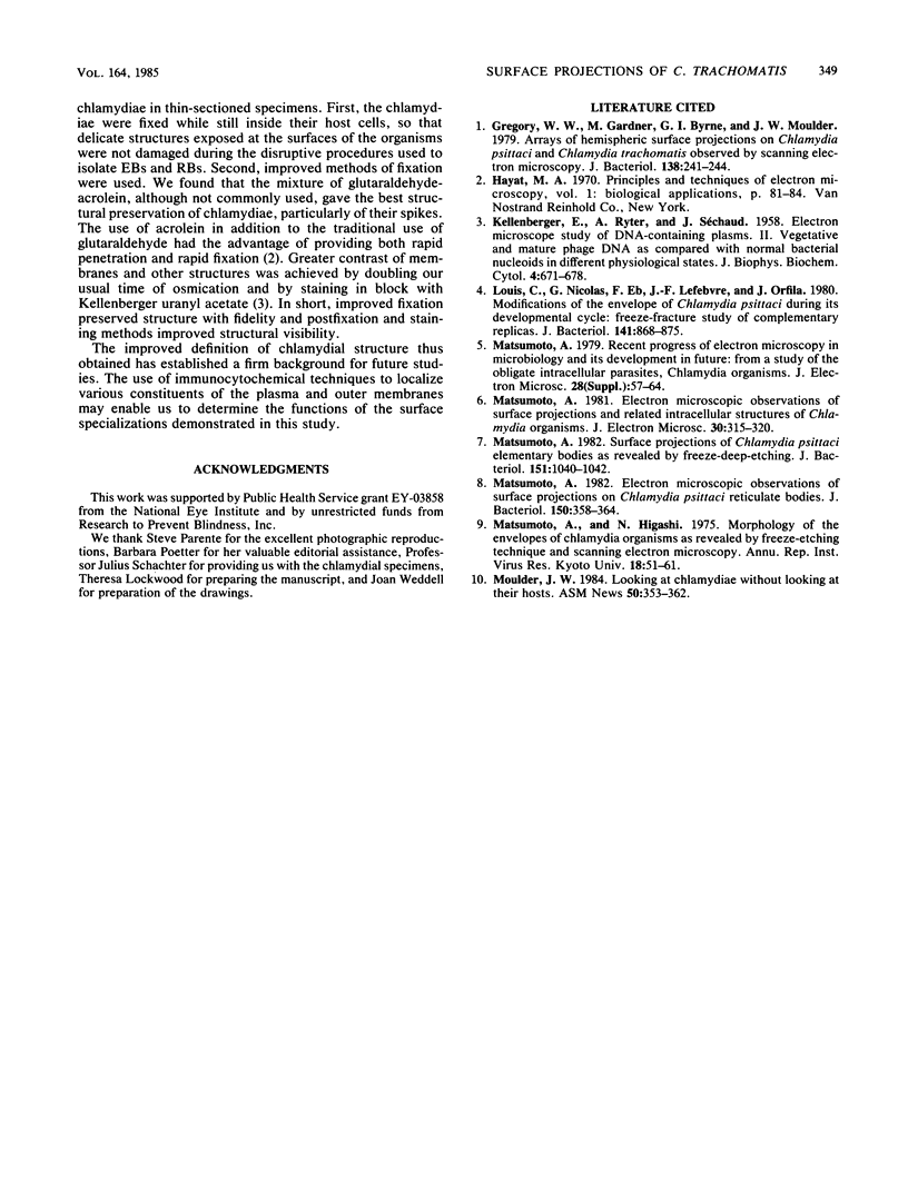
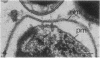
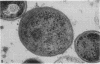
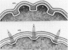
Two kinds of surface specializations of chlamydiae have been described: hemispheric projections and spikelike rods. We undertook the present studies to demonstrate chlamydial ultrastructure in greater detail in conventional thin-sectioned specimens. Chlamydia trachomatis (LGV strain L2/434/Bu), cultured for 40 h in L929 mouse fibroblasts, was fixed in glutaraldehyde-acrolein, p-formaldehyde-glutaraldehyde, or glutaraldehyde-osmium tetroxide mixtures, postfixed in osmium tetroxide, stained in uranyl acetate, dehydrated in ethanols, and embedded in Epon. By the use of fixatives that penetrate and fix rapidly, chlamydial outer and plasma membranes were clearly revealed. Our results indicate that the hemispheric projections are specializations of the plasma membrane of elementary bodies. The spikelike projections are found in intermediate forms, originate beneath depressions of the plasma membrane, and extend through the periplasmic space and outer membrane to end with pointed tips. Improved preservation of chlamydiae provides a new, informative view of their complex structure. Significant interactions between chlamydiae and host cells might be influenced by the surface structures shown in this study.
Full text
PDF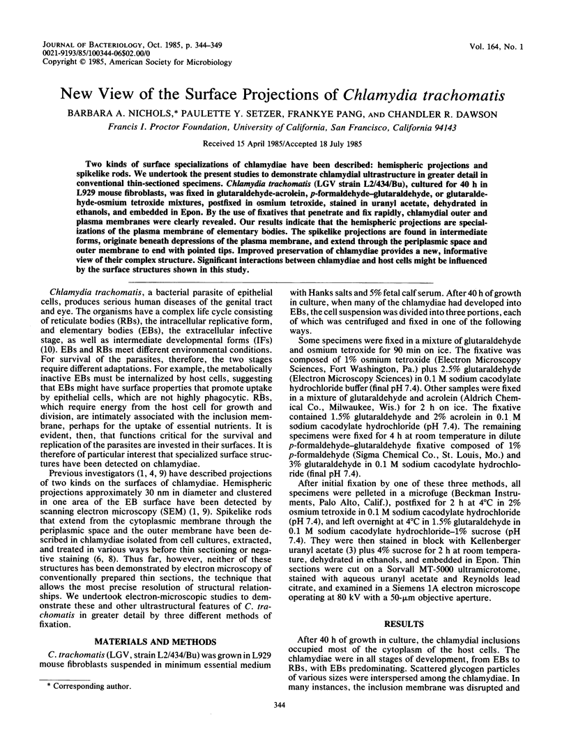
Images in this article
Selected References
These references are in PubMed. This may not be the complete list of references from this article.
- Gregory W. W., Gardner M., Byrne G. I., Moulder J. W. Arrays of hemispheric surface projections on Chlamydia psittaci and Chlamydia trachomatis observed by scanning electron microscopy. J Bacteriol. 1979 Apr;138(1):241–244. doi: 10.1128/jb.138.1.241-244.1979. [DOI] [PMC free article] [PubMed] [Google Scholar]
- KELLENBERGER E., RYTER A., SECHAUD J. Electron microscope study of DNA-containing plasms. II. Vegetative and mature phage DNA as compared with normal bacterial nucleoids in different physiological states. J Biophys Biochem Cytol. 1958 Nov 25;4(6):671–678. doi: 10.1083/jcb.4.6.671. [DOI] [PMC free article] [PubMed] [Google Scholar]
- Louis C., Nicolas G., Eb F., Lefebvre J. F., Orfila J. Modifications of the envelope of Chlamydia psittaci during its developmental cycle: freeze-fracture study of complementary replicas. J Bacteriol. 1980 Feb;141(2):868–875. doi: 10.1128/jb.141.2.868-875.1980. [DOI] [PMC free article] [PubMed] [Google Scholar]
- Matsumoto A. Electron microscopic observations of surface projections and related intracellular structures of Chlamydia organisms. J Electron Microsc (Tokyo) 1981;30(4):315–320. [PubMed] [Google Scholar]
- Matsumoto A. Electron microscopic observations of surface projections on Chlamydia psittaci reticulate bodies. J Bacteriol. 1982 Apr;150(1):358–364. doi: 10.1128/jb.150.1.358-364.1982. [DOI] [PMC free article] [PubMed] [Google Scholar]
- Matsumoto A. Surface projections of Chlamydia psittaci elementary bodies as revealed by freeze-deep-etching. J Bacteriol. 1982 Aug;151(2):1040–1042. doi: 10.1128/jb.151.2.1040-1042.1982. [DOI] [PMC free article] [PubMed] [Google Scholar]