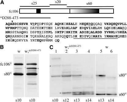
Figure 5.—
Processing of endogenous fc106 and fc106ΔV288–E473 derivatives in wild-type egg chambers carrying the fc106ΔV288–E473 transgene. (A) A schematic of fc106 highlighting an alanine/proline-rich segment in the s25 region and the tandem repeat in the s60 region. The discontinuity in the line below the schematic indicates the region that was deleted in the fc106ΔV288–E473 transgene (*D288–473). The amino acids in the s20 region are shown at the bottom; those that are identical in D. melanogaster and D. virilis are in boldface type. Amino acids previously deleted in a dec-1 mutant transgene (ΔS456–E473, Badciong et al. 2001) are underlined. The downward arrowhead denotes the beginning of s80 (S281). (B and C) Western blots incubated with the Cfc106 antiserum depicted in Figure 1. (B) Western blot of SDS-soluble proteins of stage 10 egg chambers from white females (w) and white flies carrying the fc106ΔV288–E473 transgene (w;*D288–473); proteins were separated using the Mini-Protean electrophoresis cell. The positions of the variant forms of fc106 and s80 are indicated. An aberrant fc106 proprotein produced by the fc106ΔV288–E473 transgene is indicated (*). (C) Developmental Western blot of SDS-soluble proteins from flies designated as in B. The positions of variant forms of s80 and s60 are indicated; the positions of the fc106ΔV288–E473 proprotein and its s60-like C-terminal derivative are indicated (*). Egg chamber stages are shown at the bottom of each lane.

