

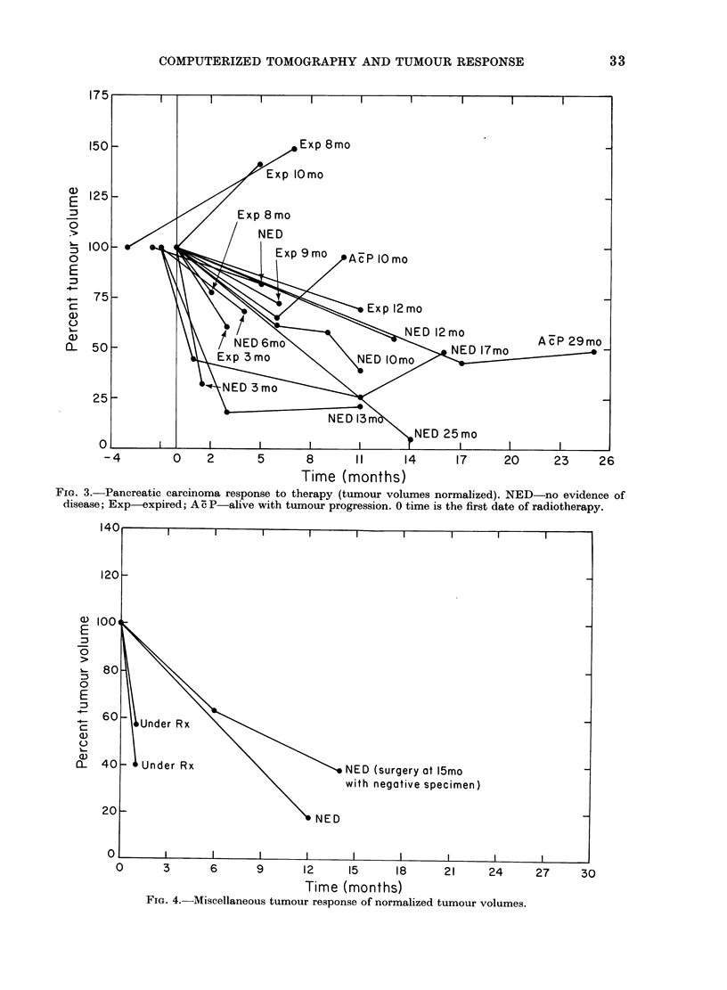
Abstract
While Computerized Tomographic (CT) scans have been a valuable asset to radiotherapy treatment planning, their role in the critical assessment of tumour response has been less well defined. Recent refinements in radiotherapy treatment techniques have intensified the need for non-invasive methods to evaluate the local control of inaccessible neoplasms. In this study, 20 cases in which both pre- and post-treatment CT scans were available up to 27 months post-therapy were reviewed. The majority of the patients had pancreatic carcinoma. In both pre- and post-therapy scans, the tumour volume was calculated from contours outlined on sequential CT slices. These interpretations were compared with surgical findings, clinical course and patient survival. The rate of the primary tumour regression had little bearing on the clinical outcome. However, there was CT scan evidence of initial local tumour stabilization or regression in > 80% of the cases. In addition, CT scan evidence of tumour progression was correlated with clinical progression in > 90% of the patients. This suggests that a growth delay analysis might be possible with the use of CT scans, at least for the evaluation of pancreatic carcinoma.
Full text
PDF


Images in this article

Selected References
These references are in PubMed. This may not be the complete list of references from this article.
- Chen G. T., Singh R. P., Castro J. R., Lyman J. T., Quivey J. M. Treatment planning for heavy ion radiotherapy. Int J Radiat Oncol Biol Phys. 1979 Oct;5(10):1809–1819. doi: 10.1016/0360-3016(79)90564-9. [DOI] [PubMed] [Google Scholar]
- van Peperzeel H. A., Breur K., Broerse J. J., Barendsen RBE values of 15 MeV neutrons for responses of pulmonary metastases in patients. Eur J Cancer. 1974 Jun;10(6):349–355. doi: 10.1016/0014-2964(74)90198-4. [DOI] [PubMed] [Google Scholar]