

Abstract
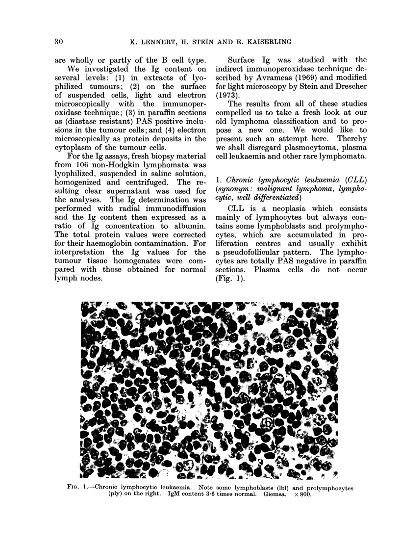
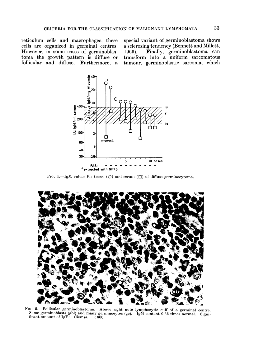
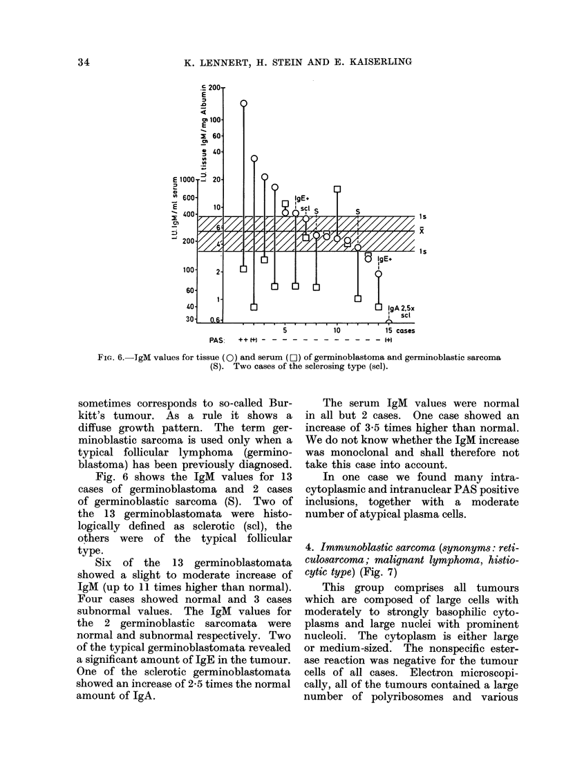
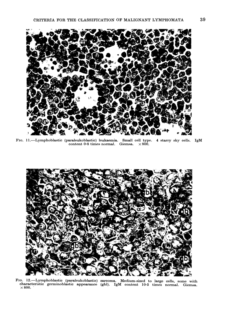
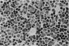
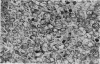
The subtle morphology and functional properties of cells are the best parameters to use for their definition. This is also true for the corresponding tumours, especially malignant lymphomata. In studies of 106 cases of malignant lymphoma we therefore applied as morphological methods haematological staining (Giemsa in sections and imprints) and electron microscopic analysis. As functional criteria we used the nonspecific esterase reaction to define tumours of histiocytes and an estimation of the immunoglobulin content of tissue extracts and single cells to define tumours of B lymphocytes and their derivatives. By combining all of these methods it was possible to propose a new classification. Whereas not one histiocytic malignant lymphoma ("reticulosarcoma") was found in the series, at least most of the malignant lymphomata investigated seemed to be derived from the B lymphocyte system. The following types are distinguished: (1) Chronic lymphocytic leukaemia; (2) diffuse germinocytoma (malignant lymphoma, lymphocytic, intermediate); (3) germinoblastoma (follicular, follicular and diffuse, diffuse; sclerotic, nonsclerotic) which can show a transition into germinoblastic sarcoma; (4) immunoblastic sarcoma of the B cell type (previously called reticulo-sarcoma); (5) lymphoplasmocytoid immunocytoma, which may be associated with Waldenstrom's macroglobulinaemia and can show a mixed cellularity; (6) lymphoblastic (paraleukoblastic) sarcoma and leukaemia, which are, at least in most cases, probably neoplasias of germinoblasts. All of these lymphomata can produce immunoglobulins. Sixty-seven cases showed an Ig increase in the tumour. This was mostly IgM, but sometimes IgG, IgA, IgD and/or IgE. A morphological equivalent of even abnormal Ig secretion is the globular positive (diastase resistant) PAS reaction in lymphoid cells (not in the histiocytes) in paraffin sections, which we found in 43 cases. Only 19 of the 63 cases with an IgM increase in the tumour showed an increase of IgM in the serum. Waldenström's macroglobulinaemia is a facultative symptom of morphologically different malignant lymphomata and should therefore be considered only as a clinical syndrome and not as a nosological entity.
Full text
PDF


Images in this article


Selected References
These references are in PubMed. This may not be the complete list of references from this article.
- Avrameas S. Coupling of enzymes to proteins with glutaraldehyde. Use of the conjugates for the detection of antigens and antibodies. Immunochemistry. 1969 Jan;6(1):43–52. doi: 10.1016/0019-2791(69)90177-3. [DOI] [PubMed] [Google Scholar]
- Bennett M. H., Millett Y. L. Nodular sclerotic lymphosarcoma: a possible new clinico-pathological entity. Clin Radiol. 1969 Jul;20(3):339–343. doi: 10.1016/s0009-9260(69)80155-8. [DOI] [PubMed] [Google Scholar]
- Kim H., Heller P., Rappaport H. Monoclonal gammopathies associated with lymphoproliferative disorders: a morphologic study. Am J Clin Pathol. 1973 Mar;59(3):282–294. doi: 10.1093/ajcp/59.3.282. [DOI] [PubMed] [Google Scholar]
- LENNERT K. Die pathologische Anatomie der Makroglobulinämie Waldenström. Frankf Z Pathol. 1955;66(3):201–226. [PubMed] [Google Scholar]
- Stein H., Drescher S. Darstellung von Oberflächen-IgM an Blutlymphozyten mit der Immuno-Peroxydase-Methode. Blut. 1973 Jan;26(1):35–42. doi: 10.1007/BF01631309. [DOI] [PubMed] [Google Scholar]