Abstract
There is an urgent need for the development of new drugs for the treatment of tropical parasitic diseases such as Chagas' disease and leishmaniasis. One potential drug target in the organisms that cause these diseases is sterol biosynthesis. This paper describes the design and synthesis of quinuclidine derivatives as potential inhibitors of a key enzyme in sterol biosynthesis, squalene synthase (SQS). A number of compounds that were inhibitors of the recombinant Leishmania major SQS at submicromolar concentrations were discovered. Some of these compounds were also selective for the parasite enzyme rather than the homologous human enzyme. The compounds inhibited the growth of and sterol biosynthesis in Leishmania parasites. In addition, we identified other quinuclidine derivatives that inhibit the growth of Trypanosoma brucei (the causative organism of human African trypanosomiasis) and Plasmodium falciparum (a causative agent of malaria), but through an unknown mode(s) of action.
Leishmaniasis and Chagas' disease are parasitic diseases caused by the protozoan parasites Leishmania spp. and Trypanosoma cruzi, respectively. Together, both diseases are responsible for high rates of mortality and morbidity, especially in tropical regions of the world. With increasing problems due to resistance and clinical efficacy, the drugs currently used to treat these diseases are becoming increasingly less effective, resulting in the urgent need for new drug candidates in this area.
A particular area of interest are the enzymes of the sterol biosynthesis pathway; these provide attractive targets because the parasites that cause these diseases have ergosterol and other 24-alkylated sterols as the principal sterols present in the plasma membrane, while humans have cholesterol. Encouragingly, a number of enzymes in the sterol biosynthetic pathway have been studied as potential drug targets in these organisms and have shown great promise. Thus, 14α-demethylase (9, 17-20, 29, 30, 38, 41, 42, 45), sterol 24-methyltransferase (9, 20-24, 32, 44, 46, 48), 3-Hydroxy-3-methyl-glutaryl coenzyme A reductase (8, 40), squalene epoxidase (18, 39), squalene synthase (SQS) (7, 31, 33, 36, 37), and farnesyl pyrophosphate synthase (25-27, 50) have been studied both individually and in combination, with various degrees of success.
The enzyme SQS, which catalyzes the condensation of two molecules of farnesyl pyrophosphate to produce squalene, has been identified as a potential drug target for the inhibition of cholesterol biosynthesis in humans (5). The activities of a variety of compounds, including bisphosphonates, benzylamines, squalestatins, and quinuclidines, against mammalian enzymes have been investigated.
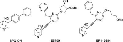
One class of compounds whose activities against mammalian SQS have been studied extensively are the arylquinuclidines. These compounds are protonated at physiological pH and are thought to mimic a high-energy carbocation intermediate in the reaction pathway. We are interested in this class of molecules and recently demonstrated that 3-biphenyl-4-yl-3-hydroxyquinuclidine (BPQ-OH) (Fig. 1) is a noncompetitive inhibitor of Leishmania mexicana and T. cruzi SQS (Kis, 12 to 62 nM), blocks sterol biosynthesis, and concomitantly inhibits the growth of L. mexicana promastigotes and T. cruzi epimastigotes (7, 33, 37). We have also shown that other biphenylquinuclidines (analogues of BPQ-OH) displayed inhibition of L. major SQS (31) and showed growth inhibition of L. mexicana promastigotes. Furthermore, quinuclidine derivatives developed by Eisai Company, Ltd. (Tokyo, Japan) as cholesterol and triglyceride-lowering agents (E5700 and ER119884; Fig. 1) have been shown to have activity against T. cruzi in vitro, and one derivative was able to prevent the development of parasitemia and induced full survival in a rodent model of acute Chagas' disease (36) (Fig. 1).
FIG. 1.
Structures of the SQS inhibitors.
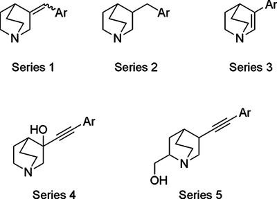
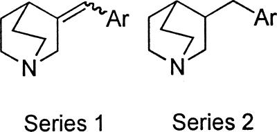
Following on from these initial structure-activity relationship studies, we have designed and synthesized five further series of quinuclidines derivatives (Fig. 2). In this paper we present our evaluation of the new derivatives as potential antiparasitics. The aim of these studies was to discover compounds which are selective for the parasite enzyme and to elucidate structure-activity relationships.
FIG. 2.
Structures of the series of quinuclidine derivatives prepared.
MATERIALS AND METHODS
Preparation of compounds.
The preparation of compounds is described at http://www.lifesci.dundee.ac.uk/groups/ian_gilbert/Supporting_Information_aac0205.pdf.
Assays against recombinant SQS.
Experimental details for the assay with Leishmania major SQS have been reported previously (31). A standard SQS activity assay preparation contained 50 mM phosphate buffer (pH 7.4), 20 mM MgCl2, 5 mM 3-[(3-cholamidopropyl)dimethylammonio]-1-propanesulfonate, 1% Tween 80, 10 mM dithiothreitol, 0.025 mg/ml bovine serum albumin, 0.25 mM NADPH, 2.1 mM glucose-6-phosphate, 0.125 mg/ml glucose-6-phosphate dehydrogenase, and 0.5 μM farnesyl pyrophosphate (10,080 dpm/pmol) as the substrate. Soluble protein extracts of Escherichia coli cells expressing a double-truncated version of L. major that lacks 16 residues at the N terminus and 40 residues at the C terminus were used as the enzyme source, as described previously (31). The reaction was started with the protein extract, and the final volume of the reaction mixture was 200 μl. After incubation at 37°C for 10 min, 40 μl of 10 M NaOH was added, followed by the addition of 10 μl of a (50:1) mixture of 70% ethanol and squalene. The resulting mixtures were vigorously mixed by vortexing, and then 20-μl aliquots were applied to channels (2.5 by 10 cm) of a silica gel thin-layer chromatogram and the newly formed squalene was separated from the unreacted substrate by chromatography in toluene-ethyl acetate (9:1). The region of the squalene band was removed, immersed in Hydrofluor liquid scintillation fluid, and assessed for radioactivity by using a Pharmacia LKB liquid scintillation counter. Negative controls were reaction mixtures containing soluble extracts of E. coli BL21(DE3) RP cells transformed with pET28a (which does not overexpress L. major SQS). No activity was observed by using this extract as an enzyme source. The 50% inhibitory concentrations (IC50s) were calculated from a hyperbolic plot of the percent inhibition versus the concentration of the inhibitor. For IC50 determinations, five different concentrations of inhibitor were tested in duplicate, and the experiment was performed twice in most cases.
Human SQS activity was determined by using the same conditions described above for the Leishmania enzyme. Extracts of E. coli BL21(DE3 pLysS) cells transformed with the expression system pHSS16 (35) were used as the enzyme source.
Growth inhibition of L. mexicana promastigotes.
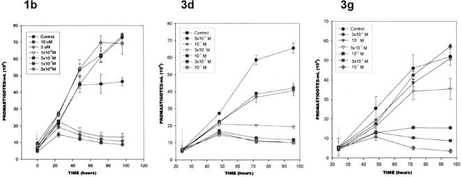
Experimental details for assays of the growth inhibition of L. mexicana promastigotes have been reported previously (31). L. mexicana amazonensis promastigotes were cultivated in liver infusion-tryptose medium supplemented with lactabulmin and 10% fetal calf serum (Gibco) (3) at 26°C, without agitation. The cultures were initiated with a cell density of 2 × 10⁶ cells per ml, and the drug was added at a cell density of 0.5 × 10⁷ to 1 × 10⁷ cells per ml. Cell densities were measured with an electronic particle counter (model ZBI; Coulter Electronics Inc., Hialeah, FL) and by direct counting with a hemocytometer. Cell viability was monitored by the detection of trypan blue exclusion by light microscopy. The growth experiments were carried out in triplicate, and the standard deviation of the cell densities at each time point are given by the error bars (see Fig. 3 and the growth curves at http://www.lifesci.dundee.ac.uk/groups/ian_gilbert/Supporting_Information_aac0205.pdf).
FIG. 3.
Inhibition of the growth of L. mexicana promastigotes in the presence of compounds 1b, 3d, and 3g.
Sterol composition analysis of L. mexicana promastigotes treated with inhibitors.
For the analysis of the effects of the drugs on the lipid compositions of the promastigotes, total lipids from control and drug-treated cells were extracted and fractionated into neutral and polar lipid fractions by silicic acid column chromatography and gas-liquid chromatography (19, 20, 42, 43). The neutral lipid fractions were first analyzed by thin-layer chromatography (on Merck 5721 silica gel plates with heptane-isopropyl ether-glacial acetic acid [60:40:4] as the developing solvent) and conventional gas-liquid chromatography (by isothermic separation in a 4-m glass column packed with 3% OV-1 on Chromosorb 100/200 mesh and nitrogen as the carrier gas at 24 ml/min and with flame ionization detection in a Varian 3700 gas chromatograph). For quantitative analysis and structural assignments, the neutral lipids were separated in a capillary high-resolution column (25 m by 0.20 mm [inner diameter] Ultra-2 column, 5% phenyl-methyl-siloxane, 0.33-μm film thickness) in a Hewlett-Packard 6890 Plus gas chromatograph equipped with an HP5973A mass-sensitive detector. The lipids were injected in chloroform, and the column was kept at 50°C for 1 min, and then the temperature was increased to 270°C at a rate of 25°C·min−1 and finally to 300°C at a rate of 1°C·min−1. The carrier gas (He) flow was kept constant at 0.5 ml·min−1. The injector temperature was 250°C, and the detector was kept at 280°C.
Growth inhibition of Leishmania donovani axenic amastigotes.
Experimental details for assays of the growth inhibition of Leishmania donovani axenic amastigotes have been reported previously (15). Amastigotes of Leishmania donovani strain MHOM/ET/67/L82 were grown in axenic culture at 37°C in synthetic minimal medium (12), at pH 5.4, supplemented with 10% heat-inactivated fetal bovine serum under an atmosphere of 5% CO2 in air. One hundred microliters of culture medium with 1 × 10⁵ amastigotes from axenic culture with or without a serial drug dilution was seeded in 96-well microtiter plates. Seven threefold dilutions covering a concentration range from 30 to 0.041 μg/ml were used. After 72 h of incubation, 10 μl of resazurin solution (12.5 mg resazurin dissolved in 100 ml phosphate-buffered saline [PBS]) was added to each well. The plates were incubated for another 2 to 4 h and read with a Spectramax Gemini XS microplate fluorometer (Molecular Devices Cooperation, Sunnyvale, CA) by using an excitation wavelength of 536 nm and an emission wavelength of 588 nm. The data were analyzed by using the software Softmax Pro (Molecular Devices Cooperation). The IC50s were calculated from the sigmoidal inhibition curves.
Growth inhibition of T. cruzi intracellular amastigotes.
Experimental details for assays of the growth inhibition of T. cruzi intracellular amastigotes have been reported previously (15). Rat skeletal myoblasts (L-6 cells) were seeded in 96-well microtiter plates at 2,000 cells/well/100 μl in RPMI 1640 medium with 10% fetal bovine serum and 2 mM l-glutamine. After 24 h, 5,000 trypomastigotes of T. cruzi (Tulahuen strain C2C4 containing the β-galactosidase [lacZ] gene) were added in aliquots of 100 μl per well with 2× of a serial drug dilution. The plates were incubated at 37°C in 5% CO2 for 4 days. Then the substrate chlorophenol red-β-d-galactopyranoside/Nonidet was added to the wells. The color reaction, which developed during the following 2 to 4 h, was read photometrically at 540 nm. IC50 values were calculated from the sigmoidal inhibition curve by using Microsoft Excel software.
Growth inhibition of bloodstream form of Trypanosoma brucei rhodesiense.
Experimental details for assays of the growth inhibition of the bloodstream form of Trypanosoma brucei rhodesiense have been reported previously (15). Minimum essential medium (50 μl), supplemented with 2-mercaptoethanol and 15% heat-inactivated horse serum as described by Baltz et al. (3), was added to each well of a 96-well microtiter plate. Serial drug dilutions were added to the wells. Then 50 μl of trypanosome suspension (T. b. rhodesiense STIB 900) was added to each well and the plate was incubated at 37°C under a 5% CO2 atmosphere for 72 h. Ten microliters of resazurin solution (12.5 mg resazurin dissolved in 100 ml PBS) was then added to each well, and incubation was continued for an additional 2 to 4 h (39). The plates were read in a microplate fluorescence scanner (Spectramax Gemini XS; Molecular Devices) by using an excitation wavelength of 536 nm and an emission wavelength of 588 nm. IC50 values were calculated from the sigmoidal inhibition curve.
Growth inhibition of Plasmodium falciparum.
Experimental details for assays of the growth inhibition of P. falciparum have been reported previously (15). Antiplasmodial activity was determined by using the K1 strain of P. falciparum (which is resistant to chloroquine and pyrimethamine). A modification of the [3H]hypoxanthine incorporation assay was used (13, 28). Briefly, infected human red blood cells in RPMI 1640 medium with 5% lipid enriched bovine serum albumin (Albumax) were exposed to serial drug dilutions in microtiter plates. After 48 h of incubation at 37°C in a reduced oxygen atmosphere, 0.5 μCi [3H]hypoxanthine was added to each well. The cultures were incubated for a further 24 h before they were harvested onto glass-fiber filters and washed with distilled water. The radioactivity was counted by using a Betaplate liquid scintillation counter (Wallac, Zurich, Switzerland). The results were recorded as the counts per minute per well at each drug concentration and are expressed as a percentage of that for the untreated controls. IC50 values were calculated from the sigmoidal inhibition curves by using Microsoft Excel software.
Cytotoxicity against mammalian cells.
Experimental details for assays for cytotoxicity against mammalian cells have been reported previously (15). Rat skeletal myoblasts (L-6 cells) were seeded in 96-well microtiter plates in RPMI 1640 medium with 10% fetal bovine serum and 2 mM l-glutamine at a density of 4 × 10⁴ cells/ml. After 24 h, the medium was removed and replaced by fresh medium containing a serial drug dilution, and the plate was incubated at 37°C under a 5% CO2 atmosphere for 72 h. Ten microliters of resazurin solution (12.5 mg resazurin dissolved in 100 ml PBS) was then added to each well, and incubation was continued for an additional 2 to 4 h. The plates were read in a microplate fluorescence scanner (Spectramax Gemini XS; Molecular Devices) by using an excitation wavelength of 536 nm and an emission wavelength of 588 nm. IC50 values were calculated from the sigmoidal inhibition curve.
The in vitro IC50 determination assays (L. donovani axenic amastigotes, T. cruzi intracellular amastigotes, bloodstream form of T. b. rhodesiense, P. falciparum, and L-6 cells) were run in duplicate, and the reference drug was always included. For compounds showing at least moderate activity (activity at approximately <15 μM), an independent replicate assay was performed. The values for the active compounds represent the averages of four determinations (two determinations of two independent experiments). The variation factor of the two independent assays was <2.
RESULTS
Enzyme assays.
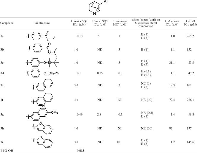
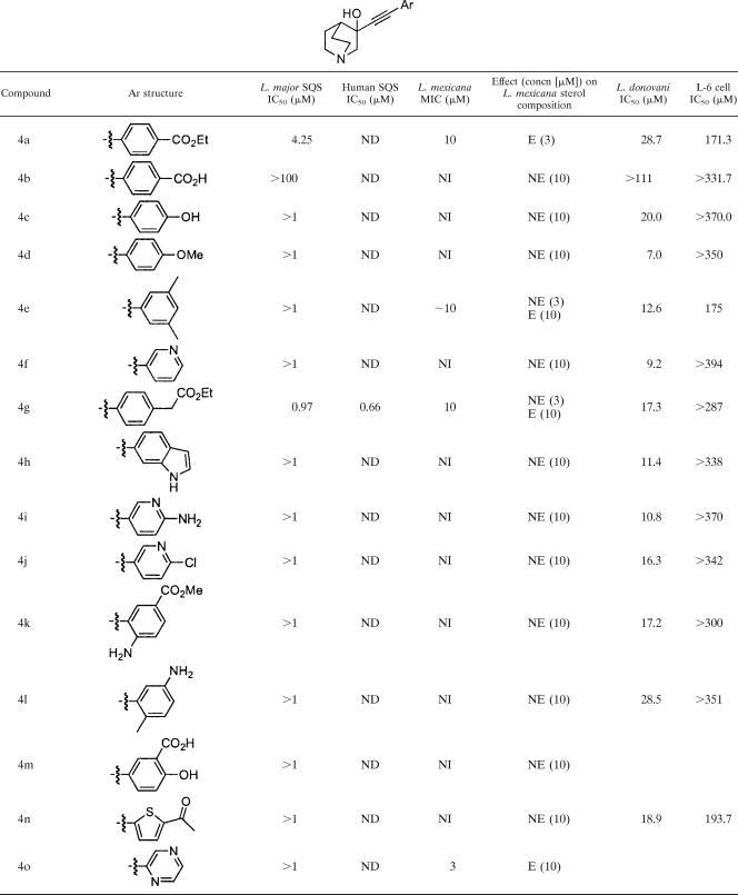
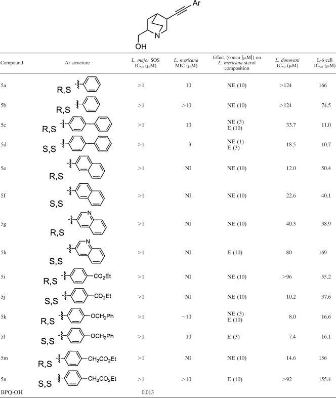
Compounds were assayed against the recombinant L. major enzyme, which was overexpressed in Escherichia coli. The assay was most readily performed with cell extracts rather than the purified enzyme, owing to stability problems with the purified enzyme (31). No SQS is present in E. coli, so there is no interference with any host enzyme. Compounds that exhibited IC50 values of less than 1 μM were also evaluated against the recombinant human enzyme to determine their selectivity. No compound in series 1 or 2 was inhibitory at a concentration of less than 1 μM. Presumably, the extra methylene group is suboptimal compared to the structure of BPQ-OH, and in the case of series 1, the double bond may hold the quinuclidine substituent in the wrong orientation for interaction with the enzyme active site (Table 1). Compounds of series 3 showed the highest activity against the L. major enzyme, with compounds 3a, 3d, and 3g giving IC50 values below 1 μM (Table 2). These all appeared to be selective for the parasite enzyme over the human enzyme. Among the compounds in series 4, compound 4g also inhibited the enzyme at submicromolar concentrations, although the compound was not selective for the parasite enzyme (Table 3). None of the compounds in series 5 showed significant inhibition of the enzyme (Table 4), probably due to the hydroxymethyl substituent undergoing unfavorable interactions with the enzyme.
TABLE 1.
Structures of compounds prepared; inhibition of recombinant SQS for compounds of series 1 and series 2; activities against L. mexicana promastigotes, axenic L. donovani amastigotes, and L-6 cells; and effects on the sterol compositions of L. mexicana promastigotesa
NI, no inhibition, with MIC >10 μM; NE, no effect on sterol composition compared to that for control at the concentration stated; E, effect on sterol composition at the concentration stated, where an effect is considered if there is a change in cholesterol levels of greater than 10 percentage points. Control compounds were miltefosin (IC50 = 0.338 μM) for L. donovani and podophyllotoxin (IC50 = 0.014 μM) for L-6 cells. The IC50s for human SQS were not determined.
TABLE 2.
Structures of compounds prepared; inhibition of recombinant SQS for compounds of series 3; activities against L. mexicana promastigotes, axenic L. donovani amastigotes, and L-6 cells; and effects on the sterol compositions of L. mexicana promastigotesa
See footnote a of Table 1 for explanations and the definitions of the abbreviations. ND, not determined; Ph, phenyl; OMe, methoxy.
TABLE 3.
Structures of compounds prepared; inhibition of recombinant SQS for compounds of series 4; activities against L. mexicana promastigotes, axenic L. donovani amastigotes, and L-6 cells; and effects on the sterol compositions of L. mexicana promastigotesa
See footnote a of Table 1 for explanations and the definitions of the abbreviations. ND, not determined; Et, ethyl; OMe, methoxy; Me, methyl.
TABLE 4.
Structures of compounds prepared; inhibition of recombinant SQS for compounds of series 5; activities against L. mexicana promastigotes, axenic L. donovani amastigotes, and L-6 cells; and effects on the sterol compositions of L. mexicana promastigotesa
See footnote a of Table 1 for explanations and the definitions of the abbreviations. The IC50s for human SQS were not determined. Et, ethyl; Ph, phenyl.
Taken together, the results suggest that the compounds that were active had a hydrophobic substituent on the aromatic ring, although not all compounds with hydrophobic substituents were active. Also, the relative orientation of the aromatic ring and the quinuclidine ring is important.
Interestingly, the Eisai Co. compounds, compounds ER119884 and E5700 (Fig. 1) (provided by Tsukuba Research Laboratories, Eisai Co., Ltd., Ibaraki, Japan), had activities comparable to that of BPQ-OH against the parasite enzyme (Table 1).
Whole-cell assays. (i) L. mexicana promastigotes.
Compounds were screened against L. mexicana promastigotes in modified liver infusion-tryptose medium (31). A number of compounds showed MICs of 10 μM or less. The most potent compounds were compounds 1b (MIC, 0.3 μM), 3a (MIC, 1 μM), 3d (MIC range, 0.1 to 0.3 μM), and 3g (MIC range, 0.3 to 1 μM). The growth curves for parasites treated with these compounds are shown in Fig. 3, and those for the other compounds are shown at http://www.lifesci.dundee.ac.uk/groups/ian_gilbert/Supporting_Information_aac0205.pdf. Series 3 gave rise to the greatest number of compounds with activities against the promastigote form of the parasite. In this series of compounds, the aromatic group is directly attached to the quinuclidine functionality. Compounds with the greatest inhibition of growth of L. mexicana tended to have nonpolar aromatic functionalities. Thus, for example, compounds 3e and 3f are very similar, except that compound 3e has an aromatic substituent, while compound 3f has a quinoline substituent, yet compound 3e had an MIC of 3 μM, while compound 3f had an MIC of >10 μM. The more polar substituent appeared to reduce the activity of the compound. This is more clearly seen in series 4, in which most of the aromatic groups contain a polar substituent, and these were almost all inactive; the exception to this was compound 4o, which has a 1,4-pyrimidine functionality and which had modest but detectable activity.
(ii) Sterol composition.
L. mexicana promastigotes were also used to study the effects of SQS inhibitors on the sterol compositions of the parasites. In these experiments, the sterol compositions of parasites grown in the presence of inhibitors were investigated by gas-liquid chromatography with mass spectrometry. The sterol composition is important information, as it gives an indication of the effects of the inhibitors on the cells and their probable cellular modes of action.
L. mexicana strains predominantly have the 24-alkylated sterols episterol and 5-dehydroepisterol in their cell membranes (Fig. 4) (36, 37). If compounds inhibit SQS, then a reduction in the proportion of these 24-alkylated sterols to (exogenous) cholesterol would be predicted at concentrations near the MIC. This would provide indirect confirmation that the mode of action of these quinuclidine analogues is through the inhibition of sterol (ergosterol) biosynthesis, although it does not necessarily indicate the inhibition of SQS; the disruption of ergosterol biosynthesis could also be due to inhibition of another enzyme in the pathway. Compounds were investigated at concentrations near their MICs.
FIG. 4.
Structures of the main sterols present in Leishmania mexicana promastigotes. Cholesterol is acquired from the growth medium.
In general, compounds that inhibited L. major SQS and the growth of L. mexicana promastigotes also reduced the levels of 24-alkylated sterols (5-dehydroepisterol and episterol) at the MIC. These results are presented in Table 5 for compounds 1b, 3d, and 3g, which were particularly active against the intact parasite; data for the other compounds are shown at http://www.lifesci.dundee.ac.uk/groups/ian_gilbert/Supporting_Information_aac0205.pdf. and are summarized in Tables 1 to 4. These data indicate that the predominant (or at the very least significant) mode of action of these compounds is inhibition of SQS. An interesting exception is compound 4s. This compound inhibited the growth of L. mexicana at a lower level (MIC, ∼1 μM) than that at which it had an effect on the sterol composition (∼10 μM), indicating that it had a mode of action other than the inhibition of sterol biosynthesis. Conversely, compound 5l had a pronounced effect on the sterol composition at 3 μM, but the MIC was 10 μM, indicating that the parasite can still remain viable with some degree of changes to the sterol composition.
TABLE 5.
Effects of BPQ-OH and compounds 1b, 3d, and 3g on the free sterol composition of Leishmania mexicana (NR) promastigotesa
| Sterol source and sterol | Composition (mass %) after treatment with the following compound at the indicated concn:
|
|||||||||||
|---|---|---|---|---|---|---|---|---|---|---|---|---|
| BPQ-OH
|
1b
|
3d
|
3g
|
|||||||||
| 0 μM | 1 μM | 3 μM | 0 μM | 0.1 μM | 0.3 μM | 0 μM | 0.1 μM | 0.3 μM | 0 μM | 0.3 μM | 1 μM | |
| Exogenous, cholesterol | 13.9 | 17.5 | 39.6 | 13.9 | 31.4 | 36.7 | 7.4 | 31.6 | 52.9 | 17.2 | 13.9 | 41.2 |
| Endogenous | ||||||||||||
| 5-Dehydro episterol | 67.3 | 65.7 | 39.6 | 67.3 | 26.4 | 18.1 | 82.7 | 53.0 | 24.2 | 70.8 | 72.7 | 43.0 |
| Episterol | 12.5 | 16.8 | 20.8 | 12.5 | 42.2 | 33.3 | 8.9 | 15.4 | 22.9 | 12.0 | 13.4 | 15.8 |
The sterols were extracted from cells exposed to the indicated drug concentration for 96 h; they were separated from polar lipids by silicic acid column chromatography and analyzed by quantitative capillary gas-liquid chromatography and mass spectrometry.
(iii) L. donovani amastigotes.
The compounds were also screened against L. donovani axenic amastigotes, which is used as a model of the clinically relevant intracellular amastigote stage of leishmaniasis. Compounds of series 1 and 2 showed little activity against L. donovani axenic amastigotes (Table 1). This is in agreement with the results of the enzyme assays, where none of these series showed significant activity at 1 μM.
For series 3, there was improved activity, with compounds 3a, 3b, 3d, 3g, and 3i showing growth inhibition (IC50) at concentrations on the order of 1 μM (Table 2). For series 4, the activity was again reduced, with only compounds 4d, 4q, 4r, and 4s showing IC50s under 10 μM (Table 3). Finally, for series 5, none of the compounds showed significant activity (Table 4).
(iv) Other parasites.
As part of routine screening, the compounds were also assayed against other parasites. The inhibition of ergosterol biosynthesis in T. cruzi amastigotes is a drug target in this organism; hence, inhibitors of sterol biosynthesis would be expected to have an effect against these parasites. Against intracellular T. cruzi amastigotes, compounds 1b, 1c, 2b, 3d, 3g, 4a, 4q, 4r, 5b, 5c, 5d, 5e, 5f, 5k, and 5l showed activities at concentrations below 10 μM (Table 6).
TABLE 6.
Activities of compounds against intracellular T. cruzi amastigotes, bloodstream form of Trypanosoma brucei rhodesiense, and Plasmodium falciparum cultured in red blood cellsa
| Compound | IC50 (μM)
|
||
|---|---|---|---|
| T. cruzi | T. b. rhodesiense | P. falciparum | |
| 1a | >151 | 16.0 | 0.8 |
| 1b | 1.8 | 0.7 | 3.1 |
| 1c | 2.8 | 0.5 | 0.9 |
| 1d | >132 | 13.1 | >22 |
| 1e | >111 | 12.5 | 14.2 |
| 1f | 30.2 | 10.3 | 10.0 |
| 1g | 46.0 | 5.5 | 12.3 |
| 2a | 130 | 60.6 | 5.4 |
| 2b | 4.7 | 0.7 | 2.8 |
| 2c | 25.4 | 2.5 | 1.8 |
| 3a | 16.0 | 35.1 | 11.6 |
| 3b | 23 | 7.2 | 14 |
| 3c | 16.3 | 2.1 | 4.1 |
| 3d | 3.1 | 1.1 | 4.8 |
| 3e | 34.1 | 24.2 | 11.0 |
| 3f | 51.1 | 22.4 | 15.0 |
| 3g | 10.2 | 2.2 | 9.0 |
| 3h | 61.3 | 17.1 | 6.2 |
| 3i | 64.2 | 5.6 | 12.0 |
| 4a | 5.6 | 3.3 | 4.0 |
| 4b | 90.2 | 146.3 | >18.4 |
| 4c | 90.4 | 53.0 | 18.7 |
| 4d | 67.2 | 18.4 | 8.2 |
| 4e | 18.9 | 9.2 | 4.3 |
| 4f | >131 | 173 | 18.9 |
| 4g | 25.6 | 257.8 | 10.0 |
| 4h | 26.2 | 19.9 | 7.1 |
| 4i | 97 | 5.7 | >21 |
| 4j | >114 | 53.3 | 9.3 |
| 4k | 44.9 | 30.8 | 10.9 |
| 4l | >117 | 62.4 | 11.7 |
| 4n | 72.6 | 30.3 | 10.2 |
| 4p | 21.2 | 5.4 | 5.9 |
| 4q | 5.6 | 0.5 | 0.7 |
| 4r | 9.6 | 1.9 | 1.9 |
| 4s | 13.4 | 3.4 | 2.0 |
| 5a | 25.6 | 14.5 | 6.5 |
| 5b | 8.2 | 12.8 | 3.3 |
| 5c | 5.8 | 0.5 | 1.4 |
| 5d | 5.6 | 1.2 | 0.6 |
| 5e | 5.3 | 1.4 | 3.0 |
| 5f | 4.7 | 1.1 | 2.1 |
| 5g | 33.8 | 10.9 | 2.7 |
| 5h | 19.1 | 7.1 | 1.7 |
| 5i | 18.8 | 3.8 | 1.1 |
| 5j | 19.4 | 1.7 | 0.6 |
| 5k | 3.1 | 1.2 | 1.1 |
| 5l | 3.4 | 1.2 | 0.6 |
| 5m | 22.9 | 32.3 | 5.2 |
| 5n | 50 | 64.7 | 5.0 |
The control compounds were benznidazole (IC50 = 1.36 μM) for T. cruzi, melarsoprol (IC50 = 0.008 μM) for T. b. rhodesiense, and chloroquine (IC50 = 0.083 μM) for P. falciparum.
The compounds were also assayed against the bloodstream form of T. b. rhodesiense and also against P. falciparum. The bloodstream form of T. b. rhodesiense is not thought to biosynthesize ergosterol but to acquire it from the human host (10, 11). Therefore, inhibitors of SQS would not be expected to have an effect on parasite growth. However, compounds 1b, 1c, 2b, 4q, and 5c showed IC50 values of <1 μM, while compounds 1g, 2c, 3b, 3c, 3d, 3g, 3i, 4a, 4e, 4i, 4p, 4r, 4s, 5d, 5e, 5f, 5 h, 5i, 5j, 5k, and 5l gave IC50 values between 1 and 10 μM (Table 6).
Finally, the compounds were screened against Plasmodium falciparum, although it is known that this parasite lacks the enzymatic machinery for sterol biosynthesis. Compounds 1a, 1c, 4q, 5d, 5j, and 5l showed IC50 values below 1 μM (Table 6).
DISCUSSION
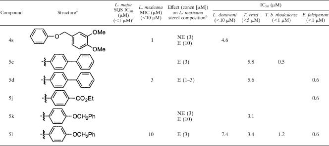
During the course of these studies, we have prepared five different series of quinuclidine derivatives. A number of different laboratories have reported that quinuclidine derivatives are inhibitors of SQS, and these compounds have been investigated as potential agents for the reduction of high cholesterol levels in humans. We have previously reported on the activities of several quinuclidine derivatives against Leishmania and T. cruzi, and here we report on the activities of a much more extensive range of analogues. The results for the most active compounds from these assays are summarized in Table 7.
TABLE 7.
Summary of activities of the active compounds in the various assays
a Ph, phenyl; OMe, methoxy; Et, ethyl.
b E, effect on sterol composition; NE, no effect on sterol composition.
c Concentrations in parentheses are those below which the compounds were active against the indicated organism or enzyme.
The compounds were evaluated for their abilities to inhibit the recombinant L. major enzyme. Relatively few compounds inhibited the enzyme at nanomolar concentrations. This is in contrast to our standard compound, BPQ-OH, which had an IC50 value of 0.013 μM. However, a number of compounds showed activities at submicromolar concentrations: compounds 3a, 3d, 3g, and 4g. These compounds were evaluated against the human enzyme; and compounds 3a, 3d, and 3g were found to exhibit selectivity for the parasite enzyme. It was also found that the sterol composition of L. mexicana promastigotes was markedly affected by compounds 3a, 3d, 3g, and 4g, again suggesting the inhibition of sterol biosynthesis as a primary mode of action. These compounds also caused growth inhibition of L. mexicana promastigotes and also of L. donovani axenic amastigotes. These data imply that inhibition of SQS leads to changes in the sterol composition of Leishmania, which leads to cellular death. However, some compounds were shown to affect the sterol composition at concentrations at which there was no inhibition of SQS, which could imply other modes of action of the compounds. This finding needs to be further investigated.
Other compounds (compounds 1b, 3b, 3c, 3i, 4q, 4r, 4s, and 5l) also affected the sterol composition of L. mexicana promastigotes and inhibited the growth of L. mexicana promastigotes and L. donovani axenic amastigotes, albeit at slightly higher concentrations. Where tested, these compounds did not inhibit the recombinant enzyme in the cell-free assays at 1 μM. This could indicate that the compounds inhibit the enzyme at a concentration only slightly higher than 1 μM, that the enzyme assay is slightly less sensitive than the sterol composition analysis, that the compounds are subject to concentration in the cells, or that the compounds are inhibiting another step in the sterol biosynthesis pathway.
Humans acquire cholesterol from the diet as well as from “de novo” biosynthesis, so it may not be necessary to have compounds which are selective for the parasite enzyme. However, the use of compounds which are selective for the parasite SQS over the human SQS would minimize any potential risks of toxicity. Thus, SQS inhibitors have been reported to increase farnesol and farnesol-derived dicarboxylic acid levels owing to the increase in farnesyl pyrophosphate levels and limited consumption by other enzymes involved in isoprenoid metabolism (2, 4, 6, 16, 47). These effects were clearly species dependent, and in certain instances very low levels of or no dioic acid excretion was observed (1, 34). More studies are required to determine the toxicities of the different classes of SQS inhibitors, but selective inhibitors of the parasite enzyme would probably avoid any potential problems.
Compounds 1b, 3a, 3d, and 3g represent lead molecules in the search for selective inhibitors of the Leishmania SQS. These compounds were also shown to have significant activities against L. mexicana promastigotes and L. donovani axenic amastigotes and were also found to cause a reduction in the levels of 24-alkylated sterols in L. mexicana promastigotes. All these data are consistent with the mode of action being inhibition of SQS. Compound 4g also inhibited the L. major SQS at submicromolar concentrations; but it was not as effective against the intact parasites, as it gave an MIC of approximately 10 μM against L. mexicana promastigotes and an IC50 of approximately 17 μM against L. donovani axenic amastigotes. The discrepancy may be related to the limited penetration of the compound into whole cells. The difference between the growth inhibition of the L. mexicana promastigotes and that of the axenic L. donovani amastigotes may be either species related or a consequence of the different metabolism of the promastigote and the axenic amastigote forms.
A number of compounds also inhibited the growth of T. cruzi intracellular amastigotes. T. cruzi, like Leishmania, has an active ergosterol biosynthetic machinery; and hence, inhibitors of SQS would be expected to show an effect on the growth inhibition of the parasite. Not all of the compounds that inhibited the growth of L. mexicana promastigotes or L. donovani axenic amastigotes were active against the intracellular T. cruzi amastigotes studied here. This may be due to the intracellular nature of the parasite, with either the access of some compounds being prevented or the compounds being metabolized before they reach T. cruzi. However, all the compounds which were active against T. cruzi also had an effect on the sterol composition of L. mexicana promastigotes (Table 7). This suggests that these compounds have the potential to inhibit L. mexicana SQS, although it cannot be ruled out that there are differences in the inhibition of the L. major and the T. cruzi SQSs or that these compounds have a different mode of action in T. cruzi.
The compounds were also evaluated against the bloodstream form of T. brucei and against P. falciparum as part of our routine screening program. The bloodstream form of T. brucei is thought to scavenge sterols from the human host, although procyclic T. brucei has an active ergosterol biosynthetic pathway that includes the enzyme SQS. Interestingly, we have recently shown that although the bloodstream form of T. brucei scavenges sterols from the human host and does not synthesize ergosterol, it does expresses sterol 24-methyltransferase, another enzyme involved in ergosterol biosynthesis (14). P. falciparum, in contrast, lacks the enzymes involved in sterol biosynthesis beyond farnesyl diphosphate synthase, as inferred from genomic data. Therefore, the activity against these organisms (T. brucei and P. falciparum) was not due to the inhibition of SQS but was due to the effect of the quinuclidines on some other molecular target. Compounds that inhibited the growth of P. falciparum were different from those that inhibited T. brucei, suggesting different mechanisms of action. However, there was a large overlap in the compounds that inhibited T. cruzi and T. b. rhodesiense, indicating that there may be similar modes of action in these parasites.
Conclusions.
We have prepared some quinuclidines which inhibit leishmanial SQS, disrupt endogenous sterol biosynthesis, and cause the inhibition of the growth of Leishmania parasites, suggesting that inhibition of sterol biosynthesis is a valid drug target in these organisms. We have now identified compounds that are selective for the parasite enzyme over the human enzyme. Should it be possible to optimize the activity further, then this may give rise to potential agents for the treatment of leishmaniasis. Quinuclidines also inhibit the growth of other parasites, although by a different mode of action.
Acknowledgments
We acknowledge the EU INCO-DEV programmme (ICA4-2000-10028), the Plan Nacional de Investigación (SAF2004-03828), and PRONEX/CNPq/FAPERJ for financial support. This investigation received financial support from the UNICEF/UNDP/World Bank/WHO Special Programme for Research and Training in Tropical Diseases and the EPSRC National Mass Spectrometry Service Centre in Swansea, United Kingdom, for accurate mass spectrometry.
We thank John F. Thompson (Pfizer) for kindly providing the expression system for human SQS.
Footnotes
Published ahead of print on 20 August 2007.
REFERENCES
- 1.Amano, Y., T. Nishimoto, R. Tozawa, E. Ishikawa, Y. Imura, and Y. Sugiyama. 2003. Lipid-lowering effects of TAK-475, a squalene synthase inhibitor, in animal models of familial hypercholesterolemia. Eur. J. Pharmacol. 466:155-161. [DOI] [PubMed] [Google Scholar]
- 2.Amin, D., R. Z. Rutledge, S. N. Needle, H. F. Galczenski, K. Neuenschwander, A. C. Scotese, M. P. Maguire, R. C. Bush, D. J. Hele, G. E. Bilder, and M. H. Perrone. 1997. RPR 107393, a potent squalene synthase inhibitor and orally effective cholesterol-lowering agent: comparison with inhibitors of HMG-CoA reductase. J. Pharmacol. Exp. Ther. 281:746-752. [PubMed] [Google Scholar]
- 3.Baltz, T., D. Baltz, C. Giroud, and J. Crockett. 1985. Cultivation in a semi-defined medium of animal infective forms of Trypanosoma brucei, T. equiperdum, T. evansi, T. rhodesiense and T. gambiense. EMBO J. 4:1273-1277. [DOI] [PMC free article] [PubMed] [Google Scholar]
- 4.Bergstrom, J. D., M. M. Kurtz, D. J. Rew, A. M. Amend, J. D. Karkas, R. G. Bostedor, V. S. Bansal, C. Dufresne, F. L. Vanmiddlesworth, O. D. Hensens, J. M. Liesch, D. L. Zink, K. E. Wilson, J. Onishi, J. A. Milligan, G. Bills, L. Kaplan, M. N. Omstead, R. G. Jenkins, L. Huang, M. S. Meinz, L. Quinn, R. W. Burg, Y. L. Kong, S. Mochales, M. Mojena, I. Martin, F. Pelaez, M. T. Diez, and A. W. Alberts. 1993. Zaragozic acids—a family of fungal metabolites that are picomolar competitive inhibitors of squalene synthase. Proc. Natl. Acad. Sci. USA 90:80-84. [DOI] [PMC free article] [PubMed] [Google Scholar]
- 5.Biller, S. A., K. Neuenschwander, M. M. Ponpipom, and C. D. Poulter. 1996. Squalene synthase inhibitors. Curr. Pharm. Design 2:1-40. [Google Scholar]
- 6.Bostedor, R. G., J. D. Karkas, B. H. Arison, V. S. Bansal, S. Vaidya, J. I. Germershausen, M. M. Kurtz, and J. D. Bergstrom. 1997. Farnesol-derived dicarboxylic acids in the urine of animals treated with zaragozic acid A or with farnesol. J. Biol. Chem. 272:9197-9203. [DOI] [PubMed] [Google Scholar]
- 7.Braga, M. V., J. A. Urbina, and W. de Souza. 2004. Effects of squalene synthase inhibitors on the growth and ultrast7ructure of Trypanosoma cruzi. Int. J. Antimicrob. Agents 24:72-78. [DOI] [PubMed] [Google Scholar]
- 8.Concepcion, J. L., D. Gonzalez-Pacanowska, and J. A. Urbina. 1998. 3-Hydroxy-3-methyl-glutaryl-CoA reductase in Trypanosoma (Schizotrypanum) cruzi: subcellular localization and kinetic properties. Arch. Biochem. Biophys. 352:114-120. [DOI] [PubMed] [Google Scholar]
- 9.Contreras, L. M., J. Vivas, and J. A. Urbina. 1997. Altered lipid composition and enzyme activities of plasma membranes from Trypanosoma (Schizotrypanum) cruzi epimastigotes grown in the presence of sterol biosynthesis inhibitors. Biochem. Pharmacol. 53:697-704. [DOI] [PubMed] [Google Scholar]
- 10.Coppens, I., and P. J. Courtoy. 2000. The adaptative mechanisms of Trypanosoma brucei for sterol homeostasis in its different life-cycle environments. Annu. Rev. Microbiol. 54:129-156. [DOI] [PubMed] [Google Scholar]
- 11.Coppens, I., and P. J. Courtoy. 1995. Exogenous and endogenous sources of sterols in the culture-adapted procyclic trypomastigotes of Trypanosoma brucei. Mol. Biochem. Parasitol. 73:179-188. [DOI] [PubMed] [Google Scholar]
- 12.Cunningham, I. 1977. New culture medium for maintenance of tsetse tissues and growth of trypanosomatids. J. Protozool. 24:325-329. [DOI] [PubMed] [Google Scholar]
- 13.Desjardins, R. E., C. J. Canfield, D. Haynes, and J. Chulay. 1979. Quantitative assessment of antimalarial activity in vitro by a semiautomated microdilution technique. Antimicrob. Agents Chemother. 16:710-718. [DOI] [PMC free article] [PubMed] [Google Scholar]
- 14.Gros, L., V. M. Castillo-Acosta, C. J. Jimenez, M. Sealey-Cardona, S. Vargas, A. M. Estevez, V. Yardley, L. Rattray, S. L. Croft, L. M. Ruiz-Perez, J. A. Urbina, I. H. Gilbert, and D. Gonzalez-Pacanowska. 2006. New azasterols against Trypanosoma brucei: role of 24-sterol methyltransferase in inhibitor action. Antimicrob. Agents Chemother. 50:2595-2601. [DOI] [PMC free article] [PubMed] [Google Scholar]
- 15.Jones, S. M., J. E. Urch, M. Kaiser, R. Brun, J. L. Harwood, C. Berry, and I. H. Gilbert. 2005. Analogues of thiolactomycin as potential antimalarial agents. J. Med. Chem. 48:5932-5941. [DOI] [PubMed] [Google Scholar]
- 16.Keller, R. K. 1996. Squalene synthase inhibition alters metabolism of nonsterols in rat liver. Biochim. Biophys. Acta Lipids Lipid Metab. 1303:169-179. [DOI] [PubMed] [Google Scholar]
- 17.Larralde, G., J. Vivas, and J. A. Urbina. 1988. Concentration and time-dependence of the effects of ketoconazole on growth and sterol synthesis by Trypanosoma (Schizotrypanum) cruzi epimastigotes. Acta Cientifica Venezolana 39:140-146. [PubMed] [Google Scholar]
- 18.Lazardi, K., J. A. Urbina, and W. Desouza. 1990. Ultrastructural alterations induced by two ergosterol biosynthesis inhibitors, ketoconazole and terbinafine, on epimastigotes and amastigotes of Trypanosoma (Schizotrypanum) cruzi. Antimicrob. Agents Chemother. 34:2097-2105. [DOI] [PMC free article] [PubMed] [Google Scholar]
- 19.Liendo, A., K. Lazardi, and J. A. Urbina. 1998. In-vitro antiproliferative effects and mechanism of action of the bis-triazole D0870 and its S(−) enantiomer against Trypanosoma cruzi. J. Antimicrob. Chemother. 41:197-205. [DOI] [PubMed] [Google Scholar]
- 20.Liendo, A., G. Visbal, M. M. Piras, R. Piras, and J. A. Urbina. 1999. Sterol composition and biosynthesis in Tryanosoma cruzi amastigotes. Mol. Biochem. Parasitol. 104:81-91. [DOI] [PubMed] [Google Scholar]
- 21.Lorente, S. O., C. J. Jimenez, L. Gros, V. Yardley, K. de Luca-Fradley, S. L. Croft, J. A. Urbina, L. M. Ruiz-Perez, D. G. Pacanowska, and I. H. Gilbert. 2005. Preparation of transition-state analogues of sterol 24-methyl transferase as potential anti-parasities. Bioorg. Med. Chem. 13:5435-5453. [DOI] [PubMed] [Google Scholar]
- 22.Lorente, S. O., J. C. F. Rodrigues, C. J. Jimenez, M. Joyce-Menekse, C. Rodrigues, S. L. Croft, V. Yardley, K. de Luca-Fradley, L. M. Ruiz-Perez, J. Urbina, W. de Souza, D. G. Pacanowska, and I. H. Gilbert. 2004. Novel azasterols as potential agents for treatment of leishmaniasis and trypanosomiasis. Antimicrob. Agents Chemother. 48:2937-2950. [DOI] [PMC free article] [PubMed] [Google Scholar]
- 23.Magaraci, F., C. Jimenez Jimenez, C. O. Rodrigues, C. F. Rodrigues, M. Vianna Braga, V. Yardley, K. de Luca-Fradley, S. L. Croft, W. de Souza, L. M. Ruiz-Perez, J. A. Urbina, D. Gonzalez Pacanowska, and I. H. Gilbert. 2003. Azasterols as inhibitors of sterol 24-methyltransferase in Leishmania and Trypanosoma cruzi. J. Med. Chem. 46:4714-4727. [DOI] [PubMed] [Google Scholar]
- 24.Maldonado, R. A., J. Molina, G. Payares, and J. A. Urbina. 1993. Experimental chemotherapy with combinations of ergosterol biosynthesis inhibitors in murine models of Chagas' disease. Antimicrob. Agents Chemother. 37:1353-1359. [DOI] [PMC free article] [PubMed] [Google Scholar]
- 25.Martin, M. B., W. Arnold, H. T. Heath, J. A. Urbina, and E. Oldfield. 1999. Nitrogen-containing bisphosphonates as carbocation transition state analogs for isoprenoid biosynthesis. Biochem. Biophys. Res. Commun. 263:754-758. [DOI] [PubMed] [Google Scholar]
- 26.Martin, M. B., J. S. Grimley, J. C. Lewis, H. T. Heath, B. N. Bailey, H. Kendrick, V. Yardley, A. Caldera, R. Lira, J. A. Urbina, S. N. J. Moreno, R. Docampo, S. L. Croft, and E. Oldfield. 2001. Bisphosphonates inhibit the growth of Trypanosoma brucei, Trypanosoma cruzi, Leishmania donovani, Toxoplasma gondii, and Plasmodium falciparum: A potential route to chemotherapy. J. Med. Chem. 44:909-916. [DOI] [PubMed] [Google Scholar]
- 27.Martin, M. B., J. M. Sanders, H. Kendrick, K. de Luca-Fradley, J. C. Lewis, J. S. Grimley, E. M. Van Brussel, J. R. Olsen, G. A. Meints, A. Burzynska, P. Kafarski, S. L. Croft, and E. Oldfield. 2002. Activity of bisphosphonates against Trypanosoma brucei rhodesiense. J. Med. Chem. 45:2904-2914. [DOI] [PubMed] [Google Scholar]
- 28.Matile, H., and J. R. L. Pink. 1990. Plasmodium falciparum malaria parasite cultures and their use in immunology, p. 221-234. In I. Lefkovits and B. Pernis (ed.), Immunological methods. Academic Press, Inc., San Diego, CA.
- 29.Molina, J., Z. Brener, A. J. Romanha, and J. A. Urbina. 2000. In vivo activity of the bis-triazole D0870 against drug-susceptible and drug-resistant strains of the protozoan parasite Trypanosoma cruzi. J. Antimicrob. Chemother. 46:137-140. [DOI] [PubMed] [Google Scholar]
- 30.Molina, J., O. Martins, Z. Brener, A. J. Romanha, D. Loebenberg, and J. A. Urbina. 2000. Activities of the triazole derivative SCH 56592 (posaconazole) against drug-resistant strains of the protozoan parasite Trypanosoma (Schizotrypanum) cruzi in immunocompetent and immunosuppressed murine hosts. Antimicrob. Agents Chemother. 44:150-155. [DOI] [PMC free article] [PubMed] [Google Scholar]
- 31.Orenes Lorente, S., R. Gomez, C. J. Jimenez, S. Cammerer, V. Yardley, K. de Luca-Fradley, S. L. Croft, L. M. Ruiz-Perez, J. Urbina, D. Gonzalez Pacanowska, and I. H. Gilbert. 2005. Biphenylquinuclidines as inhibitors of squalene synthase and growth of parasitic protozoa. Bioorg. Med. Chem. 13:3519-3529. [DOI] [PubMed] [Google Scholar]
- 32.Rodrigues, J. C. F., M. Attias, C. Rodriguez, J. A. Urbina, and W. de Souza. 2002. Ultrastructural and biochemical alterations induced by 22,26- azasterol, a delta(24(25))-sterol methyltransferase inhibitor, on promastigote and amastigote forms of Leishmania amazonensis. Antimicrob. Agents Chemother. 46:487-499. [DOI] [PMC free article] [PubMed] [Google Scholar]
- 33.Rodrigues, J. C. F., J. A. Urbina, and W. de Souza. 2005. Antiproliferative and ultrastructural effects of BPQ-OH, a specific inhibitor of squalene synthase, on Leishmania amazonensis. Exp. Parasitol. 111:230-238. [DOI] [PubMed] [Google Scholar]
- 34.Sharma, A., P. H. Slugg, J. L. Hammett, and W. J. Jusko. 1998. Clinical pharmacokinetics and pharmacodynamics of a new squalene synthase inhibitor, BMS-188494, in healthy volunteers. J. Clin. Pharmacol. 38:1116-1121. [PubMed] [Google Scholar]
- 35.Thompson, J. F., D. E. Danley, S. Mazzalupo, P. M. Milos, M. E. Lira, and H. J. J. Harwood. 1998. Truncation of human squalene synthase yields active, crystallizable protein. Arch. Biochem. Biophys. 350:283-290. [DOI] [PubMed] [Google Scholar]
- 36.Urbina, J. A., J. L. Concepcion, A. Caldera, G. Payares, C. Sanoja, T. Otomo, and H. Hiyoshi. 2004. In vitro and in vivo activities of E5700 and ER-119884, two novel orally active squalene synthase inhibitors, against Trypanosoma cruzi. Antimicrob. Agents Chemother. 48:2379-2387. [DOI] [PMC free article] [PubMed] [Google Scholar]
- 37.Urbina, J. A., J. L. Concepcion, S. Rangel, G. Visbal, and R. Lira. 2002. Squalene synthase as a chemotherapeutic target in Trypanosoma cruzi and Leishmania mexicana. Mol. Biochem. Parasitol. 125:35-45. [DOI] [PubMed] [Google Scholar]
- 38.Urbina, J. A., K. Lazardi, T. Aguirre, M. M. Piras, and R. Piras. 1991. Antiproliferative effects and mechanism of action of ICI-195,739, a novel bis-triazole derivative, on epimastigotes and amastigotes of Trypanosoma (Schizotrypanum) cruzi. Antimicrob. Agents Chemother. 35:730-735. [DOI] [PMC free article] [PubMed] [Google Scholar]
- 39.Urbina, J. A., K. Lazardi, T. Aguirre, M. M. Piras, and R. Piras. 1988. Antiproliferative synergism of the allylamine Sf-86327 and ketoconazole on epimastigotes and amastigotes of Trypanosoma (Schizotrypanum) cruzi. Antimicrob. Agents Chemother. 32:1237-1242. [DOI] [PMC free article] [PubMed] [Google Scholar]
- 40.Urbina, J. A., K. Lazardi, E. Marchan, G. Visbal, T. Aguirre, M. M. Piras, R. Piras, R. A. Maldonado, G. Payares, and W. Desouza. 1993. Mevinolin (lovastatin) potentiates the antiproliferative effects of ketoconazole and terbinafine against Trypanosoma (Schizotrypanum) cruzi: in vitro and in vivo studies. Antimicrob. Agents Chemother. 37:580-591. [DOI] [PMC free article] [PubMed] [Google Scholar]
- 41.Urbina, J. A., R. Lira, G. Visbal, and J. Bartroli. 2000. In vitro antiproliferative effects and mechanism of action of the new triazole derivative UR-9825 against the protozoan parasite Trypanosoma (Schizotrypanum) cruzi. Antimicrob. Agents Chemother. 44:2498-2502. [DOI] [PMC free article] [PubMed] [Google Scholar]
- 42.Urbina, J. A., G. Payares, L. M. Contreras, A. Liendo, C. Sanoja, J. Molina, M. Piras, R. Piras, N. Perez, P. Wincker, and D. Loebenberg. 1998. Antiproliferative effects and mechanism of action of SCH 56592 against Trypanosoma (Schizotrypanum) cruzi: in vitro and in vivo studies. Antimicrob. Agents Chemother. 42:1771-1777. [DOI] [PMC free article] [PubMed] [Google Scholar]
- 43.Urbina, J. A., G. Payares, J. Molina, C. Sanoja, A. Liendo, K. Lazardi, M. M. Piras, R. Piras, N. Perez, P. Wincker, and J. F. Ryley. 1996. Cure of short- and long-term experimental Chagas' disease using D0870. Science 273:969-971. [DOI] [PubMed] [Google Scholar]
- 44.Urbina, J. A., J. Vivas, K. Lazardi, J. Molina, G. Payares, M. M. Piras, and R. Piras. 1996. Antiproliferative effects of delta 24(25) sterol methyl transferase inhibitors on Trypanosoma (Schizotrypanum) cruzi: in vitro and in vivo studies. Chemotherapy 42:294-307. [DOI] [PubMed] [Google Scholar]
- 45.Urbina, J. A., J. Vivas, H. Ramos, G. Larralde, Z. Aguilar, and L. Avilan. 1988. Alteration of lipid order profile and permeability of plasma- membranes from Trypanosoma cruzi epimastigotes grown in the presence of ketoconazole. Mol. Biochem. Parasitol. 30:185-195. [DOI] [PubMed] [Google Scholar]
- 46.Urbina, J. A., J. Vivas, G. Visbal, and L. M. Contreras. 1995. Modification of the sterol composition of Trypanosoma (Schizotrypanum) cruzi epimastigotes by delta(24(25))-sterol methyl transferase inhibitors and their combinations with ketoconazole. Mol. Biochem. Parasitol. 73:199-210. [DOI] [PubMed] [Google Scholar]
- 47.Vaidya, S., R. Bostedor, M. M. Kurtz, J. D. Bergstrom, and V. S. Bansal. 1998. Massive production of farnesol-derived dicarboxylic acids in mice treated with the squalene synthase inhibitor zaragozic acid A. Arch. Biochem. Biophys. 355:84-92. [DOI] [PubMed] [Google Scholar]
- 48.Vivas, J., J. A. Urbina, and W. deSouza. 1997. Ultrastructural alterations in Trypanosoma (Schizotrypanum) cruzi induced by delta(24(25)) sterol methyl transferase inhibitors and their combinations with ketoconazole. Int. J. Antimicrob. Agents 8:1-6. [DOI] [PubMed] [Google Scholar]
- 49.Reference deleted.
- 50.Yardley, V., A. A. Khan, M. B. Martin, T. R. Slifer, F. G. Araujo, S. N. J. Moreno, R. Docampo, S. L. Croft, and E. Oldfield. 2002. In vivo activities of farnesyl pyrophosphate synthase inhibitors against Leishmania donovani and Toxoplasma gondii. Antimicrob. Agents Chemother. 46:929-931. [DOI] [PMC free article] [PubMed] [Google Scholar]


