Abstract
In vertebrate rods, photoisomerization of the 11-cis retinal chromophore of rhodopsin to the all-trans conformation initiates a biochemical cascade that closes cGMP-gated channels and hyperpolarizes the cell. All-trans retinal is reduced to retinol and then removed to the pigment epithelium. The pigment epithelium supplies fresh 11-cis retinal to regenerate rhodopsin. The recent discovery that tens of nanomolar retinal inhibits cloned cGMP-gated channels at low [cGMP] raised the question of whether retinoid traffic across the plasma membrane of the rod might participate in the signaling of light. Native channels in excised patches from rods were very sensitive to retinoid inhibition. Perfusion of intact rods with exogenous 9- or 11-cis retinal closed cGMP-gated channels but required higher than expected concentrations. Channels reopened after perfusing the rod with cellular retinoid binding protein II. PDE activity, flash response kinetics, and relative sensitivity were unchanged, ruling out pharmacological activation of the phototransduction cascade. Bleaching of rhodopsin to create all-trans retinal and retinol inside the rod did not produce any measurable channel inhibition. Exposure of a bleached rod to 9- or 11-cis retinal did not elicit channel inhibition during the period of rhodopsin regeneration. Microspectrophotometric measurements showed that exogenous 9- or 11-cis retinal rapidly cross the plasma membrane of bleached rods and regenerate their rhodopsin. Although dark-adapted rods could also take up large quantities of 9-cis retinal, which they converted to retinol, the time course was slow. Apparently cGMP-gated channels in intact rods are protected from the inhibitory effects of retinoids that cross the plasma membrane by a large-capacity buffer. Opsin, with its chromophore binding pocket occupied (rhodopsin) or vacant, may be an important component. Exceptionally high retinoid levels, e.g., associated with some retinal degenerations, could overcome the buffer, however, and impair sensitivity or delay the recovery after exposure to bright light.
INTRODUCTION
In vertebrate rod photoreceptors, light isomerizes the 11-cis retinal chromophore of rhodopsin to begin the act of vision. Photoexcited rhodopsin activates transducin, which stimulates cGMP hydrolysis by PDE. The fall in cGMP closes cyclic-nucleotide-gated (CNG) channels, terminates the influx of Na+ and Ca2+, and hyperpolarizes the rod. The system begins to recover with the phosphorylation of rhodopsin and the binding of arrestin. Transducin and PDE shut off after hydrolysis of GTP due to the intrinsic GTPase activity of transducin. In addition, the light-induced fall in intracellular Ca2+ stimulates cGMP synthesis, which facilitates channel reopening. This collective scheme of reactions, termed the phototransduction cascade, provides for highly amplified, reproducible responses to single photons (for reviews see Pugh and Lamb, 2000; Fain et al., 2001).
Isomerization of the chromophore by light destabilizes the visual pigment, causing it to dissociate into opsin and all-trans retinal (ATR). Retinal is reduced to retinol and shuttled to the adjacent pigment epithelium, where it is converted back into 11-cis retinal. 11-Cis retinal then is translocated to the rod to regenerate visual pigment, completing the visual cycle (for reviews see McBee et al., 2001, Rando, 2001; Lamb and Pugh, 2004). Since rod outer segments contain ∼3 mM visual pigment (e.g., Harosi, 1975), bleaching can result in millimolar concentrations of retinal and retinol within the outer segment. Thus, the recent discovery that submicromolar concentrations of retinal and retinol inhibit cGMP-gated channels in membrane patches (Dean et al., 2002) raises the possibility that light absorbed by the visual pigment could bypass the phototransduction cascade and close channels directly. In the human retina rhodopsin regeneration after exposure to bright light proceeds with a time constant of ∼360 s. This corresponds to an accumulation of 11-cis retinal inside the rod at a rate of several micromolar per second. If a fraction of this retinal were to bind to the channels in the plasma membrane, then 11-cis retinal might paradoxically mediate an action that opposes dark adaptation. In this study, we explored the conditions required to observe retinoid inhibition of CNG channels in intact rods in order to assess its role in light and dark adaptation.
A preliminary report has appeared in abstract form (McCabe, S.L., P. Calvert, C.L. Makino, and A.L. Zimmerman. 2004. Biophys. J. 86:292a; McCabe, S.L., Q. He, P.D. Calvert, C.L. Makino, and A.L. Zimmerman. 2005. Biophys. J. 88:508a).
MATERIALS AND METHODS
Tissue Preparation
Tiger salamanders, Ambystoma tigrinum (Sullivan), were dark adapted overnight before use. Salamanders were anesthetized in cold water and decapitated; the brain and spinal cord were pithed and the eyes removed. The eyes were then hemisected and the retinas isolated and stored on ice in Ringer's solution. These manipulations were performed in infrared light for suction electrode experiments and in room light for patch clamp experiments. Ringer's solution contained (in mM) 108 NaCl, 2.5 KCl, 1.0 MgCl2, 1.5 CaCl2, 0.02 EDTA, 10 glucose, 10 HEPES, pH 7.6, and was bubbled with 95% O2/5% CO2. EDTA was omitted in some experiments with 11-cis retinal and in the measurements of PDE and guanylate cyclase activities. For patch clamp experiments, the Ringer's solution contained (in mM) 111 NaCl, 2.5 KCl, 1.5 MgCl2, 1.0 CaCl2, 0.01 EDTA, 10 glucose, 3 HEPES and was not bubbled with 95% O2/5% CO2.
For suction electrode experiments, retinal samples were minced, transferred to a 130-μl experimental chamber made from plexiglass and sapphire, and perfused continuously at room temperature. For patch clamp experiments, isolated rods or detached rod outer segments were obtained by repeatedly dipping small pieces of retina into the cell chamber (a glass Petri dish), which had been precoated with poly-d-lysine. This large, 20-ml chamber was not continuously perfused.
Patch Clamp Experiments
Standard patch clamp methods (Sakmann and Neher, 1983) were used to record currents from excised, inside-out membrane patches pulled from outer segments (either from isolated rod outer segments or from the outer segments of intact isolated rods). Pipette openings were typically 0.5–1.5 μm in diameter before fire polishing, with resistances of 5–20 MΩ. All recordings were obtained at room temperature. To prevent pore block by divalent cations (Haynes et al., 1986; Zimmerman and Baylor, 1986; Matthews and Watanabe, 1987) and thereby maximize currents, both sides of the patches (pipette and bath) were bathed in a low-divalent sodium solution consisting of 130 mM NaCl, 500 μM EDTA, and 2 mM HEPES, pH 7.2. The CNG channels were activated by adding 2 mM cGMP to the solution bathing the intracellular surface of the patch.
Patch currents were recorded in response to 100-ms voltage pulses to −100, −50, +50, and +100 mV from a holding potential of 0 mV. Leak currents obtained in the absence of cGMP were subtracted from each record. Recordings were made with an Axopatch 1B or an Axopatch 200 patch clamp amplifier (Axon Instruments, Inc.). The output of the amplifier was connected to a Macintosh Quadra or G4 computer running Pulse software (Instrutech). The data were low-pass filtered with an 8-pole Bessel filter (Frequency Devices) with a cutoff frequency (−3 dB point) of 2 kHz, and digitized at 10 kHz to prevent aliasing. Data were analyzed with IgorPro software (WaveMetrics).
All currents were measured in the steady state after completion of voltage-dependent gating (Karpen et al., 1988) and before significant ion depletion effects had occurred (Zimmerman et al., 1988). ATR was applied to patches only after completion of the spontaneous increase in apparent cGMP affinity of the channel due to dephosphorylation by endogenous patch-associated phosphatases (Gordon et al., 1992). This increase in apparent cGMP affinity took tens of minutes and was monitored by sampling the current periodically at a subsaturating cGMP concentration (10 μM), while incubating the patch the rest of the time in saturating cGMP (2 mM).
ATR was obtained from Sigma-Aldrich. Stock solutions (100 or 500 μM) were made in 100% ethanol and kept in amber glass vials in a light tight container at −80 or −20°C until use. The purity and stability of the ATR stock were checked periodically by measuring its absorption spectrum (200–800 nm) with a Beckman DU640 spectrophotometer. ATR was applied to the intracellular surface of patches by removing 50% of the bath volume, vigorously mixing the retinoid stock into this solution using a glass Pasteur pipette in a glass beaker, and then transferring this solution back into the remaining bath and mixing again. The highest concentration of ethanol, 0.1%, applied to any patch had no effect on cGMP-activated current or on the seal resistance. Petri dishes and agar bridges were replaced after each ATR experiment to eliminate the possibility of contamination by previous solutions. ATR was applied to patches under dim room lighting. Spectroscopic measurements showed that no degradation of ATR occurred under these conditions; however, degradation was apparent in brighter room light. After each addition of ATR, the current was monitored for about an hour to ensure that a steady state of channel activity had been attained. The long time course of these experiments precluded measurements of more than two ATR concentrations per patch. The seal resistance between the membrane and the electrode was rechecked at the end of each experiment by applying the low divalent solution to the patch through a glass capillary tube anchored in the bath.
Suction Electrode Recordings of Single Rods
Isolation of intact rod photoreceptors was performed as described above. Rods were drawn, inner segment first, into a fire-polished micropipette (suction electrode) attached to the head stage of a patch clamp amplifier (Axopatch 200A; Axon Instruments). The output of the amplifier was low pass filtered at 30 Hz (−3 dB, 8-pole Bessel filter, Frequency Devices), digitized at 400 Hz using Pulse software, and stored for later analysis on a MacIntosh computer as described previously (Makino et al., 1990b). No corrections were made for the delay introduced by low pass filtering. Flashes were produced by light from a xenon arc lamp passed through an electronic shutter (Uniblitz), calibrated interference and neutral density filters (Omega Optical), and an electronic shutter. The light was calibrated with a digital photometer (UDT 350; Graseby) through a 200-μm diameter pinhole, placed at the level of the preparation. At the beginning of each recording, the rod was exposed to flashes at 440 and at 500 nm to determine the spectral type. All experiments were performed on “red rods.” Test flashes and bleaching exposures thereafter were restricted to 500 nm.
In some experiments, 11-cis retinal (a gift from R. Crouch and the National Eye Institute, Bethesda, MD) in a 0.1% ethanolic solution was injected into the chamber to give a final concentration of ∼25 μM. In most experiments, however, rods were perfused with 9-cis retinal (Sigma-Aldrich), 11-cis retinal, or cellular retinoid binding protein (CRBP) II from glass syringes through delivery tubes using a motorized drive (Motor-Mike; Oriel) in order to improve control over and to extend the duration of retinoid exposure. Ringer's solution with retinal also contained 0.1 mg/ml fatty acid–free BSA (Sigma-Aldrich). After it became clear that long perfusions were necessary, we began to chill the syringes with freezer packs to discourage thermal isomerization or breakdown of the retinoids. The delivery tubes were not chilled so the retinoid and CRBP II solutions warmed to room temperature before entering the experimental chamber. For a nominal concentration of 30 μM loaded into the perfusion syringe, the measured concentration emerging from the tube that delivered retinoid to the bath ranged from 1 to 7 μM. Retinoid concentrations were determined using extinction coefficients given for 9-cis and 11-cis retinal in ethanol: 36,100 and 24,935 M−1cm−1, respectively (Robeson et al., 1955b; Brown and Wald, 1956). The true concentrations may have been about twofold higher because the extinction coefficients of retinoids decrease with hydration (Szuts and Harosi, 1991). Degradation of retinal (compare Szuts and Harosi, 1991) and adsorption to the syringe and tubing (Klaassen et al., 1999) may have been additional sources of discrepancies. Recombinant CRBP II was prepared as described previously (Dew et al., 1993) and stored in 0.1 M imidazole acetate, pH 6–7 at −20°C. It was diluted with Ringer's solution before use and was applied extracellularly.
An estimate of the fractional pigment bleach was made using the relationship:
 |
(1) |
where I is the light intensity at 500 nm, p (6.9 × 10⁻⁹μm2 per photon) is the transverse photosensitivity of the rod to unpolarized light at 500 nm (Makino et al., 1991), and t is the duration of light exposure. IgorPro software (version 5.0.3, Wavemetrics) was used for data analysis.
Measurements of PDE and Guanylate Cyclase Activities
The enzymatic rates of cGMP phosphodiesterase (PDE) and guanylate cyclase (GC), were determined using a technique first developed by Hodgkin and Nunn (1988) and later employed by Cornwall and Fain (1994) (see also Kefalov et al., 1999; Cornwall et al., 2000). In brief, the concentration of cGMP in the cell is controlled by a balance between its synthesis by GC (with rate α) and its hydrolysis by PDE (with rate β) and is given by
 |
(2) |
The cGMP-dependent current, j, is given by
 |
(3) |
where jm is the maximal current, [cGMP] is the concentration of free cGMP, K 1/2 is the cGMP concentration required to activate half of the channels, and n is the Hill coefficient, assumed to be 3. Combining Eqs. 2 and 3 and taking into account that for jm ≫ j, j/(jm − j) ≈ j/jm, the relationship between the change in current and the rates of GC and PDE activities is given by
 |
(4) |
The rate of GC, α, was estimated by rapidly jumping the cell into a solution containing 500 μM 3-isobuty1-1-methylxanthine (IBMX), which directly inhibited PDE (β ≅ 0). Assuming that jm, K 1/2, and n remain constant immediately after a solution change (Hodgkin and Nunn, 1988; Cornwall and Fain, 1994; Kefalov et al., 1999), then the relative activity of GC is given by
 |
(5) |
where j and α represent circulating current and GC activity, respectively, after retinoid treatment or rhodopsin bleach, relative to the dark-adapted, control condition (denoted by the subscript “c”).
In a similar way, and using the same assumptions, the relative rate of PDE was estimated by rapidly jumping the cell into a solution in which Li+ was substituted for Na+. Li+ permeates the CNG channel and supports the circulating current, but it disables Ca2+ extrusion via the Na+/Ca2+-K+ exchanger (Yau et al., 1981; Yau and Nakatani, 1984). As a result, intracellular Ca2+ increases rapidly and inhibits GC (Hodgkin et al., 1985). The relative activity of PDE in this case is given by
 |
(6) |
where J = j/j 0 and j 0 is the circulating current present before the jump into Li+, and β represents the PDE activity after retinoid treatment or rhodopsin bleach, relative to the dark-adapted control condition (denoted by the subscript “c”). The microperfusion system for rapidly delivering test solutions of IBMX or Li+ solution to cells is described by Kefalov et al. (1999), Cornwall et al. (2000), and Corson et al. (2000). 11-cis retinal was added to rods by injecting a bolus into the chamber with the bulk perfusion shut off.
Microspectrophotometry
The microspectrophotometer (MSP) was originally constructed for T.P. Williams at Florida State University. It was designed after the single beam, photon counting apparatus of MacNichol (1978) with modifications to enable scanning into the ultraviolet (Makino et al., 1990a). The objective and condenser were replaced with quartz optics and an automated, piezo electric–driven focusing mechanism was installed to compensate for chromatic aberration.
A small sample of retina was dissociated mechanically on a 25 × 40-mm #1 quartz coverslip (Electron Microscopy Services). Coverslips were precoated with poly-l-lysine to make the rods adhere (Mazia et al., 1975). Double-sided tape was placed lengthwise on the two edges of the coverslip and a 25 × 25-mm coverslip placed on top to create a “sandwich chamber.” This chamber, which was ∼100 μm thick, allowed a drop of solution containing cellular retinal fragments and intact cells to be stabilized between coverslips. After a preparation was mounted in this way, the chamber was placed on the stage of the MSP.
The probe beam used for measurements had dimensions at the level of the preparation of a 0.5 × 4-μm rectangle or a 2 × 2-μm square. A Nicol prism polarized the incident light. The beam will be referred to as perpendicular when the electric vector was oriented orthogonal to the long axis of the outer segment. This configuration gave the highest absorbance for measurements of rhodopsin. Parallel polarization gave the highest absorbances for retinal and retinol.
Since the MSP is a single-beam instrument, spectral absorbance measurements were first made in a cell-free area. Spectral scanning was made at 2-nm intervals over the range 251–699 nm. Additional measurements were made with the beam focused through the outer segment of a single cell. The total spectrum (log(IB/IS)) was calculated automatically as the log of the ratio of photons counted that had passed through the cell-free zone (IB) to those counted that passed through the outer segment (IS) at each wavelength. Complete spectra measured and calculated in this way were stored on a personal computer for subsequent analysis. Individual spectral scans bleached <1% of the rhodopsin. Retinal and CRBP II in Ringer's were delivered to the cells by placing a drop on one side of the experimental chamber. The fluid was drawn through the chamber by touching an absorbent Kimwipe to the opposite side. The chamber was perfused twice with retinoid in Ringer's solution and then again at intervals of 20 min or more. After an hour or more, the chamber was perfused once with Ringer's solution alone followed by Ringer's solution containing CRBP II. Perfusion with CRBP II was repeated as often as every 25–30 min. In control experiments, two exchanges with water successfully removed >99% of a dye from the chamber, suggesting that this procedure resulted in virtually complete solution replacement within the chamber.
RESULTS
Retinoid Inhibition of Excised Native Channels
Cyclic GMP-gated channels expressed in Xenopus oocytes are subject to powerful inhibition by submicromolar concentrations of retinoids (Dean et al., 2002; McCabe et al., 2004; Horrigan et al., 2005). However, the behavior of native channels may not match that of expressed channels due to differences in their exact subunit stoichiometry, posttranslational modifications, interactions with other proteins or other factors (for review see Kaupp and Seifert, 2002). In addition, there could be species differences. Since we planned experiments on intact amphibian rods whereas the heterologously expressed channels were mammalian, it was important to test native amphibian channels for retinoid inhibition.
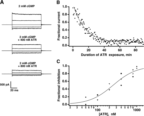
Membrane patches were excised from salamander rods in the inside-out configuration, and currents across the membrane were recorded in response to voltage steps with and without cGMP. As reported previously, sensitivity of the channels to cGMP gradually increased after excision, due to ongoing dephosphorylation of residues by phosphatase activity associated with the patch (Gordon et al., 1992; Molokanova et al., 1997). Therefore, ATR was withheld for 15–60 min, until cGMP sensitivity stabilized. In the presence of saturating cGMP, addition of submicromolar amounts of ATR caused a dose-dependent reduction in current at all voltages tested (Fig. 1 A). The onset of inhibition was variable and slow, requiring tens of minutes to reach steady state (Fig. 1 B). On average, half the current was lost in the presence of 250 nM ATR (Fig. 1 C). Thus, in saturating cGMP, the inhibitory effect of ATR applied to the intracellular face was similar for membrane patches containing native salamander channels or cloned mammalian channels (Dean et al., 2002; McCabe et al., 2004; Horrigan et al., 2005).
Figure 1.
Inhibition of native rod channels by ATR. (A) Decrement in current in a membrane patch excised from a salamander rod outer segment after exposure to ATR. The minimum exposure was ∼1 h at each concentration to allow inhibition to reach steady state. Membrane voltage was stepped from a holding potential of 0 to ±100 mV in 50-mV increments at a saturating cGMP concentration. Currents measured in the absence of cGMP were subtracted from each trace. (B) Time course of inhibition. In another membrane patch, the fractional current in saturating cGMP declined exponentially with a time constant of 15 min due to the presence of 1.0 μM ATR. (C) The dose–response relation for ATR. Symbols plot the fractional inhibition of the steady-state, cGMP-activated currents at 100 mV from nine patches. A fit of the results with 1 − r/rmax = [ATR]n/(IC50 n + [ATR]n) yielded an IC50 of 250 nM and a Hill coefficient of 1.5.
Retinoid-induced Loss of the Circulating Current in Single Rods
In darkness, Na+ enters the rod through CNG channels in the outer segment. An equal number of K+ ions exit the rod through voltage-gated channels in the inner segment. We used a suction electrode to record the circulating current, and hence the activity of the cGMP-gated channels, from the rod inner segment, during perfusion of the exposed outer segment with retinoid. At intervals, a saturating flash was given to transiently close all the CNG channels. The amplitude of the response then provided a measure of the number of CNG channels that were open just before the flash. The flashes were not bright enough to bleach any significant amount of pigment, and in control experiments without retinal treatment, the number of open channels remained constant when test flashes were given once per minute for over an hour.
Although heterologously expressed channels in excised patches are most sensitive to ATR, 11-cis retinal and all-trans retinol are nearly as effective (Dean et al., 2002). Presumably, native channels share this characteristic. We used 9- and 11-cis retinal because they would quench the cascade activity of any free opsin (Cornwall and Fain, 1994; Surya et al., 1995; Matthews et al., 1996; Melia et al., 1997) that might still be present after dark adaptation overnight (compare Xiong and Yau, 2002; Kefalov et al., 2005). Potentially then, 9-cis retinal could slightly increase the number of open cGMP-gated channels on the short term. Nevertheless, channel closure due to retinoid inhibition should prevail on the long term, after regeneration of the rod's entire complement of visual pigment.
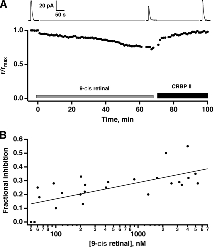
Perfusion of rods with micromolar concentrations of 9-cis retinal slowly diminished their circulating current (Fig. 2 A). The time course varied considerably from cell to cell. Complete inhibition was never achieved because it developed so slowly. The greatest fractional inhibition observed in 26 cells was 0.55. 10 experiments with bath application of 11-cis retinal yielded similar results (unpublished data). This time course of inhibition was much slower than that measured in oocyte patches with ATR applied to the intracellular surface in the presence of saturating cGMP, but was consistent with that seen upon ATR application to the extracellular surface (McCabe et al., 2004). It is important to point out, however, that CNG channels in excised patches are much more sensitive to ATR inhibition at physiological levels of cGMP, which are far from saturating. Yet, the concentrations of retinal used on intact rods in these experiments were as much as an order of magnitude greater than that used on patches. With the exception of the two cells at the lowest retinal concentration that lacked an effect, the current never leveled off by 1 h, suggesting that inhibition had not reached steady state at any of the 9-cis retinal concentrations. Because of the slow time course of the inhibition, it was not possible to obtain a steady-state, dose–response relation, so Fig. 2 B presents the fractional inhibition at 1 h as a function of 9-cis retinal concentration. The concentration dependence is shallow, with a 0.12 fractional loss in current per log unit of retinal concentration.
Figure 2.
9-cis retinal–induced closure of channels in intact rods. (A) Slow decline in circulating current of a rod induced by 3 μM 9-cis retinal. Most of the circulating current was restored relatively rapidly after removal of the retinoids by extracellular perfusion with 0.3 μM CRBP II. Representative responses to saturating flashes are shown above (left to right): before treatment, after perfusion with 9-cis retinal for 1 h, and after washing with CRBP II. (B) Dependence of channel inhibition on [9-cis retinal]. Inhibition was assessed in 27 rods after a 60-min exposure to 9-cis retinal where each rod was only exposed to a single concentration of retinal. Linear regression of fractional inhibition against the logarithm of [9-cis retinal] (continuous line, r = 0.71) suggested a 0.12 loss in current per concentration decade.
Channel closure was not simply the result of rod demise, because the effect was reversed when retinoids were removed with a binding protein. CRBP II has a low affinity for 9-cis retinal (MacDonald and Ong, 1987), but it restored the current to 0.97 ± 0.01 of the starting value (mean ± SEM, n = 8), e.g., Fig. 2 A. The time required for channel inhibition to recover by 1/e was 31 ± 9 min (n = 7), much quicker than the onset of inhibition. In a control experiment, CRBP II had no effect on the circulating current of a rod that was not treated with retinoid. Thus cGMP-gated channels in intact rods were indeed susceptible to inhibition by retinoids applied externally. However, even with an exceedingly high dose of retinoid, inhibition followed a very slow time course.
Evidence Against Activation of Cascade Activity
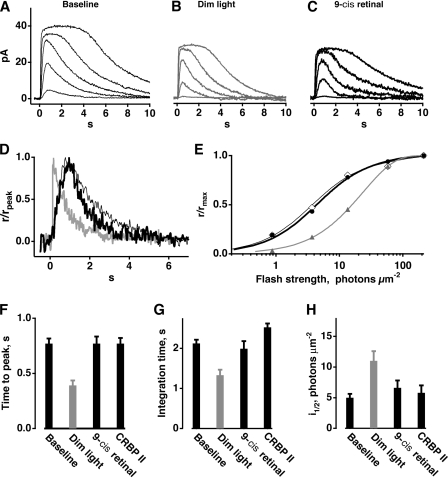
It can be argued that very high doses of 9-cis and 11-cis retinal closed channels indirectly, by stimulating transduction cascade activity and lowering cGMP (compare Kefalov et al., 2001). Two lines of evidence rule out this possibility. First, there were no changes in flash response kinetics or in relative sensitivity. Second, there was no change in PDE activity. Rods were exposed to a background light that generated cascade activity and suppressed ∼0.30 of the circulating current. An example is shown in Fig. 3 (A–E). Dim flashes, superimposed on this background light, elicited responses with faster times to peak and shorter integration time (Fig. 3 D). Relative sensitivity, defined as the multiplicative inverse of the flash strength eliciting a half-maximal response, was reduced fourfold for this rod. On average, time to peak was twofold faster, integration time was 1.6-fold briefer, and relative sensitivity was twofold lower (Fig. 3, F–H). In contrast, perfusion of rods with 9-cis retinal to achieve the same fractional loss of circulating current had no effect on relative sensitivity (Fig. 3, C, E, and H). Although there were no significant changes in the mean values for time to peak and integration time of the dim flash response by an analysis of variance (Fig. 3, F and G), we noticed that in five of seven rods, integration time decreased slightly with 9-cis retinal (e.g., Fig. 3 D) and then increased again with CRBP II. The importance of a decreased integration time will be discussed below. Flash response kinetics were sometimes slower after perfusion with CRBP II than at the beginning of the experiment, probably due to the long recording duration (>3 h) and a gradual decline in the condition of the cells.
Figure 3.
Averaged responses of a rod to flashes A, in darkness; B, in dim steady light; and C, in darkness after perfusion with 9-cis retinal to achieve the same reduction in circulating current as produced by background light. Responses to trials before and after exposure to a background light were indistinguishable so they were combined. The intensity of the background light was 0.024 photons μm−2 at 500 nm. Flashes were given at time = 0 s. (D) Little effect of 9-cis retinal on dim flash response kinetics. Flashes eliciting responses that were <20% of the maximal response were considered to be dim. Response kinetics were accelerated markedly by background light (gray trace) compared with the baseline condition (thin black trace), but not by retinoid treatment (bold, black trace). Responses were normalized by their peak amplitudes. (E) Lack of sensitivity change after 9-cis retinal treatment (baseline, open diamonds; 9-cis retinal treatment, black circles) in the rod whose responses are shown in A–C. Values for i1/2, the flash strength giving rise to a half-maximal response, were 4 and 5 photons μm−2 for baseline and retinal-treated conditions, respectively. Dim background light did reduce sensitivity several-fold, shifting the stimulus–response relation (gray triangles) to the right. Continuous lines show fits with an empirical function: r/rmax = 1 − exp(−k1 + k2exp)−k3i))i) where k1, k2, and k3 are constants, as described in Ma et al. (2001). Averaged values from seven rods for F, dim flash response time to peak; G, dim flash response integration time; and H, i1/2. Relative sensitivity is inversely proportional to i1/2. Error bars show the SEM.
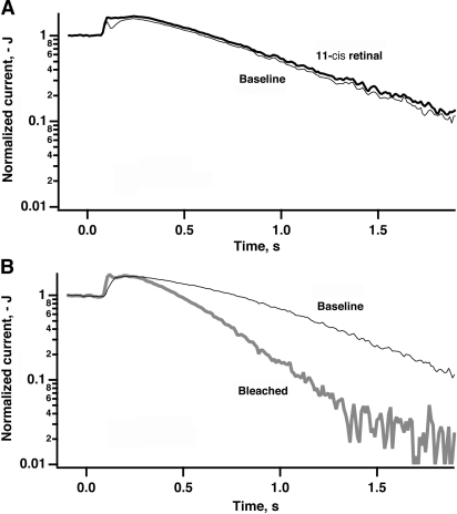
Next, we investigated the effect of retinoid application on PDE rate (see Materials and methods). Five rods were exposed to 11-cis retinal for 30–60 min. Even though the circulating current declined by a factor of 0.13–0.52, the PDE activity relative to the pretreatment level measured in these cells remained between 0.94 and 1.01, with a mean value of 0.96 ± 0.01. Fig. 4 A shows an example of PDE measurements made from one rod before (thin line) and after (thick line) a 60-min exposure to 25 μM 11-cis retinal. Although the retinoid caused a 0.18 reduction in its circulating current, no change in PDE activity was observed. For comparison, a fraction of the rhodopsin was bleached in another rod to produce a burst of cascade activity that gradually subsided to a steady, elevated level due to the persistent activity of opsin (Fig. 4 B). In this case, circulating current was reduced by a factor of 0.19 and PDE activity increased 2.2-fold. Treatment of the bleached rod with 11-cis retinal quenched opsin's activity by regenerating rhodopsin and returned the PDE activity to the prebleached level (unpublished data). These experiments demonstrate that the loss of circulating current during exposure of unbleached rods to retinal did not result from activation of the transduction cascade, as would be expected from exposure to light, but were instead consistent with direct channel inhibition.
Figure 4.
Lack of an increase in PDE activity after 11-cis retinal–induced loss of circulating current. Normalized current during steps into Li+ solution is plotted on semilogarithmic coordinates. The relative PDE rates are given by the ratio of the slopes of the falling phases of current decline. Time zero in the plot is the time at which the movement of the cell toward the Li+ solution interface was initiated. The delay between this time and the start of the current change is accounted for by the time necessary for the outer segment to reach this solution interface. (A) Measurements of PDE rates made before (thin line) and after (thick line) a 60-min exposure to 25 μM 11-cis retinal that reduced circulating current by a factor of 0.18. (B) Increased PDE rate in a different rod after exposure to bright light that bleached a fraction of the rhodopsin and reduced the circulating current by a factor of 0.19 (thick gray line), compared with the prebleached condition (thin black line).
Partial Compensation for Channel Inhibition by Feedback onto Guanylate Cyclase
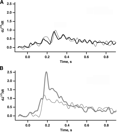
It was of interest to measure the rate of cGMP synthesis, because channel closure lowers intracellular Ca2+ that then feeds back onto guanylate cyclase, leading to the reopening of CNG channels. A shortened integration time after treatment with 9-cis retinal would be consistent with enhanced guanylate cyclase activity (see above). Thus, our method would underestimate the degree of channel inhibition by retinoids if the steady-state cGMP concentration were assumed to be constant. For the measurement of cGMP synthetic activity, rods were rapidly perfused with IBMX to inhibit the PDE (Fig. 5). Circulating current increased with cGMP concentration as guanylate cyclase activity continued unopposed.
Figure 5.
Estimation of guanylate cyclase activity. The maximum of the first derivative of the cube root of the normalized current, d(J1/3)/dt from a rod during steps into 0.5 mM IBMX solution provides an estimate of the relative GC rates. The solution change was initiated as described in the legend of Fig. 4. (A) Similar rates of cGMP synthesis before (thin line) and after (thick line) 60 min of treatment with 25 μM 11-cis retinal that resulted in 0.15 inhibition of the circulating current. (B) 2.5-fold increase in the rate of cGMP synthesis in another rod after a fractional bleach of its rhodopsin that reduced the circulating current at steady state by a factor of 0.23 (prebleach, thin black line; postbleach, thick gray line).
After establishing 0.16 ± 0.01 channel inhibition by exposure to 11-cis retinal, the rate of cGMP synthesis relative to the pretreatment value was 1.16 ± 0.15, n = 5 rods, i.e., not significantly different (e.g., Fig. 5 A). For this amount of channel inhibition, any increase in the rate of cGMP synthesis was below the resolution of the method. However, Fig. 5 B shows that a fractional reduction in dark current of 0.23 by background light increased guanylate cyclase activity 2.5-fold over its control value.
Lack of Channel Inhibition by Endogenous, Intracellular Retinoids
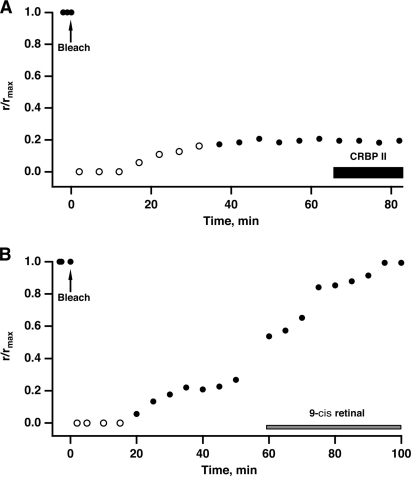
Retinal is thought to inhibit the CNG channel upon binding to an intracellular site (McCabe et al., 2004; Horrigan et al., 2005). So to facilitate channel inhibition, we exposed rods to bright light to bleach a large fraction of the rhodopsin and produce millimolar concentrations of ATR inside the rod outer segment. Retinol dehydrogenase reduces retinal to retinol on a time scale of tens of minutes (e.g., Tsina et al., 2004), but for the purpose of this experiment, the conversion is inconsequential because both types of retinoids are potent channel inhibitors. Experiments on two rods are shown in Fig. 6. Massive activation of the phototransduction cascade by the bleach kept all the cGMP-gated channels closed for many minutes. Eventually cascade activity subsided to a lower level (for reviews see McBee et al., 2001; Lamb and Pugh, 2004) and the rod recovered to a steady state with ∼0.30 of the circulating current present in darkness. If ATR released from bleached rhodopsin inhibited the channels, then removal of retinoids after the bleach should have increased the circulating current. The lack of change in the circulating current upon perfusion of eight rods with 0.3 μM CRBP II (e.g., Fig. 6 A) was therefore evidence that channels were not inhibited.
Figure 6.
Absence of channel inhibition by retinoid after pigment bleaching. (A) The ordinate plots the amplitude of the response to a fixed flash strength, relative to the amplitude at the beginning of the recording. Saturating responses are plotted with filled symbols, while subsaturating responses or trials in which no response was observed were plotted with open symbols. Exposure to 3.5 × 10⁸ photons μm−2 at 500 nm at time = 0 min to bleach 91% of the rhodopsin eliminated the circulating current and desensitized the rod. Circulating current and sensitivity partially recovered after ∼40 min. No additional circulating current was obtained upon perfusion with 0.3 μM CRBP II to remove retinoids. (B) A second rod was exposed to 2.7 × 10⁸ photons μm−2 at 500 nm at time = 0 min to bleach 84% of the rhodopsin. 59.3 min later, the rod was perfused with 4 μM 9-cis retinal to regenerate the rhodopsin. The circulating current recovered completely to the prebleached level.
In a second set of experiments we bleached 70–80% of the rhodopsin in rods and then regenerated their visual pigment with 9-cis retinal (n = 5) or 11-cis retinal (n = 2) to reopen channels that were closed due to cascade activation by opsin (e.g., Fig. 6 B). The fractional level of circulating current was restored to 0.99 ± 0.01, indicating that the channels were not inhibited by the millimolar levels of ATR formed by bleaching rhodopsin nor by the 20–55-min perfusion with micromolar retinal provided for rhodopsin regeneration. Strictly speaking, salamander rods use a mixture of retinal (A1) and 3-dehydro retinal (A2) chromophores, with the latter predominating in aquatic animals (see below), therefore bleaching created ATR of both types. Since the effect of 3-dehydro ATR on the CNG channel has not yet been characterized, three rods from terrestrial salamanders, which contain predominantly A1-based rhodopsins, were subjected to ∼80% bleach of their visual pigment. In two rods, 9-cis retinal perfusion was initiated immediately after bleaching, while perfusion of the third rod was delayed for 45 min. In all three cases, the circulating current was restored completely within 40 min after treatment with 9-cis retinal (unpublished data), ruling out channel inhibition by photoisomerized 11-cis A1. Although a contribution of retinoid-mediated inhibition to channel closure during the period of intense cascade activation just after the bleach cannot be dismissed completely, it would seem unlikely given that any effect should grow with time, as the photointermediates of rhodopsin decay fully and release additional ATR. Bleaching rhodopsin only temporarily spared channels from inhibition by exogenously applied 9-cis retinal. Continued perfusion of one rod with 9-cis retinal after full restoration of the circulating current eventually caused 0.40 channel inhibition that was reversed at least in part with CRBP II before the end of the recording (unpublished data). Thus, neither all-trans A1 nor A2 produced at millimolar levels by bleaching rhodopsin resulted in channel inhibition.
Uptake of Retinal into the Outer Segment
The intracellular accumulation of retinoids in single cells was monitored by changes in absorbance with a microspectrophotometer (MSP). Untreated, dark-adapted rods from aquatic salamanders absorbed maximally near 512 nm, in general agreement with reports by others (Harosi, 1975; Cornwall et al., 1984). In our sample of 102 rods from 15 salamanders, there was some variation in the spectral position of the absorbance maximum of rods across animals, but very little across rods from an individual animal. The variation in measured maxima can be accounted for by individual differences in the proportion of A2- and A1-based visual pigments in salamander (Ernst et al., 1978; Makino and Dodd, 1996; Govardovskii et al., 2000). After fitting the main absorption band with pigment templates (Govardovskii et al., 2000) and adjusting for the difference in extinction coefficients (Brown et al., 1963; Matthews et al., 1963), the mixture was found to consist of 73% A2 and 27% A1. In 20 rods from a terrestrial animal, the distribution was 26% A2 and 74% A1.
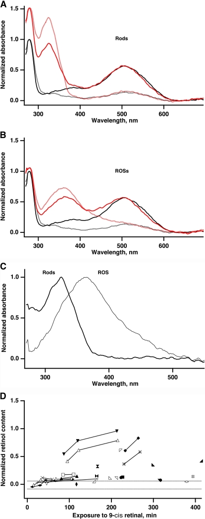
When presented with 9-cis retinal, uptake by dark-adapted rods was variable even for neighboring cells in the same preparation (Fig. 7, A–D). After 1 h, retinoid accumulation inside the outer segment in the majority of rods was typically below the level of detection, ∼500 μM. But by 2 h or more, some rods amassed quite high concentrations of retinoid. Absorbance was optimal for light polarized parallel to the long axis of the rod, opposite to the situation with rhodopsin. Interestingly, the absorbance increase was maximal near 325 nm, rather than at 373–375 nm, where ATR and 9-cis retinal absorb maximally in ethanol. In 9 of 12 rod outer segments lacking their ellipsoids (ROSs), absorbance did increase maximally near 373 nm. Similar results were obtained with 11-cis retinal. Rods and ROSs with particularly high uptake are shown in Fig. 7 (A–C). The spectral position of the absorption band at 325 nm (Robeson et al., 1955a) and its dependence upon the presence of the inner segment implicate reduction of the retinal to retinol in metabolically active rods (Tsina et al., 2004). Reduction could occur directly from cis retinals because they are substrates for the rod dehydrogenase, albeit poor ones (Daemen et al., 1974; Ishiguro et al., 1991; Palczewski et al., 1994), or after thermal isomerization to ATR (compare Daemen et al., 1974). From the ratio of absorbances at 325 nm and 520 nm, measured with optimal polarization of the probe beam, and assuming similar extinction coefficients for 9-cis retinol and rhodopsin, we estimate that rods with the highest uptake contained as many as two retinol molecules per rhodopsin, i.e., ∼6 mM. Harosi (1984) reported considerable accumulation of 11-cis retinal in dark-adapted rods with very little retinol formation. Probably his 10-fold higher ethanolic concentrations and use of solutions that were saturated with nitrogen to stabilize the retinal promoted uptake but precluded NADPH production, which is requisite for the reduction of retinal to retinol.
Figure 7.
Microspectrophotometric measurements of retinoid uptake into outer segments that were attached to (n = 2 rods), A, or had detached from (n = 3 ROSs), B, their inner segments.Measurements were made before (black lines) and after (red lines) treatment with 9-cis retinal for ∼4 h with the probe beam polarized parallel (dashed lines) and perpendicular (continuous lines) to the outer segment axis. The ROSs in B were also treated with CRBPII for 2.5 h. Nevertheless, their absorbance at 370 nm was the highest of any ROS measured. Spectra from untreated outer segments were scaled by their absorbance at 280 nm. Spectra from treated outer segments were scaled so that the absorbance at 520 nm would match that of the corresponding untreated spectra. Absorbance increased at 325 nm in outer segments retaining an inner segment and at 370 nm in outer segments lacking an inner segment. (C) Normalized spectra from rods and ROSs, taken as the difference between the measurement with and without exposure to 9-cis retinal for the probe beam polarized parallel to the rod axis. (D) Slow, variable uptake of 9-cis retinal by rods. Measurements on 25 intact rods were made with the probe beam polarized perpendicular to the long axis of the outer segment. Normalized retinol content was determined by decomposition of the spectra of individual rods into A1 and A2 pigment components (Govardovskii et al., 2000), retinal, retinol, and the protein band absorbing at 502, 521, 373, 325, and 280 nm, respectively. Retinoid and protein absorption bands were assumed to be Gaussian in form. The amplitude of the retinol band was divided by that of the protein band to adjust for outer segment diameter. Serial results from the same rod were connected with a continuous line. Dashed lines demarcate the mean ± one standard deviation for 34 untreated rods.
Although 0.3 μM CRBP II was very effective at reversing channel inhibition in physiological recordings from intact rods, it had little effect on the absorbance at 370 nm in treated ROSs as measured in MSP experiments. Only one ROS out of six showed any decline in retinal content after perfusion with CRBP II for nearly 1 h. Two other ROSs eventually lost ∼10% of their retinal content after >3 h with CRBP II. These results are consistent with the existence of two separate retinoid pools, one accessible to the CNG channels in the plasma membrane and another not available to the channels, perhaps located at buffer sites in the interior of the rod outer segment, such as associated with opsin and/or disc membranes. Removal of retinol from intact rods in MSP experiments was difficult to evaluate, due to their generally low levels of retinoid uptake (i.e., <500 μM, our approximate MSP detection limit).
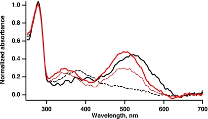
Compared with dark-adapted rods, uptake of 9-cis and 11-cis retinal was fast in partially bleached rods, judging by the rapid initial regeneration of pigments absorbing at 484 or 502 nm, respectively (Fig. 8). Well over half of the bleached visual pigment (>1 mM) regenerated within 40 min and was complete within ∼1.5 h, similar to the results reported by Harosi (1984).
Figure 8.
Rapid regeneration of rhodopsin in a bleached rod with 11-cis retinal. Absorption spectra are shown before bleaching (continuous black line), after bleaching with bright light at 500 nm (dotted black line), and after the addition of 30 μM 11-cis retinal (thin red line, 22 min treatment; thick red line, 42 min treatment).
DISCUSSION
The time course of 9-cis and 11-cis retinal inhibition of CNG channels in intact rods resembled that seen with application of ATR to the extracellular side of expressed channels (McCabe et al., 2004). In both cases, ∼20% inhibition was attained after exposure to a few hundred nanomolar retinal for 1 h. However, experiments on expressed channels were performed in the presence of saturating cGMP, whereas in dark adapted rods, the free [cGMP] is only a few micromolar (Yau and Nakatani, 1985). Recordings of outside-out ROS membrane patches at low cGMP were not attempted in this study because the small currents and exceedingly slow onset of retinoid inhibition would have prevented us from achieving a steady-state condition or determining an accurate time course of channel closure (compare McCabe et al., 2004). The K1/2 of the channels for cGMP lies between 10 and >50 μM, (for review see Yau and Baylor, 1989), so under physiological conditions, only 1–2% of channels in an intact rod are open. Since retinoids are closed state inhibitors (McCabe et al., 2004), the IC50 for CNG channels in the intact rod should drop to tens of nanomolar. On those grounds, CNG channels in intact salamander rods proved to be remarkably resistant to inhibition by retinoids.
Differences in species or subunit stoichiometry cannot explain this apparent resistance, because native channels in membrane patches excised from salamander rods exhibited an IC50 of 250 nM for ATR applied to the cytoplasmic side in saturating cGMP, nearly the same as for expressed bovine channels (Dean et al., 2002; McCabe et al., 2004). Channel closure reduces the influx of Ca2+ into the rod and the lowered intracellular Ca2+ stimulates guanylate cyclase to synthesize cGMP at a faster rate. This feedback onto guanylate cyclase would partially compensate for channel inhibition. For the level of channel inhibition attained in our sample of rods, the magnitude of the feedback apparently was not great enough to produce significant differences in flash response integration time or in guanylate cyclase activity. Had cGMP synthesis increased by 1.16-fold (see Results), the cGMP concentration would have increased proportionately, and in the absence of channel inhibition, circulating current would have been 1.6-fold greater (Eq. 3). Then a measured fractional channel inhibition of 0.30 would have corresponded to a true inhibition of (1.6)(0.30) = 0.48. Even so, this fractional inhibition was still quite low given that the retinoid concentration was orders of magnitude greater than the IC50 for channels in excised patches in the presence of low levels of cGMP.
Perhaps cGMP-gated channels in intact rods are protected in some way. Channels are subject to phosphorylation by a serine/threonine kinase in rods and by a tyrosine kinase in oocytes (Gordon et al., 1992; Molokanova et al., 1997). All of our experiments on expressed channels and native channels in excised patches were performed after channel dephosphorylation was substantially complete. Phosphorylated channels may have a reduced affinity for retinoids, or alternatively, retinoids might promote the dephosphorylated state of the channel, which opens at lower levels of cGMP. The soluble channel binding partner, Ca2+/calmodulin or a related protein (Hsu and Molday, 1993, 1994; Gordon et al., 1995), might interfere similarly with retinoid inhibition in intact rods.
The sluggish onset of inhibition did not result from an inability of retinal to enter the rod, because retinoids traverse and exchange between membranes rapidly (Rando and Bangerter, 1982; Fex and Johannesson, 1988; Ho et al., 1989). Moreover, in our MSP experiments, application of 9-cis retinal to bleached rods regenerated millimolar quantities of rhodopsin inside the rod in a few tens of minutes. It may be that after crossing the plasma membrane, retinal must accumulate in the aqueous cytosol in order to bind and inhibit the channel. Although retinal and retinol do possess some solubility in water (Szuts and Harosi, 1991), there is a strong tendency for them to partition into the membrane (Noy et al., 1995). Some retinal binds to phosphatidylethanolamine covalently as a Schiff's base (e.g., Van Breugel et al., 1979). Thus the large disk membrane surface area inside the outer segment could minimize the aqueous concentration. The reduced membranous area in the bleb of an excised patch could explain why channel inhibition is produced by lower retinoid concentrations than in intact outer segments. Yet the affinity for membranes alone would seem inadequate because retinoids rapidly equilibrate between membrane compartments across an aqueous space (Ho et al., 1989). Instead, there may exist in rods a more stable, high capacity retinoid buffer.
Opsin/rhodopsin appears to be a promising candidate for the major component of the buffer. Crude estimates suggest that opsins outnumber channels in a salamander rod outer segment by 6,000:1 (for review see Pugh and Lamb, 2000). Perfusion of a rod with tens of micromolar 9-cis or 11-cis retinal for 1 h decreased the fractional circulating current by ∼0.30 in unbleached rods yet failed to do so in partially bleached rods. Instead of binding to the channel, retinal avidly bound to opsin and regenerated rhodopsin. ATR also binds opsin, enhancing its catalytic activity (Fukada et al., 1982; Cohen et al., 1992) and phosphorylation (Hofmann et al., 1992; Palczewski et al., 1994). However, ATR binds to a site that is distinct from the pigment-forming pocket for 11-cis retinal, because the presence of ATR actually slightly facilitates the regeneration of rhodopsin with 11-cis retinal (Sachs et al., 2000). Moreover, removal of the paired palmitates attached to cysteines 322 and 323 on opsin diminishes ATR-induced opsin activity but has little effect on light-dependent rhodopsin activity (Sachs et al., 2000). Thus after a bleach, opsin may retain ATR, even after reduction to retinol (for review see Lamb and Pugh, 2004). If retinoid binding sites are also available on rhodopsin, then it could sequester retinoids and prevent channel inhibition.
CRBP II removed very little retinal from the ROSs, even after several hours. In contrast, CRBP II mediated recovery from retinal-induced channel inhibition within tens of minutes in every rod tested in single cell recordings. The development of channel inhibition by retinoid and its reversal by CRBP II without measurable changes in the outer segment retinoid content of rods and ROSs could indicate that channel behavior was governed by a small pool of free retinoid while MSP measurements registered a large pool of bound retinoid and that the two pools equilibrated slowly. Retinal bound to opsin rapidly and irreversibly, hence channel inhibition did not occur. But slower binding to rhodopsin supported a larger fraction of free retinoid that did close some channels.
The ability of the rod to withstand exposure to large amounts of retinoids without significant channel inhibition makes it unlikely that closure of the channel by retinoids constitutes any meaningful signaling pathway under normal physiological conditions. Even exposure to very bright light that bleached a substantial fraction of the millimolar rhodopsin content in the rod outer segment failed to elicit any significant retinoid inhibition of the CNG channel. It should be noted, however, that our experiments involved isolated rods. The presence of retinoid binding proteins in the interplexiform matrix and proximity to large stores of retinoids in the retinal pigment epithelium are important factors that need to be assessed for rods in the intact eye. Channel inhibition may also contribute to the slowed time course of dark adaptation characteristic of diseases such as Stargardt's (Fishman et al., 1991), wherein mutations in ABCR, the retina-specific, ATP-binding cassette transporter (Allikmets et al., 1997), cause an abnormally high accumulation of ATR in rods (Weng et al., 1999).
Acknowledgments
We thank Drs. T.P. and R.A. Williams and Florida State University (Tallahassee, FL) for granting us custody of the MSP and Mr. and Mrs. J.P. Webbers and Mr. I. Kirillov for technical assistance in setting it up. We also thank Dr. R.K. Crouch and the National Eye Institute for providing 11-cis retinal.
This research was funded by the Lion's of Massachusetts, the Knights Templar Foundation, and the National Institutes of Health: EY012944, EY014104, EY01157, EY07774, and DK32642.
Olaf S. Andersen served as editor.
S.L. McCabe's present address is Harvard Clinical Research Institute, Boston, MA 02215.
P.D. Calvert's present address is Department of Ophthalmology, State University of New York Upstate Medical University, Syracuse, NY 13210.
Abbreviations used in this paper: ATR, all-trans retinal; CNG, cyclic nucleotide-gated; CRBP, cellular retinoid binding protein; GC, guanylyl cyclase; IBMX, 3-isobutyl-1-methylxanthine; MSP, microspectrophotometer, PDE, phosphodiesterase; ROS, rod outer segment.
References
- Allikmets, R., N. Singh, H. Sun, N.F. Shroyer, A. Hutchinson, A. Chidambaram, B. Gerrard, L. Baird, D. Stauffer, A. Peiffer, et al. 1997. A photoreceptor cell-specific ATP-binding transporter gene (ABCR) is mutated in recessive Stargardt macular dystrophy. Nat. Genet. 15:236–246. [DOI] [PubMed] [Google Scholar]
- Brown, P.K., and G. Wald. 1956. The neo-b isomer of vitamin A and retinene. J. Biol. Chem. 222:865–877. [PubMed] [Google Scholar]
- Brown, P.K., I.R. Gibbons, and G. Wald. 1963. The visual cells and visual pigment of the mudpuppy, Necturus. J. Cell Biol. 19:79–106. [DOI] [PMC free article] [PubMed] [Google Scholar]
- Cohen, G.B., D.D. Oprian, and P.R. Robinson. 1992. Mechanism of activation and inactivation of opsin: role of Glu113 and Lys296. Biochemistry. 31:12592–12601. [DOI] [PubMed] [Google Scholar]
- Cornwall, M.C., G.J. Jones, V.J. Kefalov, G.L. Fain, and H.R. Matthews. 2000. Electrophysiological methods for measurement of activation of phototransduction by bleached visual pigment in salamander photoreceptors. Methods Enzymol. 316:224–252. [DOI] [PubMed] [Google Scholar]
- Cornwall, M.C., and G.L. Fain. 1994. Bleached pigment activates transduction in isolated rods of the salamander retina. J. Physiol. 480:261–279. [DOI] [PMC free article] [PubMed] [Google Scholar]
- Cornwall, M.C., E.F. MacNichol Jr., and A. Fein. 1984. Absorptance and spectral sensitivity measurements of rod photoreceptors of the tiger salamander, Ambystoma tigrinum. Vision Res. 24:1651–1659. [DOI] [PubMed] [Google Scholar]
- Corson, D.W., V.J. Kefalov, M.C. Cornwall, and R.K. Crouch. 2000. Effect of 11-cis 13-demethylretinal on phototransduction in bleach-adapted rod and cone photoreceptors. J. Gen. Physiol. 116:283–297. [DOI] [PMC free article] [PubMed] [Google Scholar]
- Daemen, F.J.M., J.P. Rotmans, and S.L. Bonting. 1974. On the rhodopsin cycle. Exp. Eye Res. 18:97–103. [DOI] [PubMed] [Google Scholar]
- Dean, D.M., W. Nguitragool, A. Miri, S.L. McCabe, and A.L. Zimmerman. 2002. All-trans-retinal shuts down rod cyclic nucleotide-gated ion channels: a novel role for photoreceptor retinoids in the response to bright light? Proc. Natl. Acad. Sci. USA. 99:8372–8377. [DOI] [PMC free article] [PubMed] [Google Scholar]
- Dew, S.E., S.A. Wardlaw, and D.E. Ong. 1993. Effects of pharmacological retinoids on several vitamin A-metabolizing enzymes. Cancer Res. 53:2965–2969. [PubMed] [Google Scholar]
- Ernst, W., C.M. Kemp, and D.E. Price. 1978. Studies on the effects of bleaching amphibian rod pigments in situ. I. The absorbance spectra of axolotl and tiger salamander rhodopsin and porphyropsin. Exp. Eye Res. 26:329–336. [DOI] [PubMed] [Google Scholar]
- Fain, G.L., H.R. Matthews, M.C. Cornwall, and Y. Koutalos. 2001. Adaptation in vertebrate photoreceptors. Physiol. Rev. 81:117–151. [DOI] [PubMed] [Google Scholar]
- Fex, G., and G. Johannesson. 1988. Retinol transfer across and between phospholipid bilayer membranes. Biochim. Biophys. Acta. 944:249–255. [DOI] [PubMed] [Google Scholar]
- Fishman, G.A., J.S. Farbman, and K.R. Alexander. 1991. Delayed rod dark adaptation in patients with Stargardt's disease. Ophthalmology. 98:957–962. [DOI] [PubMed] [Google Scholar]
- Fukada, Y., T. Yoshizawa, M. Ito, and K. Tsukida. 1982. Activation of phosphodiesterase in frog rod outer segments by rhodopsin analogues. Biochim. Biophys. Acta. 708:112–117. [DOI] [PubMed] [Google Scholar]
- Gordon, S.E., D.L. Brautigan, and A.L. Zimmerman. 1992. Protein phosphatases modulate the apparent agonist affinity of the light-regulated ion channel in retinal rods. Neuron. 9:739–748. [DOI] [PubMed] [Google Scholar]
- Gordon, S.E., J. Downing-Park, and A.L. Zimmerman. 1995. Modulation of the cGMP-gated ion channel in frog rods by calmodulin and an endogenous inhibitory factor. J. Physiol. 486:533–546. [DOI] [PMC free article] [PubMed] [Google Scholar]
- Govardovskii, V.I., N. Fyhrquist, T. Reuter, D.G. Kuzmin, and K. Donner. 2000. In search of the visual pigment template. Vis. Neurosci. 17:509–528. [DOI] [PubMed] [Google Scholar]
- Harosi, F.I. 1975. Absorption spectra and linear dichroism of some amphibian photoreceptors. J. Gen. Physiol. 66:357–382. [DOI] [PMC free article] [PubMed] [Google Scholar]
- Harosi, F.I. 1984. In vitro regeneration of visual pigment in isolated vertebrate photoreceptors. In Photoreceptors. A. Borsellino and L. Cervetto, editors. Plenum Press, New York. 41–63.
- Haynes, L.W., A.R. Kay, and K.-W. Yau. 1986. Single cyclic GMP-activated channel activity in excised patches of rod outer segment membrane. Nature. 321:66–70. [DOI] [PubMed] [Google Scholar]
- Ho, M.-T., J.B. Massey, H.J. Pownall, R.E. Anderson, and J.G. Hollyfield. 1989. Mechanism of vitamin A movement between rod outer segments, interphotoreceptor retinoid-binding protein, and liposomes. J. Biol. Chem. 264:928–935. [PubMed] [Google Scholar]
- Hodgkin, A.L., and B.J. Nunn. 1988. Control of light-sensitive current in salamander rods. J. Physiol. 403:439–471. [DOI] [PMC free article] [PubMed] [Google Scholar]
- Hodgkin, A.L., P.A. McNaughton, and B.J. Nunn. 1985. The ionic selectivity and calcium dependence of the light-sensitive pathway in toad rods. J. Physiol. 358:447–468. [DOI] [PMC free article] [PubMed] [Google Scholar]
- Hofmann, K.P., A. Pulvermuller, J. Buczylko, P. Van Hooser, and K. Palczewski. 1992. The role of arrestin and retinoids in the regeneration pathway of rhodopsin. J. Biol. Chem. 267:15701–15706. [PubMed] [Google Scholar]
- Horrigan, D.M., M.L. Tetreault, N. Tsomaia, C. Vasileiou, B. Borhan, D.F. Mierke, R.K. Crouch, and A.L. Zimmerman. 2005. Defining the retinoid binding site in the rod cyclic nucleotide-gated channel. J. Gen. Physiol. 126:453–460. [DOI] [PMC free article] [PubMed] [Google Scholar]
- Hsu, Y.T., and R.S. Molday. 1993. Modulation of the cGMP-gated channel of rod photoreceptor cells by calmodulin. Nature. 361:76–79. [DOI] [PubMed] [Google Scholar]
- Hsu, Y.T., and R.S. Molday. 1994. Interaction of calmodulin with the cyclic GMP-gated channel of rod photoreceptor cells. Modulation of activity, affinity purification, and localization. J. Biol. Chem. 269:29765–29770. [PubMed] [Google Scholar]
- Ishiguro, S., Y. Suzuki, M. Tamai, and K. Mizuno. 1991. Purification of retinol dehydrogenase from bovine retinal rod outer segments. J. Biol. Chem. 266:15520–15524. [PubMed] [Google Scholar]
- Karpen, J.W., A.L. Zimmerman, L. Stryer, and D.A. Baylor. 1988. Gating kinetics of the cyclic-GMP-activated channel of retinal rods: flash photolysis and voltage-jump studies. Proc. Natl. Acad. Sci. USA. 85:1287–1291. [DOI] [PMC free article] [PubMed] [Google Scholar]
- Kaupp, U.B., and R. Seifert. 2002. Cyclic nucleotide-gated ion channels. Physiol. Rev. 82:769–824. [DOI] [PubMed] [Google Scholar]
- Kefalov, V.J., M.C. Cornwall, and R.K. Crouch. 1999. Occupancy of the chromophore binding site of opsin activates visual transduction in rod photoreceptors. J. Gen. Physiol. 113:491–503. [DOI] [PMC free article] [PubMed] [Google Scholar]
- Kefalov, V.J., R.K. Crouch, and M.C. Cornwall. 2001. Role of noncovalent binding of 11-cis-retinal to opsin in dark adaptation of rod and cone photoreceptors. Neuron. 29:749–755. [DOI] [PubMed] [Google Scholar]
- Kefalov, V.J., M.E. Estevez, M. Kono, P.W. Goletz, R.K. Crouch, M.C. Cornwall, and K.-W. Yau. 2005. Breaking the covalent bond-a pigment property that contributes to desensitization in cones. Neuron. 46:879–890. [DOI] [PMC free article] [PubMed] [Google Scholar]
- Klaassen, I., R.H. Brakenhoff, S.J. Smeets, G.B. Snow, and B.J.M. Braakhuis. 1999. Considerations for in vitro retinoid experiments: importance of protein interaction. Biochim. Biophys. Acta. 1427:265–275. [DOI] [PubMed] [Google Scholar]
- Lamb, T.D., and E.N. Pugh Jr. 2004. Dark adaptation and the retinoid cycle of vision. Prog. Retin. Eye Res. 23:307–380. [DOI] [PubMed] [Google Scholar]
- Ma, J., S. Znoiko, K.L. Othersen, J.C. Ryan, J. Das, T. Isayama, M. Kono, D.D. Oprian, D.W. Corson, M.C. Cornwall, et al. 2001. A visual pigment expressed in both rod and cone photoreceptors. Neuron. 32:451–461. [DOI] [PubMed] [Google Scholar]
- MacDonald, P.N., and D.E. Ong. 1987. Binding specificities of cellular retinol-binding protein and cellular retinol-binding protein, type II. J. Biol. Chem. 262:10550–10556. [PubMed] [Google Scholar]
- MacNichol, E.F., Jr. 1978. A photon counting microspectrophotometer for the study of single vertebrate photoreceptor cells. In Frontiers in Visual Science. S.J. Cool and E. Smith, editors. Springer-Verlag New York Inc., New York. 194–208.
- Makino, C.L., and R.L. Dodd. 1996. Multiple visual pigments in a photoreceptor of the salamander retina. J. Gen. Physiol. 108:27–34. [DOI] [PMC free article] [PubMed] [Google Scholar]
- Makino, C.L., L.N. Howard, and T.P. Williams. 1990. a. Axial gradients of rhodopsin in light-exposed retinal rods of the toad. J. Gen. Physiol. 96:1199–1220. [DOI] [PMC free article] [PubMed] [Google Scholar]
- Makino, C.L., T.W. Kraft, R.A. Mathies, J. Lugtenburg, M.E. Miley, R. van der Steen, and D.A. Baylor. 1990. b. Effects of modified chromophores on the spectral sensitivity of salamander, squirrel and macaque cones. J. Physiol. 424:545–560. [DOI] [PMC free article] [PubMed] [Google Scholar]
- Makino, C.L., W.R. Taylor, and D.A. Baylor. 1991. Rapid charge movements and photosensitivity of visual pigments in salamander rods and cones. J. Physiol. 442:761–780. [DOI] [PMC free article] [PubMed] [Google Scholar]
- Matthews, G., and S.-I. Watanabe. 1987. Properties of ion channels closed by light and opened by guanosine 3',5'-cyclic monophosphate in toad retinal rods. J. Physiol. 389:691–715. [DOI] [PMC free article] [PubMed] [Google Scholar]
- Matthews, H.R., M.C. Cornwall, and G.L. Fain. 1996. Persistent activation of transducin by bleached rhodopsin in salamander rods. J. Gen. Physiol. 108:557–563. [DOI] [PMC free article] [PubMed] [Google Scholar]
- Matthews, R.G., R. Hubbard, P.K. Brown, and G. Wald. 1963. Tautomeric forms of metarhodopsin. J. Gen. Physiol. 47:215–240. [DOI] [PMC free article] [PubMed] [Google Scholar]
- Mazia, D., G. Schatten, and W. Sale. 1975. Adhesion of cells to surfaces coated with polylysine. J. Cell Biol. 66:198–200. [DOI] [PMC free article] [PubMed] [Google Scholar]
- McBee, J.K., K. Palczewski, W. Baehr, and D.R. Pepperberg. 2001. Confronting complexity: the interlink of phototransduction and retinoid metabolism in the vertebrate retina. Prog. Retin. Eye Res. 20:469–529. [DOI] [PubMed] [Google Scholar]
- McCabe, S.L., D.M. Pelosi, M. Tetreault, A. Miri, W. Nguitragool, P. Kovithvathanaphong, R. Mahajan, and A.L. Zimmerman. 2004. All-trans-retinal is a closed-state inhibitor of rod cyclic nucleotide-gated ion channels. J. Gen. Physiol. 123:521–531. [DOI] [PMC free article] [PubMed] [Google Scholar]
- Melia, T.J., Jr., C.W. Cowan, J.K. Angleson, and T.G. Wensel. 1997. A comparison of the efficiency of G protein activation by ligand-free and light-activated forms of rhodopsin. Biophys. J. 73:3182–3191. [DOI] [PMC free article] [PubMed] [Google Scholar]
- Molokanova, E., B. Trivedi, A. Savchenko, and R.H. Kramer. 1997. Modulation of rod photoreceptor cyclic nucleotide-gated channels by tyrosine phosphorylation. J. Neurosci. 17:9068–9076. [DOI] [PMC free article] [PubMed] [Google Scholar]
- Noy, N., D.J. Kelleher, and A.W. Scotto. 1995. Interactions of retinol with lipid bilayers: studies with vesicles of different radii. J. Lipid Res. 36:375–382. [PubMed] [Google Scholar]
- Palczewski, K., S. Jager, J. Buczylko, R.K. Crouch, D.L. Bredberg, K.P. Hofmann, M.A. Asson-Batres, and J.C. Saari. 1994. Rod outer segment retinol dehydrogenase: substrate specificity and role in phototransduction. Biochemistry. 33:13741–13750. [DOI] [PubMed] [Google Scholar]
- Pugh, E.N. Jr., and T.D. Lamb. 2000. Phototransduction in vertebrate rods and cones: molecular mechanisms of amplification, recovery and light adaptation. In Handbook of Biological Physics. Volume 3. D.G. Stavenga, W.J. DeGrip, and E.N. Pugh Jr., editors. Elsevier Science Publishing Co. Inc., New York. 183–255.
- Rando, R.R. 2001. The biochemistry of visual cycle. Chem. Rev. 101:1881–1896. [DOI] [PubMed] [Google Scholar]
- Rando, R.R., and F.W. Bangerter. 1982. The rapid intermembranous transfer of retinoids. Biochem. Biophys. Res. Commun. 104:430–436. [DOI] [PubMed] [Google Scholar]
- Robeson, C.D., J.D. Cawley, L. Weisler, M.H. Stern, C.C. Eddinger, and A.J. Chechak. 1955. a. Chemistry of vitamin A. XXIV. The synthesis of geometric isomers of vitamin A via methyl β-methylglutaconate. J. Am. Chem. Soc. 77:4111–4119. [Google Scholar]
- Robeson, C.D., W.P. Blum, J.M. Dieterle, J.D. Cawley, and J.G. Baxter. 1955. b. Chemistry of vitamin A. XXV. Geometrical isomers of vitamin A aldehyde and an isomer of its α-analog. J. Am. Chem. Soc. 77:4120–4125. [Google Scholar]
- Sachs, K., D. Maretzki, C.K. Meyer, and K.P. Hofmann. 2000. Diffusible ligand all-trans-retinal activates opsin via a palmitoylation-dependent mechanism. J. Biol. Chem. 275:6189–6194. [DOI] [PubMed] [Google Scholar]
- Sakmann, B., and E. Neher. 1983. Single-channel Recording. Plenum Publishing Corp., New York. 503 pp.
- Surya, A., K.W. Foster, and B.E. Knox. 1995. Transducin activation by the bovine opsin apoprotein. J. Biol. Chem. 270:5024–5031. [DOI] [PubMed] [Google Scholar]
- Szuts, E.Z., and F.I. Harosi. 1991. Solubility of retinoids in water. Arch. Biochem. Biophys. 287:297–304. [DOI] [PubMed] [Google Scholar]
- Tsina, E., C. Chen, Y. Koutalos, P. Ala-Laurila, M. Tsacopoulos, B. Wiggert, R.K. Crouch, and M.C. Cornwall. 2004. Physiological and microfluorometric studies of reduction and clearance of retinal in bleached rod photoreceptors. J. Gen. Physiol. 124:429–443. [DOI] [PMC free article] [PubMed] [Google Scholar]
- Van Breugel, P.J.G.M., P.H.M. Bovee-Geurts, S.L. Bonting, and F.J.M. Daemen. 1979. Biochemical aspects of the visual process. XL. Spectral and chemical analysis of metarhodopsin III in photoreceptor membrane suspensions. Biochim. Biophys. Acta. 557:188–198. [DOI] [PubMed] [Google Scholar]
- Weng, J., N.L. Mata, S.M. Azarian, R.T. Tzekov, D.G. Birch, and G.H. Travis. 1999. Insights into the function of rim protein in photoreceptors and etiology of Stargardt's disease from the phenotype in abcr knockout mice. Cell. 98:13–23. [DOI] [PubMed] [Google Scholar]
- Xiong, W.-H., and K.-W. Yau. 2002. Rod sensitivity during Xenopus development. J. Gen. Physiol. 120:817–827. [DOI] [PMC free article] [PubMed] [Google Scholar]
- Yau, K.-W., and D.A. Baylor. 1989. Cyclic GMP-activated conductance of retinal photoreceptor cells. Annu. Rev. Neurosci. 12:289–327. [DOI] [PubMed] [Google Scholar]
- Yau, K.-W., and K. Nakatani. 1984. Electrogenic Na-Ca exchange in retinal rod outer segment. Nature. 311:661–663. [DOI] [PubMed] [Google Scholar]
- Yau, K.-W., and K. Nakatani. 1985. Light-suppressible, cyclic GMP-sensitive conductance in the plasma membrane of a truncated rod outer segment. Nature. 317:252–255. [DOI] [PubMed] [Google Scholar]
- Yau, K.W., P.A. McNaughton, and A.L. Hodgkin. 1981. Effect of ions on the light-sensitive current in retinal rods. Nature. 292:502–505. [DOI] [PubMed] [Google Scholar]
- Zimmerman, A.L., and D.A. Baylor. 1986. Cyclic GMP-sensitive conductance of retinal rods consists of aqueous pores. Nature. 321:70–72. [DOI] [PubMed] [Google Scholar]
- Zimmerman, A.L., J.W. Karpen, and D.A. Baylor. 1988. Hindered diffusion in excised membrane patches from retinal rod outer segments. Biophys. J. 54:351–355. [DOI] [PMC free article] [PubMed] [Google Scholar]


