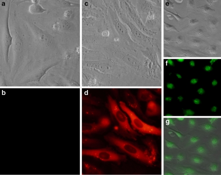
Fig. 1.
The intracellular delivery of fluorescent proteins (R-PE and Histone-H1) mediated by a non-peptide based reagent. Two μg of R-PE was diluted in 20 mM Hepes buffer and added with HeLa cells in a 24-well. The plate was incubated for 16 h at 37 °C and cells were analysed by phase-contrast (a) and fluorescence microscopy (b) An amount of 2 μg of R-PE was delivered into HeLa cells after formation of complexes with 4 μL of delivery reagent. Intracellular protein delivery was analyzed after 16 h incubation at 37 °C by phase-contrast (c) and fluorescence microscopy (d) An amount of 4 μg of Histone-H1-AF®488 was delivered into HeLa cells after formation of complexes with 3 μl of delivery reagent. Intracellular protein delivery was analyzed 20 h later. Superimposition (g) of images obtained by phase-contrast (e) and fluorescence microscopy (f) was obtained using Adobe Photoshop software

