Abstract
Heterotrimeric G proteins transduce multiple growth-factor-receptor-initiated and intracellular signals that may lead to activation of the mitogen-activated or stress-activated protein kinases. Herein we report on the identification of a novel p53 target gene (A28-RGS14) that is induced in response to genotoxic stress and encodes a novel member of a family of regulators of G protein signaling (RGS) proteins with proposed GTPase-activating protein activity. Overexpression of A28-RGS14p protein inhibits both Gi- and Gq-coupled growth-factor-receptor-mediated activation of the mitogen-activated protein kinase signaling pathway in mammalian cells. Thus, through the induction of A28-RGS14, p53 may regulate cellular sensitivity to growth and/or survival factors acting through G protein-coupled receptor pathways.
Inactivation of the p53 tumor suppressor protein is the most common aberration known to occur in human cancers (1). As a consequence of loss of wild-type p53 functions, cells are defective in critical cell cycle checkpoints as well as intracellular and extracellular pathways regulating cellular growth and programmed cell death (2–5). Several p53-induced target genes that encode a complex spectrum of regulators of such pathways have been identified. For instance, p21WAF1 (6) mediates p53-induced cell cycle arrest and may exert protective effects against apoptosis (7), whereas bax (8) encodes a positive effector of cell death. Induction of IGF-BP3, an inhibitor of insulin-like growth factors, provides a mechanism whereby p53 may interfere with the mitogenic and survival functions of insulin-like growth factors, thereby further sensitizing cells to apoptotic stimuli (5). Cell-specific integration of the activity of such and yet to be identified p53-regulated pathways is intimately associated with cell fate of normal and tumorigenic cells. To gain further insight into p53 signaling pathways, we undertook a screen to clone novel p53 target genes. Herein we report the identification of a novel factor induced by p53 that can inhibit G protein-coupled mitogenic signal transduction and activation of the mitogen-activated protein kinase (MAPK) signaling cascade implicated in cellular proliferation, transformation, and oncogenesis.
MATERIALS AND METHODS
Cell Culture.
EB1 colon carcinoma cells (9) were cultured as described (5). RKO and RKO E6 colon carcinoma cells were cultured at 37°C and 5% CO2/95% air in modified Eagle’s medium supplemented with 10% fetal bovine serum (FBS) and penicillin–streptomycin (GIBCO/BRL). NIH 3T3 M1 and M2 cells were cultured as described (10). T98G glioblastoma, U-87 astrocytoma, HL-60 promyelocytic leukemia, and MCF7 breast carcinoma cells were obtained from American Type Culture Collection and maintained at 37°C and 5% CO2/95% air in RPMI 1640 medium supplemented with 10% FBS and penicillin–streptomycin (100 units/ml) (GIBCO/BRL). MCF7 Adr (11) and MCF 7 clone 6 (clonal population derived from the parental cells) were cultured as the parental MCF7 cells were cultured.
RNA and Northern Blot Analysis.
RNA preparation and Northern blot analysis were as described (12). Quantitation of Northern blots was performed with laser densitometry (Molecular Dynamics) of the autoradiograms or by exposing the blots to phosphorimaging plates followed by analysis on a phosphorimager (Fuji).
cDNA Isolation and Cloning.
A PCR-based library subtraction procedure was used to enrich for cDNA fragments representing RNAs induced by p53 (12). One fragment, A28, detected an ≈2.5-kb p53-regulated transcript and was used as a probe to screen a human brain cDNA library in λ ZAPII (Stratagene). Several independent clones were identified and isolated as pBluescript plasmids by phagemid rescue (Stratagene). A28–15B, the longest clone, was sequenced in both directions by automated DNA sequencing (Applied Biosystems) using vector- and gene-specific primers. A28–15B was 1969 nt and all other clones were found to be 5′ truncated versions of this sequence. Thus, none of the identified clones appeared to be full-length. Additional upstream sequence was obtained by using 5′ rapid amplification of cDNA ends (CLONTECH) and RNA obtained from cadmium chloride-stimulated (10 h) EB1 cells (12). This additional 416 nt of cDNA sequence was confirmed by sequencing the corresponding genomic region from a cosmid clone (L.B., R.T., N.K., and L.G., unpublished results).
Plasmid Construction.
The 5′ fragment obtained from rapid amplification of cDNA ends was subcloned into a unique BglII restriction site near the 5′ end of A28–15B, yielding a recombinant full-length clone (2383 nt). The entire A28-RGS14 sequence (where RGS is regulators of G protein signaling) was excised by EcoRI digestion and subcloned into the in vivo/in vitro expression vector (pCDNA3), yielding pIGI1.4 (sense) or pAS3 (antisense).
In Vitro Interaction of A28-RGS14 with Gα Protein.
A28-RGS14 was expressed in baculovirus as a polyhistidine fusion protein (pBlueBacHis, Invitrogen) and purified by chromatography using nickel-agarose (Qiagen, Chatsworth, CA). Gα proteins (13) were expressed and labeled with [35S]methionine by using a coupled in vitro transcription–translation system (TnT, Promega). Conditions for in vitro interaction and coimmunoprecipitation were as described (ref. 16 and Y.C., L.B., and N.K., unpublished results).
Muscarinic Receptor Signaling and MAPK Activity.
Plasmids encoding the M1 or M2 muscarinic receptors (1 μg per plate) were cotransfected with a pcDNA3-A28-RGS14 cDNA expression vector and a plasmid encoding hemagglutinin (HA)-tagged MAPK (ERK2). Total transfected DNA was maintained constant with an empty expression vector (pCDNA3). After treatment with Carbachol (100 μM) or phorbol 12-myristate 13-acetate (100 ng/ml), cells were lysed and ERK activity was assayed on anti-HA immunoprecipitates as described (13). Radioactivity incorporated into myelin basic protein was quantitated with a Molecular Dynamics PhosphorImager. Total input MAPK was monitored by Western blot analysis using anti-HA (Boehringer Mannheim) monoclonal antibody as described (13). A28-RGS14 expression was monitored with a monoclonal antibody (Y.C., L.B., and N.K., unpublished results).
RESULTS AND DISCUSSION
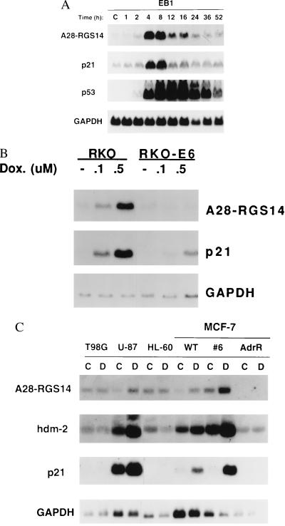
As described (12), a differential cDNA cloning approach was used to identify novel p53 target genes potentially implicated in cellular growth control. This led to the isolation of a cDNA fragment derived from a novel gene induced by both exogenous and endogenous wild-type p53. Fig. 1 describes studies related to the regulation of the expression of this gene, called A28-RGS14, by wild-type p53. A28-RGS14 transcript expression is rapidly induced upon activation of an inducible p53 transgene in human EB1 colon carcinoma cells (Fig. 1A). The magnitude and time course of induction is comparable to that observed for other known p53 response genes, such as p21WAF1, and similar to that observed in other human cells types carrying inducible p53 transgenes (12). Subsequent studies addressed the inducibility of the A28-RGS14 gene by endogenous p53. Consistent with the A28-RGS14 gene being induced upon activation of endogenous wild-type p53, and thus also in response to genotoxic stress, treatment of RKO colon carcinoma cells with the anticancer and DNA damaging agent doxorubicin leads to induction of A28-RGS14 transcripts (Fig. 1B). No such induction was observed in RKO-E6 cells in which p53 signaling is defective as a result of the human papillomavirus E6 viral protein promoting degradation of p53 (Fig. 1B). Importantly, experiments using an anti-A28-RGS14p monoclonal antibody have shown that A28-RGS14p protein is only detected in RKO and EB1 cells after p53 induction (Y.C., L.B., and N.K., unpublished results). As observed for other known p53 target genes, such as the bax and p21WAF1 genes, the basal levels of A28-RGS14 mRNA were found to vary somewhat with cell type, tissue type, and cell culture conditions (Figs. 1C, 2C, and 3B), which is likely to reflect the complex regulation of this gene by p53-dependent and p53-independent mechanisms. However, consistent with results obtained with RKO cells (Fig. 1B), A28-RGS14 transcripts were found to be induced by doxorubicin only in cells expressing wild-type p53 and not in p53 mutant or null cells (Fig. 1C). Thus, these findings suggest a role for endogenous p53 in the induction of the A28-RGS14 gene in response to genotoxic stress in human cells. Curiously, no induction of A28-RGS14 mRNA was observed in mouse embryonic fibroblasts in response to p53 induction. Whether this reflects a cell-specific or species-specific phenomenon remains to be determined.
Figure 1.
Regulation of A28-RGS14 gene expression by p53 and genotoxic stress. (A) Regulation by exogenous p53. The metallothionein-promoter driven p53 transgene in EB1 colon carcinoma cells (9) was induced by the addition of CdCl2 (6 μM) to the cell culture medium. Total RNA or protein was prepared from cells at the times (in hours) indicated. Northern blots (A) were prepared and analyzed by hybridization with 32P-labeled cDNA probes corresponding to A28-RGS14, p21WAF1, p53, or GAPDH. (B) Induction of A28-RGS14 expression by endogenous p53. Northern blot analysis of RKO (colon carcinoma cells, wild-type p53) and clonal RKO-E6 cells (express human papillomavirus E6 viral protein and consequently do not express significant levels of p53) treated with increasing concentrations of doxorubicin for 16 h. (C) Induction of A28-RGS14 transcripts in human cells that are wild type (WT) (MCF-7, breast carcinoma cells; MCF-7 subclone 6; U87, astrocytoma cells) but not mutant (T98G; glioblastoma cells, MCF-7Adr.; doxorubicin-resistant MCF7 subclone) or null (HL-60, promyelocytic leukemia cells) for p53. Cell lines were treated with (lanes D) or without (lanes C) doxorubicin (1 μM) for 16 h. RNA was prepared and analyzed by Northern blot as described in A.
Figure 2.
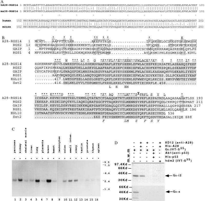
A28-RGS14 gene encodes a novel conserved RGS protein. (A) Predicted protein sequences of human and mouse A28-RGS14p protein. (B) Alignment of A28-RGS14p amino acid sequence and that of related mammalian, yeast, and C. elegans RGS protein family members. Shaded areas indicate a 130-amino acid core domain with highest sequence conservation and a predicted α-helical structure. (C) Tissue expression pattern of the A28-RGS14 gene. A human multitissue Northern blot (CLONTECH) was hybridized with a 32P-labeled A28-RGS14 cDNA probe as described in Fig. 1A. (D) In vitro interaction of A28-RGS14p with Gαi2, but not Gαs subunits of heterotrimeric G proteins. The in vitro-translated 35S-labeled Gα proteins were coimmunoprecipitated with purified His-tagged A28-RGS14p (His-A28) and anti-A28-RGS14p antibody 8D12 (Ab-8D12; Y.C., L.B., and N.K., unpublished results). Lanes: 1, in vitro-translated Gα; 2, coimmunoprecipitation of Gα with His-A28-RGS14p using Ab-8D12; 3, coimmunoprecipitation of Gα without His-A28-RGS14p; 4, coimmunoprecipitation of in vitro-translated Gα with purified His-tagged p53 (His-p53) and its specific antibody (Ab-1, Oncogene Science); 5, coimmunoprecipitation of in vitro-translated hdm2 protein with His-A28-RGS14p and Ab-8D12. m.w.m., Molecular mass markers.
Figure 3.
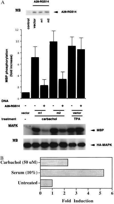
(A) A28-RGS14 coexpression blocks agonist-induced activation of ERK2 in M1- or M2-transfected COS-7 cells. Plasmids encoding M1 or M2 muscarinic receptors were cotransfected with a pcDNA3-A28-RGS14 cDNA vector or empty expression vector, as indicated, and a plasmid encoding HA-tagged MAPK (ERK2). Cells were exposed to Carbachol (100 μM) or phorbol 12-myristate 13-acetate (100 ng/ml) for 5 min and lysed, and ERK activity was assayed on anti-HA immunoprecipitates. After autoradiography, radioactivity incorporated into myelin basic protein (MBP) was quantitated with a Molecular Dynamics PhosphorImager. Data represent the mean ± SEM of six to eight experiments, expressed as fold increase with respect to vector-transfected cells (control). Fifty micrograms of total lysate proteins was subjected to Western blot analysis using anti-HA or anti-A28-RGS14 mouse monoclonal antibodies (Ab-1C5; Y.C., L.B., and N.K., unpublished results). (B) Induction of A28-RGS14 expression in response to mitogenic signals—a potential role as negative feedback regulator. NIH 3T3 cells expressing the muscarinic M1 receptor (10) were grown to confluence, transferred to serum-free medium (DMEM containing 0.1% BSA) for 16 h, and then stimulated with or without Carbachol (50 μM) or fetal bovine serum (10%) for 6 h. RNA prepared from the samples was examined by Northern blot analysis using 32P-labeled probes for mouse A28-RGS14 and GAPDH (as in Fig. 1A). Autoradiograms were analyzed by laser densitometric scanning (Applied Biosystems) and the signal was normalized to GAPDH control.
The following observations indicate that the A28-RGS14 gene might be a direct p53 response gene: (i) A28-RGS14 transcripts are rapidly induced in response to p53 expression (Fig. 1A); (ii) A28-RGS14 transcripts are induced by the conditional activation of a temperature-sensitive mutant of p53 in a human osteosarcoma cell line, even in the absence of ongoing protein synthesis, as we have shown (12); and (iii) the induction of A28-RGS14 transcripts by p53 is not a general consequence of cells arresting in a particular stage of the cell cycle, as demonstrated by the lack of induction in cells blocked in G1 (serum deprived), S (thymidine treated) or G2/M (nocodozole treated) phases of the cell cycle (data not shown). Thus, induction of A28-RGS14 is an early p53 response. To determine whether this might be mediated via direct activation by p53, genomic analysis of the A28-RGS14 gene was performed.
Purified p53 was found to bind to DNA elements resembling the consensus p53-binding sequence (14) and located in intron 3 (not shown) and the 3′ untranslated region (5′-TGGGCTAGCCCAGAGTCCCTtAGCTTGTaC-3′, where underlined type represents p53 half sites and lowercase type represents divergent nucleotides) of the 7.5-kb A28-RGS14 gene, although it is as yet unclear whether these mediate p53 responsiveness in vivo. Computer analysis did not reveal any other putative p53 binding sites in sequences extending to ∼1.5 kb upstream of the putative transcription start site (L.G., L.B., N.K., unpublished data). Future studies should assess whether putative p53-binding sites are encoded by sequences even further upstream of the transcription start site, as reported for the p21WAF1 gene (6), and whether any putative binding site may cooperate in p53-mediated induction of A28-RGS14 gene expression.
Screening of cDNA libraries, 5′ rapid amplification of cDNA ends (RACE) analyses, and genomic analyses revealed that this novel gene encodes a new member of an evolutionarily highly conserved gene family that encodes proteins implicated in the regulation of G protein signaling (regulators of G protein signaling or RGS proteins; refs. 15–17). It is in keeping with the numbering system for predicted RGS proteins that this gene was named A28-RGS14 and the protein product be named A28-RGS14p. On the basis of translation of cloned cDNA sequences, both human and mouse A28-RGS14 genes encode predicted proteins of 202 amino acids with 86% amino acid identity and 90% similarity (Fig. 2A). These share high homology over a core region of 130 amino acids to other members of this family found in species as distant as Aspergillus nidulans (18), yeast (19), and Caenorhabditis elegans (17) (Fig. 2B). The predicted A28-RGS14p protein is closely related to the human RGS1 (BL34) and RGS2 (GOS8) proteins, encoded by genes induced in activated B cells and peripheral blood mononuclear cells, respectively (20, 21). However, RNA expression studies indicate that the A28-RGS14 gene is widely expressed in human tissues (Fig. 2C), in contrast to some other members of this family, including RGS1 and RGS2, which show more tissue-specific expression patterns. As reported for RGS1 (20), expression of A28-RGS14 was also detected in activated B cells (data not shown), but of several RGS genes analyzed to date only A28-RGS14 was found to be induced by p53 (L.B. and N.K, unpublished data).
First indications as to a potential role of this new class of proteins in cellular signaling came from studies in yeast (19). In Saccharomyces cerevisiae, pheromone signaling is mediated through a seven-transmembrane-domain receptor and is coupled via a G protein to downstream events leading to activation of the MAPK pathway. It is the βγ moiety of the G protein that activates downstream signaling leading to growth arrest in the G1 phase of the cell cycle. However, pheromone signaling also promotes expression of the RGS protein Sst2p, which in turn mediates subsequent desensitization to pheromone and recovery from growth arrest. Genetic and biochemical studies indicate that Sst2p controls pheromone signaling through direct interaction with the coupling G protein Gpa1p (19, 22). Importantly, recently reported studies showed that the mammalian RGS1 and RGS4 proteins can complement the function of the yeast homologue Sst2p in a yeast pheromone desensitization assay. Alternatively, they regulate signaling by G protein-coupled receptors in human B cells (15), suggesting that these proteins encode structurally and functionally conserved regulators of G protein-linked signaling pathways. These may, however, not necessarily involve plasma-membrane receptor-coupled signaling pathways. Thus, the α subunit of Gi3, a G protein involved in intracellular trafficking through the Golgi, directly interacts with GAIP in yeast and in vitro (16). GAIP does not appear to interact with Gαq, indicating specificity for interaction with Gαi3 (16). As demonstrated for the yeast Sst2p protein, these findings also indicate that mammalian RGS proteins may operate by directly interacting with α subunits of specific G proteins, disrupting receptor–G protein interaction or acting at the level of the G protein itself. Recent biochemical studies with GAIP and RGS4 have shown that RGS proteins can act as GTPase activating proteins, accelerating the rate of hydrolysis of all tested members of the Gi subfamily of G protein α subunits (23). Preliminary studies indicate that A28-RGS14p can also interact with members of the Gi/Go subfamily of G proteins, including Gαi2, but not Gαs in vitro (Fig. 2D and Y.C., L.B., and N.K., unpublished results). Thus, A28-RGS14p may possibly act as a more general inhibitor of Gi/Go transduced signaling.
Accordingly, we tested whether A28-RGS14p could regulate plasma-membrane receptor-initiated and G protein-transduced signaling in intact cells. We chose to study a well characterized system in which activation of Gi-coupled M2 muscarinic receptor results in G protein βγ-subunit-mediated Ras-dependent activation of the MAPK pathway (13) (the mammalian pathway related to the yeast pheromone response pathway). In addition, we tested the effects on Gq-coupled M1 muscarinic receptor signaling, which is also associated with induction of growth signals and activation of the MAPK pathway. COS-7 cells were transiently cotransfected with mammalian expression constructs encoding M1 or M2 receptors, an HA-tagged-MAPK fusion protein, and a pcDNA3 plasmid expressing A28-RGS14p or pcDNA3 control vector. Cells were stimulated with the muscarinic agonist Carbachol and MAPK activity was assayed in anti-HA immunoprecipitates from cell lysates (Fig. 3A). Pronounced activation of MAPK was observed upon activation of either M1 or M2 receptors. This activation was markedly reduced (∼70%) in cells coexpressing exogenous A28-RGS14p (Fig. 3A). Immunoblot data indicate that the inhibition of MAPK activity by A28-RGS14p did not result from a change in the level of expressed HA-MAPK. Furthermore, the inability of A28-RGS14p to inhibit phorbol 12-myristate 13-acetate-induced activation of MAPK (Fig. 3A) indicates that inhibition is specific and occurs upstream of Raf kinase, consistent with a proposed role for RGS proteins as direct regulators of G proteins. The findings that A28-RGS14p interacts with (Fig. 2D and data not shown) and regulates signal transduction implicating both Gi-like and Gq proteins, suggest that it may encode an important negative regulator of extra and intracellular mitogenic signals associated with the activation of diverse G protein-coupled signaling cascades (among which the MAPK pathway would only be one example). These may include βγ- and α-subunit-activated pathways. In this context, it is also of interest to note that Gi2α, which also interacts with A28-RGS14p in vitro (Fig. 2D), is encoded by the gip2 protooncogene found mutated in certain human tumors (24).
In its capacity as an RGS protein, A28-RGS14p may not only act as a mechanism for p53 to exert cellular growth control by acting upstream of the ras–raf–MAPK pathway but also as a negative feedback regulator in response to mitogenic signals. Thus, similar to regulation of the Sst2p RGS protein upon pheromone stimulation in yeast, A28-RGS14 gene expression is induced by serum growth factors and activation of G protein-coupled receptors (Fig. 3B). This activation is conserved between human and mouse and appears to be p53-independent (S.V., L.B., and N.K., unpublished data). Thus, these data implicate A28-RGS14p in a mammalian desensitization response and as a new signaling molecule whereby p53 may regulate cellular sensitivity to mitogenic and possibly apoptotic signals in human cells. Additional studies should address in more detail the integrated role of A28-RGS14 in p53 signaling and specificities of interactions of various members of the RGS family of proteins with heterotrimeric G proteins.
Acknowledgments
The RKO cell lines were kindly provided by Dr. Michael B. Kastan. EB and EB1 colon carcinoma cells were the gift of Dr. P. Shaw. We thank C. Molloy for insightful discussions and X. Villarreal for automated DNA sequencing assistance.
ABBREVIATIONS
- MAPK
mitogen-activated protein kinase
- RGS
regulators of G protein signaling
- HA
hemagglutinin
Footnotes
References
- 1.Hollstein M, Sidransky D, Vogelstein B, Harris C. Science. 1991;253:49–53. doi: 10.1126/science.1905840. [DOI] [PubMed] [Google Scholar]
- 2.Hartwell L H, Kastan M B. Science. 1994;266:1821–1828. doi: 10.1126/science.7997877. [DOI] [PubMed] [Google Scholar]
- 3.Ko L J, Prives C. Genes Dev. 1996;10:1054–1072. doi: 10.1101/gad.10.9.1054. [DOI] [PubMed] [Google Scholar]
- 4.Dameron K M, Volpert O V, Tainsky M A, Bounk N. Science. 1994;265:1582–1584. doi: 10.1126/science.7521539. [DOI] [PubMed] [Google Scholar]
- 5.Buckbinder L, Talbott R, Velasco-Miguel S, Takenaka I, Faha B, Seizinger B R, Kley N. Nature (London) 1995;377:646–649. doi: 10.1038/377646a0. [DOI] [PubMed] [Google Scholar]
- 6.El-Deiry W S, Tokino T, Velculescu V E, Levy D B, Parsons R, Trent J M, Lim D, Mercer N E, Kinzler K W, Vogelstein B. Cell. 1993;75:817–825. doi: 10.1016/0092-8674(93)90500-p. [DOI] [PubMed] [Google Scholar]
- 7.Polyak K, Waldman T, He T-C, Kinzler K W, Vogelstein B. Genes Dev. 1996;10:1945–1952. doi: 10.1101/gad.10.15.1945. [DOI] [PubMed] [Google Scholar]
- 8.Miyashita T, Reed J C. Cell. 1995;80:293–299. doi: 10.1016/0092-8674(95)90412-3. [DOI] [PubMed] [Google Scholar]
- 9.Shaw P, Bovey R, Tardy S, Sahli R, Sordat B, Costa J. Proc Natl Acad Sci USA. 1992;89:4495–4499. doi: 10.1073/pnas.89.10.4495. [DOI] [PMC free article] [PubMed] [Google Scholar]
- 10.Gutkind J S, Novotny E A, Brann M R, Robbins K C. Proc Natl Acad Sci USA. 1991;88:4703–4707. doi: 10.1073/pnas.88.11.4703. [DOI] [PMC free article] [PubMed] [Google Scholar]
- 11.Cowan K H, Batist G, Tulpule A, Sinha B K, Myers C E. Proc Natl Acad Sci USA. 1986;83:9328–9332. doi: 10.1073/pnas.83.24.9328. ,. [DOI] [PMC free article] [PubMed] [Google Scholar]
- 12.Buckbinder L, Talbott R, Seizinger B R, Kley N. Proc Natl Acad Sci USA. 1994;91:10640–10644. doi: 10.1073/pnas.91.22.10640. [DOI] [PMC free article] [PubMed] [Google Scholar]
- 13.Crespo P, Xu N, Simonds W F, Gutkind J S. Nature (London) 1994;369:418–420. doi: 10.1038/369418a0. [DOI] [PubMed] [Google Scholar]
- 14.El-Deiry W S, Kern S E, Pietenpol J A, Kinzler K W, Vogelstein B. Nat Genet. 1992;1:45–49. doi: 10.1038/ng0492-45. [DOI] [PubMed] [Google Scholar]
- 15.Druey K M, Blumer K J, Kang V H, Kehrl J H. Nature (London) 1996;379:742–746. doi: 10.1038/379742a0. [DOI] [PubMed] [Google Scholar]
- 16.De Vries L, Mousli M, Wurmser A, Farquhar M G. Proc Natl Acad Sci USA. 1995;92:11916–11920. doi: 10.1073/pnas.92.25.11916. [DOI] [PMC free article] [PubMed] [Google Scholar]
- 17.Koelle M R, Horvitz H R. Cell. 1996;84:115–125. doi: 10.1016/s0092-8674(00)80998-8. [DOI] [PubMed] [Google Scholar]
- 18.Adams T H, Hide W A, Yager L N, Lee B N. Mol Cell Biol. 1992;12:3827–3833. doi: 10.1128/mcb.12.9.3827. [DOI] [PMC free article] [PubMed] [Google Scholar]
- 19.Dohlman H G, Apaniesk D, Chen Y, Song J, Nusskern D. Mol Cell Biol. 1995;15:3635–3643. doi: 10.1128/mcb.15.7.3635. [DOI] [PMC free article] [PubMed] [Google Scholar]
- 20.Hong J X, Wilson G L, Fox C H, Kehrl J H. J Immunol. 1993;150:3895–3904. [PubMed] [Google Scholar]
- 21.Siderovski D P, Heximer S P, Forsdyke D R. DNA Cell Biol. 1994;13:125–147. doi: 10.1089/dna.1994.13.125. [DOI] [PubMed] [Google Scholar]
- 22.Dohlman H G, Song J, Ma D, Courchesne W E, Thorner J. Mol Cell Biol. 1996;16:5194–5209. doi: 10.1128/mcb.16.9.5194. [DOI] [PMC free article] [PubMed] [Google Scholar]
- 23.Berman D M, Wilkie T M, Gilman A G. Cell. 1996;86:445–452. doi: 10.1016/s0092-8674(00)80117-8. [DOI] [PubMed] [Google Scholar]
- 24.Lyons J, Landis C A, Marsh G, Vallar L, Grunewald K, et al. Science. 1990;249:655–659. doi: 10.1126/science.2116665. [DOI] [PubMed] [Google Scholar]


