Abstract
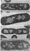
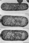
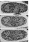
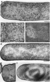
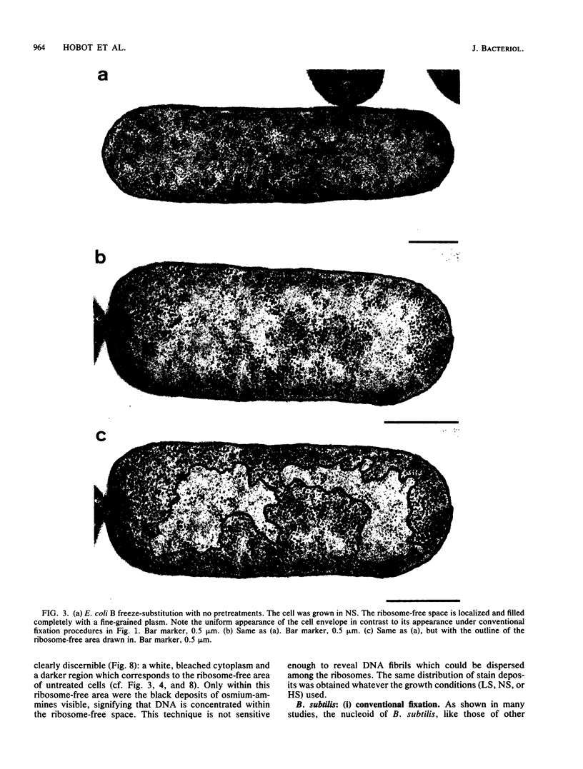
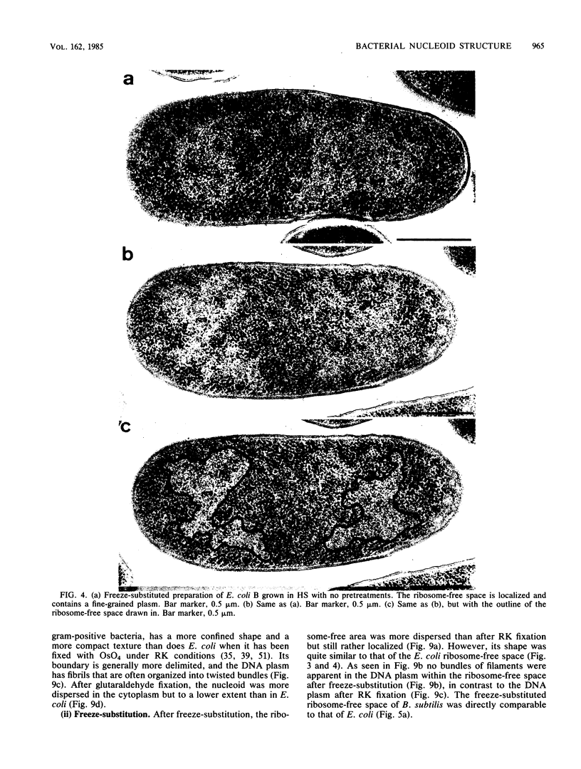
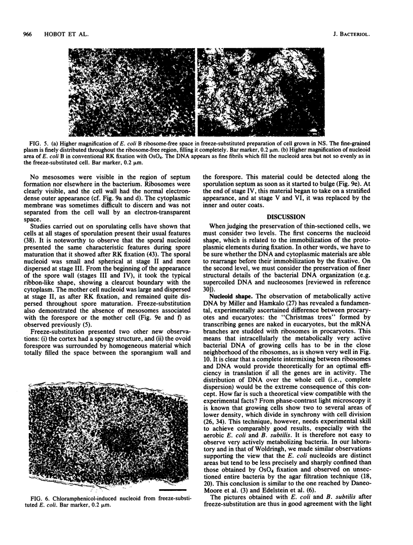
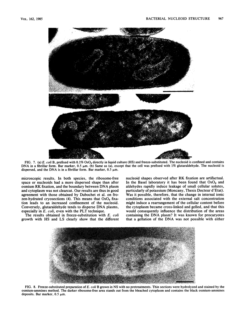
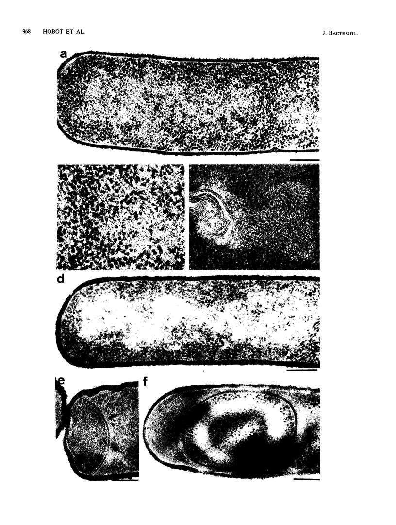
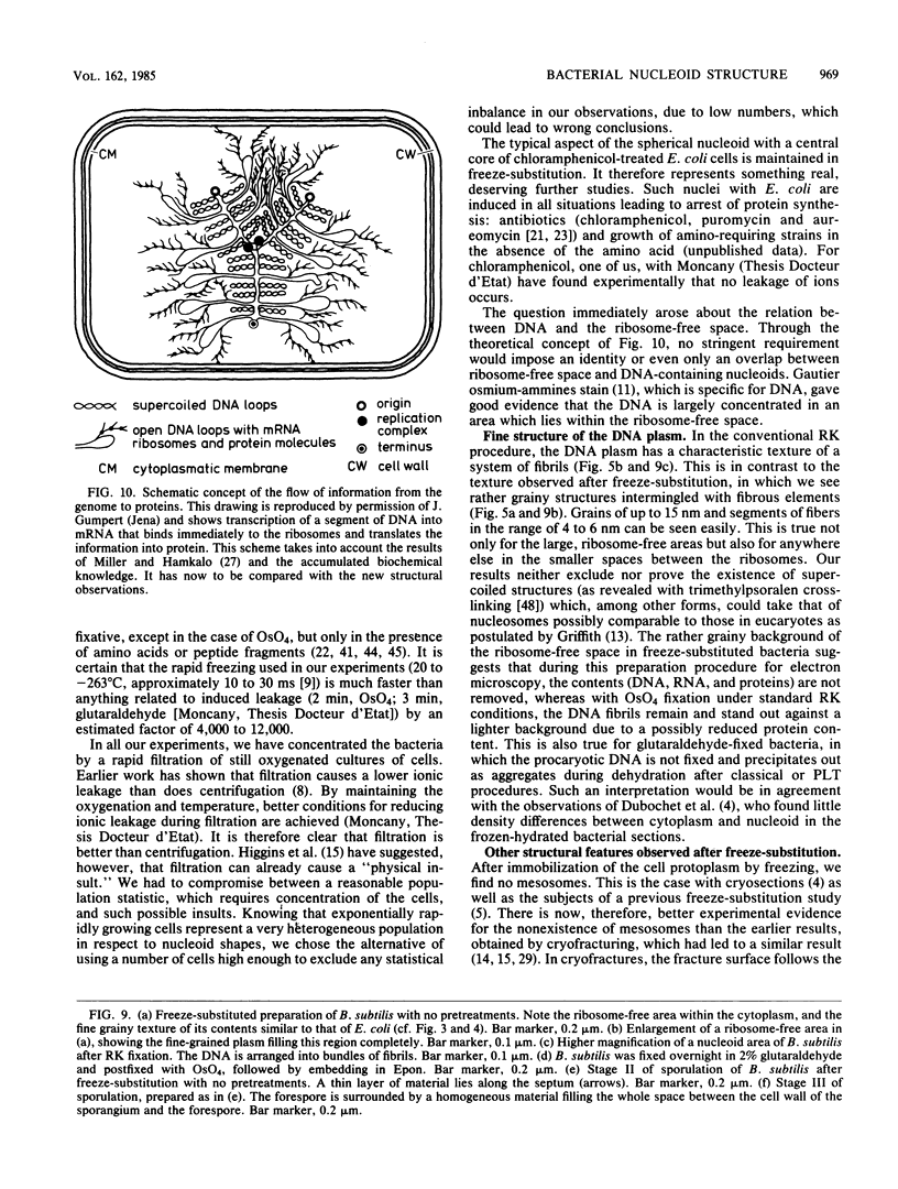
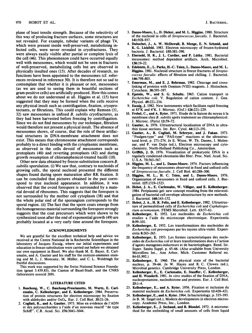
Very rapidly frozen cells of Escherichia coli and Bacillus subtilis were substituted at low temperature into acetone with 1% OsO4 and embedded in Epon. They showed ribosome-free spaces filled with globular and fibrillar material of up to 15 nm. The sizes of structures seen do not exclude DNA superstructures such as supercoils, aggregates, and nucleosomes. With the Feulgen analog osmium-ammines stain, DNA was localized within the ribosome-free space. The bulk of DNA, the nucleoid, is therefore a major part of, or identical to, the main ribosome-free space. The ribosome-free space would correspond directly to the light microscopy phase-contrast image of nucleoids in living bacteria. The shape of the ribosome-free space does not reflect intracellular salt concentrations, nor do the Feulgen-positive areas. The previously observed dependency on the salt concentration of the growth medium seems to be due to permeabilization induced by the chemical fixative at room temperature. The ribosome-free space is more cleft in appearance than the nucleoid obtained by fixation with OsO4 but more confined than its very dispersed form found after aldehyde fixation.
Full text
PDF
Images in this article
Selected References
These references are in PubMed. This may not be the complete list of references from this article.
- Cogliati R., Gautier A. Mise en évidence de l'ADN et des polysaccharides à l'aide d'un nouveau réactif "de type Schiff". C R Acad Sci Hebd Seances Acad Sci D. 1973 Jun 4;276(23):3041–3044. [PubMed] [Google Scholar]
- Daneo-Moore L., Dicker D., Higgins M. L. Structure of the nucleoid in cells of Streptococcus faecalis. J Bacteriol. 1980 Feb;141(2):928–937. doi: 10.1128/jb.141.2.928-937.1980. [DOI] [PMC free article] [PubMed] [Google Scholar]
- Dubochet J., McDowall A. W., Menge B., Schmid E. N., Lickfeld K. G. Electron microscopy of frozen-hydrated bacteria. J Bacteriol. 1983 Jul;155(1):381–390. doi: 10.1128/jb.155.1.381-390.1983. [DOI] [PMC free article] [PubMed] [Google Scholar]
- Ebersold H. R., Cordier J. L., Lüthy P. Bacterial mesosomes: method dependent artifacts. Arch Microbiol. 1981 Sep;130(1):19–22. doi: 10.1007/BF00527066. [DOI] [PubMed] [Google Scholar]
- Edelstein E., Parks L., Tsien H. C., Daneo-Moore L., Higgins M. L. Nucleoid structure in freeze fractures of Streptococcus faecalis: effects of filtration and chilling. J Bacteriol. 1981 May;146(2):798–803. doi: 10.1128/jb.146.2.798-803.1981. [DOI] [PMC free article] [PubMed] [Google Scholar]
- Emerman M., Behrman E. J. Cleavage and cross-linking of proteins with osmium (VII) reagents. J Histochem Cytochem. 1982 Apr;30(4):395–397. doi: 10.1177/30.4.7061831. [DOI] [PubMed] [Google Scholar]
- Gautier A. Ultrastructural localization of DNA in ultrathin tissue sections. Int Rev Cytol. 1976;44:113–191. doi: 10.1016/s0074-7696(08)61649-6. [DOI] [PubMed] [Google Scholar]
- Griffith J. D. Visualization of prokaryotic DNA in a regularly condensed chromatin-like fiber. Proc Natl Acad Sci U S A. 1976 Feb;73(2):563–567. doi: 10.1073/pnas.73.2.563. [DOI] [PMC free article] [PubMed] [Google Scholar]
- Higgins M. L., Daneo-Moore L. Factors influencing the frequency of mesosomes observed in fixed and unfixed cells of Streptococcus faecalis. J Cell Biol. 1974 May;61(2):288–300. doi: 10.1083/jcb.61.2.288. [DOI] [PMC free article] [PubMed] [Google Scholar]
- Higgins M. L., Tsien H. C., Daneo-Moore L. Organization of mesosomes in fixed and unfixed cells. J Bacteriol. 1976 Sep;127(3):1519–1523. doi: 10.1128/jb.127.3.1519-1523.1976. [DOI] [PMC free article] [PubMed] [Google Scholar]
- Hobot J. A., Carlemalm E., Villiger W., Kellenberger E. Periplasmic gel: new concept resulting from the reinvestigation of bacterial cell envelope ultrastructure by new methods. J Bacteriol. 1984 Oct;160(1):143–152. doi: 10.1128/jb.160.1.143-152.1984. [DOI] [PMC free article] [PubMed] [Google Scholar]
- KELLENBERGER E. Les nucléoides de Escherichia coli étudiés à l'aide du microscope électronique. Experientia. 1952 Mar 15;8(3):99–101. doi: 10.1007/BF02301441. [DOI] [PubMed] [Google Scholar]
- KELLENBERGER E. Les transformations des nucléoïdes de Escherichia coli provoquées par les rayons ultra-violets. Experientia. 1952 Jul 15;8(7):263–265. doi: 10.1007/BF02301386. [DOI] [PubMed] [Google Scholar]
- KELLENBERGER E., RYTER A. Fixation et inclusion du matériel nucléaire de Escherichia coli. Experientia. 1956 Nov 15;12(11):420–421. doi: 10.1007/BF02157362. [DOI] [PubMed] [Google Scholar]
- Kellenberger E., Carlemalm E., Stauffer E., Kellenberger C., Wunderli H. In vitro studies of the fixation of DNA, nucleoprotamine, nucleohistone and proteins. Eur J Cell Biol. 1981 Aug;25(1):1–4. [PubMed] [Google Scholar]
- MASON D. J., POWELSON D. M. Nuclear division as observed in live bacteria by a new technique. J Bacteriol. 1956 Apr;71(4):474–479. doi: 10.1128/jb.71.4.474-479.1956. [DOI] [PMC free article] [PubMed] [Google Scholar]
- Miller O. L., Jr, Hamkalo B. A. Visualization of RNA synthesis on chromosomes. Int Rev Cytol. 1972;33:1–25. doi: 10.1016/s0074-7696(08)61446-1. [DOI] [PubMed] [Google Scholar]
- Moncany M. L., Kellenberger E. High magnesium content of Escherichia coli B. Experientia. 1981;37(8):846–847. doi: 10.1007/BF01985672. [DOI] [PubMed] [Google Scholar]
- Nanninga N. The mesosome of Bacillus subtilis as affected by chemical and physical fixation. J Cell Biol. 1971 Jan;48(1):219–224. doi: 10.1083/jcb.48.1.219. [DOI] [PMC free article] [PubMed] [Google Scholar]
- Pettijohn D. E. Structure and properties of the bacterial nucleoid. Cell. 1982 Oct;30(3):667–669. doi: 10.1016/0092-8674(82)90269-0. [DOI] [PubMed] [Google Scholar]
- REYNOLDS E. S. The use of lead citrate at high pH as an electron-opaque stain in electron microscopy. J Cell Biol. 1963 Apr;17:208–212. doi: 10.1083/jcb.17.1.208. [DOI] [PMC free article] [PubMed] [Google Scholar]
- ROBINOW C. F. Morphology of the bacterial nucleus. Br Med Bull. 1962 Jan;18:31–35. doi: 10.1093/oxfordjournals.bmb.a069931. [DOI] [PubMed] [Google Scholar]
- ROBINOW C. F. The chromatin bodies of bacteria. Bacteriol Rev. 1956 Dec;20(4):207–242. doi: 10.1128/br.20.4.207-242.1956. [DOI] [PMC free article] [PubMed] [Google Scholar]
- RYTER A. ETUDE MORPHOLOGIQUE DE LA SPORULATION DE BACILLUS SUBTILIS. Ann Inst Pasteur (Paris) 1965 Jan;108:40–60. [PubMed] [Google Scholar]
- RYTER A., KELLENBERGER E., BIRCHANDERSEN A., MAALOE O. Etude au microscope électronique de plasmas contenant de l'acide désoxyribonucliéique. I. Les nucléoides des bactéries en croissance active. Z Naturforsch B. 1958 Sep;13B(9):597–605. [PubMed] [Google Scholar]
- RYTER A. [Electron microscopic study of the nuclear transformations 05 E. coli K12S and K12S (lambda 26) after irradiation with ultraviolet rays and x-rays]. J Biophys Biochem Cytol. 1960 Oct;8:399–412. [PMC free article] [PubMed] [Google Scholar]
- Remsen C. C. Fine structure of the mesosome and nucleoid in frozen-etched Bacillus subtilis. Arch Mikrobiol. 1968;61(1):40–47. doi: 10.1007/BF00704290. [DOI] [PubMed] [Google Scholar]
- Remsen C. C. The fine structure of frozen-etched Bacillus cereus spores. Arch Mikrobiol. 1966 Sep 8;54(3):266–275. doi: 10.1007/BF00408999. [DOI] [PubMed] [Google Scholar]
- Rouvière-Yaniv J., Yaniv M., Germond J. E. E. coli DNA binding protein HU forms nucleosomelike structure with circular double-stranded DNA. Cell. 1979 Jun;17(2):265–274. doi: 10.1016/0092-8674(79)90152-1. [DOI] [PubMed] [Google Scholar]
- Ryter A., Chang A. Localization of transcribing genes in the bacterial cell by means of high resolution autoradiography. J Mol Biol. 1975 Nov 15;98(4):797–810. doi: 10.1016/s0022-2836(75)80011-8. [DOI] [PubMed] [Google Scholar]
- Ryter A., Jacob F. Etude morphologique de la liaison du noyau à la membrane chez E. coli et chez les protoplastes de B. subtilis. Ann Inst Pasteur (Paris) 1966 Jun;110(6):801–812. [PubMed] [Google Scholar]
- Ryter A., Whitehouse R. Uracil incorporation in the forespore and the mother cell during spore development in Bacillus subtilis. Autoradiographic electron microscopic study. Arch Microbiol. 1978 Jul;118(1):27–34. doi: 10.1007/BF00406070. [DOI] [PubMed] [Google Scholar]
- SCHREIL W. H. STUDIES ON THE FIXATION OF ARTIFICIAL AND BACTERIAL DNA PLASMS FOR THE ELECTRON MICROSCOPY OF THIN SECTIONS. J Cell Biol. 1964 Jul;22:1–20. doi: 10.1083/jcb.22.1.1. [DOI] [PMC free article] [PubMed] [Google Scholar]
- Scraba D. G., Raska I., Kellenberger E. Electron microscopy of polyheads of bacteriophage T4 prepared by freeze-etching. J Ultrastruct Res. 1973 Jul;44(1):27–40. doi: 10.1016/s0022-5320(73)90038-5. [DOI] [PubMed] [Google Scholar]
- Sinden R. R., Carlson J. O., Pettijohn D. E. Torsional tension in the DNA double helix measured with trimethylpsoralen in living E. coli cells: analogous measurements in insect and human cells. Cell. 1980 Oct;21(3):773–783. doi: 10.1016/0092-8674(80)90440-7. [DOI] [PubMed] [Google Scholar]
- Spudich J. A. Symposium on bacterial spores: 3. Biochemical studies of spore core and coat protein synthesis. J Appl Bacteriol. 1970 Mar;33(1):25–33. doi: 10.1111/j.1365-2672.1970.tb05231.x. [DOI] [PubMed] [Google Scholar]
- Séchaud J., Kellenberger E. Electron microscopy of DNA-containing plasms. IV. Glutaraldehyde-uranyl acetate fixation of virus-infected bacteria for thin sectioning. J Ultrastruct Res. 1972 Jun;39(5):598–607. doi: 10.1016/s0022-5320(72)90124-4. [DOI] [PubMed] [Google Scholar]
- Séchaud J., Kellenberger E. Electron microscopy of DNA-containing plasms. IV. Glutaraldehyde-uranyl acetate fixation of virus-infected bacteria for thin sectioning. J Ultrastruct Res. 1972 Jun;39(5):598–607. doi: 10.1016/s0022-5320(72)90124-4. [DOI] [PubMed] [Google Scholar]
- VENABLE J. H., COGGESHALL R. A SIMPLIFIED LEAD CITRATE STAIN FOR USE IN ELECTRON MICROSCOPY. J Cell Biol. 1965 May;25:407–408. doi: 10.1083/jcb.25.2.407. [DOI] [PMC free article] [PubMed] [Google Scholar]
- Van Iterson W., Michels P. A., Vyth-Dreese F., Aten J. A. Nuclear and cell division in Bacillus subtilis: dormant nucleoids in stationary-phase cells and their activation. J Bacteriol. 1975 Mar;121(3):1189–1199. doi: 10.1128/jb.121.3.1189-1199.1975. [DOI] [PMC free article] [PubMed] [Google Scholar]
- WHITFIELD J. F., MURRAY R. G. The effects of the ionic environment on the chromatin structures of bacteria. Can J Microbiol. 1956 May;2(3):245–260. doi: 10.1139/m56-029. [DOI] [PubMed] [Google Scholar]
- Woldringh C. L., Nanninga N. Organization of the nucleoplasm in Escherichia coli visualized by phase-contrast light microscopy, freeze fracturing, and thin sectioning. J Bacteriol. 1976 Sep;127(3):1455–1464. doi: 10.1128/jb.127.3.1455-1464.1976. [DOI] [PMC free article] [PubMed] [Google Scholar]


