

Abstract
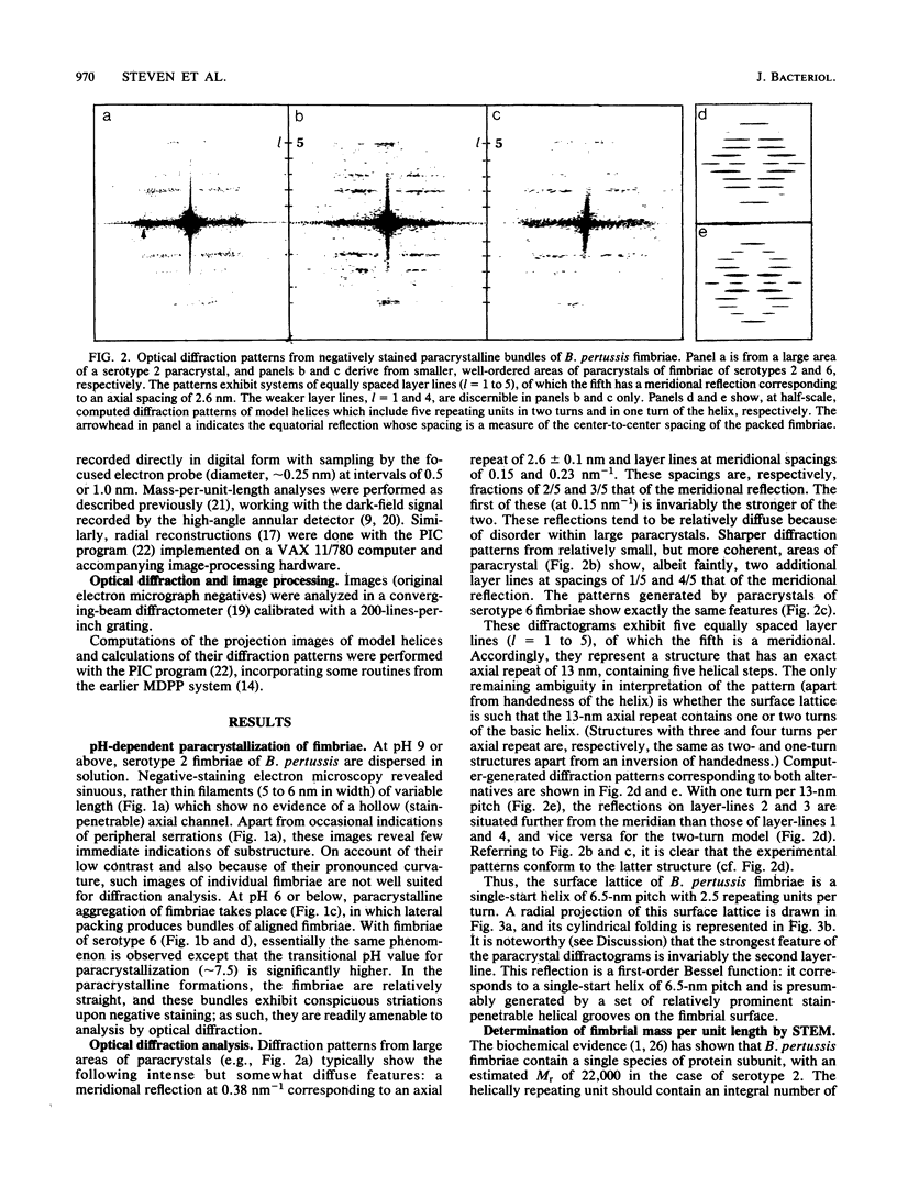
The helical structures of Bordetella pertussis fimbriae of serotypes 2 and 6 were determined by optical diffraction analysis of electron micrographs of negatively stained paracrystalline bundles of purified fimbriae. The fimbrial structure is based on an axial repeat of 13 nm that contains five repeating units in two complete turns of a single-start helix. This structure was confirmed by direct measurements of mass per unit length for individual fimbriae performed by dark-field scanning transmission electron microscopy of unstained specimens. These data further established that the helically repeating unit is a monomer of fimbrial protein (Mr congruent to 22,000 for type 2 and Mr congruent to 21,500 for type 6). Radial density profiles calculated from the scanning transmission electron micrographs showed that the fimbria has peak density at its center, i.e., no axial channel, consistent with the results of conventional negative-staining electron microscopy. The radial profile gives an outermost diameter of approximately 7.5 nm, although the peripheral density is, on average, diffuse, allowing sufficient intercalation between adjacent fimbriae to give a center-to-center spacing of approximately 5.5 nm in the paracrystals. Despite serological and biochemical differences between type 2 and type 6 fimbriae, the packing arrangements of their fimbrial subunits are identical. From this observation, we infer that the respective subunits may have in common conserved regions whose packing dictates the helical geometry of the fimbria. It is plausible that a similar mechanism may underlie the phenomenon of phase variations in other systems of bacterial fimbriae.
Full text
PDF


Images in this article


Selected References
These references are in PubMed. This may not be the complete list of references from this article.
- Ashworth L. A., Irons L. I., Dowsett A. B. Antigenic relationship between serotype-specific agglutinogen and fimbriae of Bordetella pertussis. Infect Immun. 1982 Sep;37(3):1278–1281. doi: 10.1128/iai.37.3.1278-1281.1982. [DOI] [PMC free article] [PubMed] [Google Scholar]
- Blom J., Hansen G. A., Poulsen F. M. Morphology of cells and hemagglutinogens of Bordetella species: resolution of substructural units in fimbriae of Bordetella pertussis. Infect Immun. 1983 Oct;42(1):308–317. doi: 10.1128/iai.42.1.308-317.1983. [DOI] [PMC free article] [PubMed] [Google Scholar]
- Brinton C. C., Jr The structure, function, synthesis and genetic control of bacterial pili and a molecular model for DNA and RNA transport in gram negative bacteria. Trans N Y Acad Sci. 1965 Jun;27(8):1003–1054. doi: 10.1111/j.2164-0947.1965.tb02342.x. [DOI] [PubMed] [Google Scholar]
- Folkhard W., Leonard K. R., Malsey S., Marvin D. A., Dubochet J., Engel A., Achtman M., Helmuth R. X-ray diffraction and electron microscope studies on the structure of bacterial F pili. J Mol Biol. 1979 May 15;130(2):145–160. doi: 10.1016/0022-2836(79)90423-6. [DOI] [PubMed] [Google Scholar]
- Hewlett E., Wolff J. Soluble adenylate cyclase from the culture medium of Bordetella pertussis: purification and characterization. J Bacteriol. 1976 Aug;127(2):890–898. doi: 10.1128/jb.127.2.890-898.1976. [DOI] [PMC free article] [PubMed] [Google Scholar]
- Labaw L. W., Padlan E. A., Segal D. M., Davies D. R. An em study of phosphorylcholine-binding fab immunoglobulin fragment crystals. J Ultrastruct Res. 1975 Jun;51(3):326–339. doi: 10.1016/s0022-5320(75)80097-9. [DOI] [PubMed] [Google Scholar]
- Mosesson M. W., Hainfeld J., Wall J., Haschemeyer R. H. Identification and mass analysis of human fibrinogen molecules and their domains by scanning transmission electron microscopy. J Mol Biol. 1981 Dec 15;153(3):695–718. doi: 10.1016/0022-2836(81)90414-9. [DOI] [PubMed] [Google Scholar]
- Sastry P. A., Finlay B. B., Pasloske B. L., Paranchych W., Pearlstone J. R., Smillie L. B. Comparative studies of the amino acid and nucleotide sequences of pilin derived from Pseudomonas aeruginosa PAK and PAO. J Bacteriol. 1985 Nov;164(2):571–577. doi: 10.1128/jb.164.2.571-577.1985. [DOI] [PMC free article] [PubMed] [Google Scholar]
- Schoolnik G. K., Fernandez R., Tai J. Y., Rothbard J., Gotschlich E. C. Gonococcal pili. Primary structure and receptor binding domain. J Exp Med. 1984 May 1;159(5):1351–1370. doi: 10.1084/jem.159.5.1351. [DOI] [PMC free article] [PubMed] [Google Scholar]
- Smith P. R. An integrated set of computer programs for processing electron micrographs of biological structures. Ultramicroscopy. 1978;3(2):153–160. doi: 10.1016/s0304-3991(78)80021-7. [DOI] [PubMed] [Google Scholar]
- Steinert P. M., Steven A. C., Roop D. R. The molecular biology of intermediate filaments. Cell. 1985 Sep;42(2):411–420. doi: 10.1016/0092-8674(85)90098-4. [DOI] [PubMed] [Google Scholar]
- Steven A. C., Hainfeld J. F., Trus B. L., Steinert P. M., Wall J. S. Radial distributions of density within macromolecular complexes determined from dark-field electron micrographs. Proc Natl Acad Sci U S A. 1984 Oct;81(20):6363–6367. doi: 10.1073/pnas.81.20.6363. [DOI] [PMC free article] [PubMed] [Google Scholar]
- Steven A. C., Hainfeld J. F., Trus B. L., Wall J. S., Steinert P. M. Epidermal keratin filaments assembled in vitro have masses-per-unit-length that scale according to average subunit mass: structural basis for homologous packing of subunits in intermediate filaments. J Cell Biol. 1983 Dec;97(6):1939–1944. doi: 10.1083/jcb.97.6.1939. [DOI] [PMC free article] [PubMed] [Google Scholar]
- Steven A. C., Navia M. A. Fidelity of structure representation in electron micrographs of negatively stained protein molecules. Proc Natl Acad Sci U S A. 1980 Aug;77(8):4721–4725. doi: 10.1073/pnas.77.8.4721. [DOI] [PMC free article] [PubMed] [Google Scholar]
- Steven A. C., Wall J., Hainfeld J., Steinert P. M. Structure of fibroblastic intermediate filaments: analysis of scanning transmission electron microscopy. Proc Natl Acad Sci U S A. 1982 May;79(10):3101–3105. doi: 10.1073/pnas.79.10.3101. [DOI] [PMC free article] [PubMed] [Google Scholar]
- Thomas D., Newcomb W. W., Brown J. C., Wall J. S., Hainfeld J. F., Trus B. L., Steven A. C. Mass and molecular composition of vesicular stomatitis virus: a scanning transmission electron microscopy analysis. J Virol. 1985 May;54(2):598–607. doi: 10.1128/jvi.54.2.598-607.1985. [DOI] [PMC free article] [PubMed] [Google Scholar]
- Watts T. H., Kay C. M., Paranchych W. Spectral properties of three quaternary arrangements of Pseudomonas pilin. Biochemistry. 1983 Jul 19;22(15):3640–3646. doi: 10.1021/bi00284a016. [DOI] [PubMed] [Google Scholar]
- Wyckoff M., Rodbard D., Chrambach A. Polyacrylamide gel electrophoresis in sodium dodecyl sulfate-containing buffers using multiphasic buffer systems: properties of the stack, valid Rf- measurement, and optimized procedure. Anal Biochem. 1977 Apr;78(2):459–482. doi: 10.1016/0003-2697(77)90107-5. [DOI] [PubMed] [Google Scholar]
- Zhang J. M., Cowell J. L., Steven A. C., Carter P. H., McGrath P. P., Manclark C. R. Purification and characterization of fimbriae isolated from Bordetella pertussis. Infect Immun. 1985 May;48(2):422–427. doi: 10.1128/iai.48.2.422-427.1985. [DOI] [PMC free article] [PubMed] [Google Scholar]