

Abstract
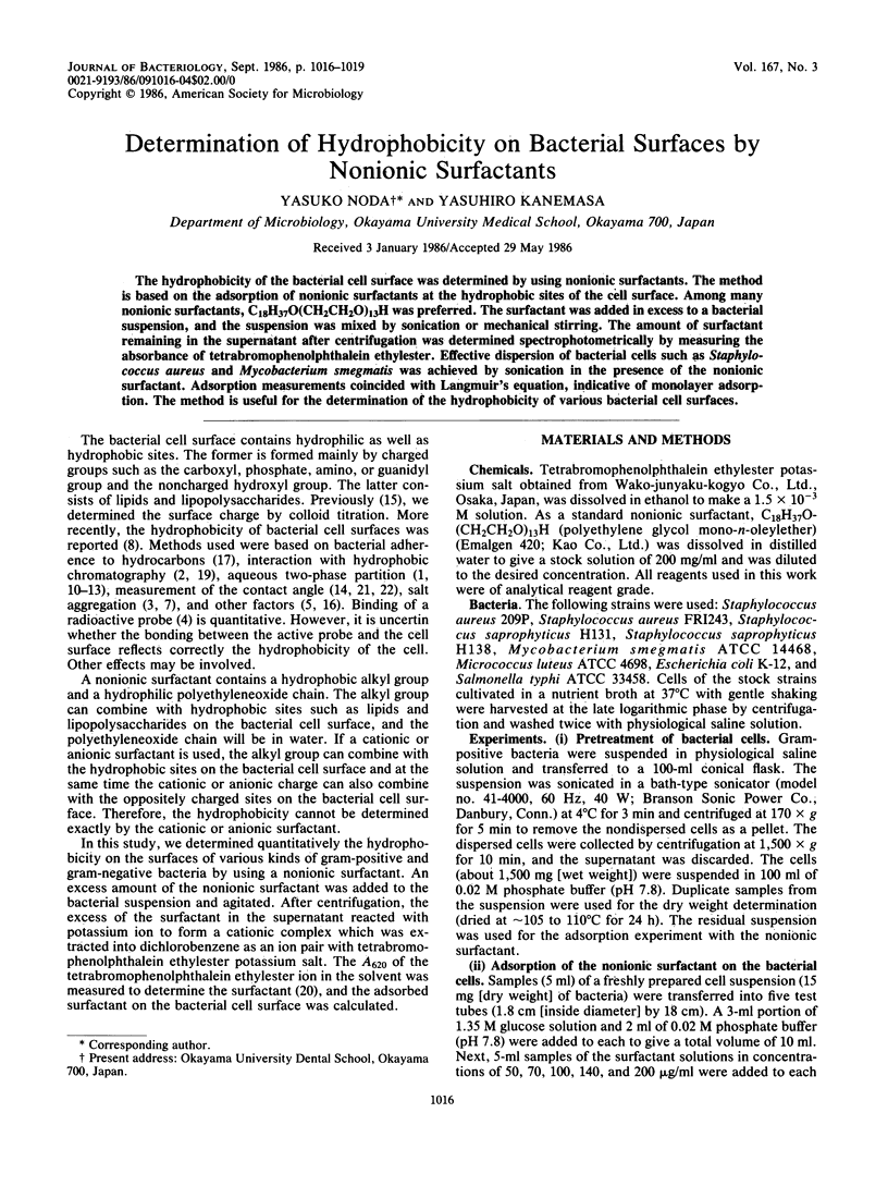
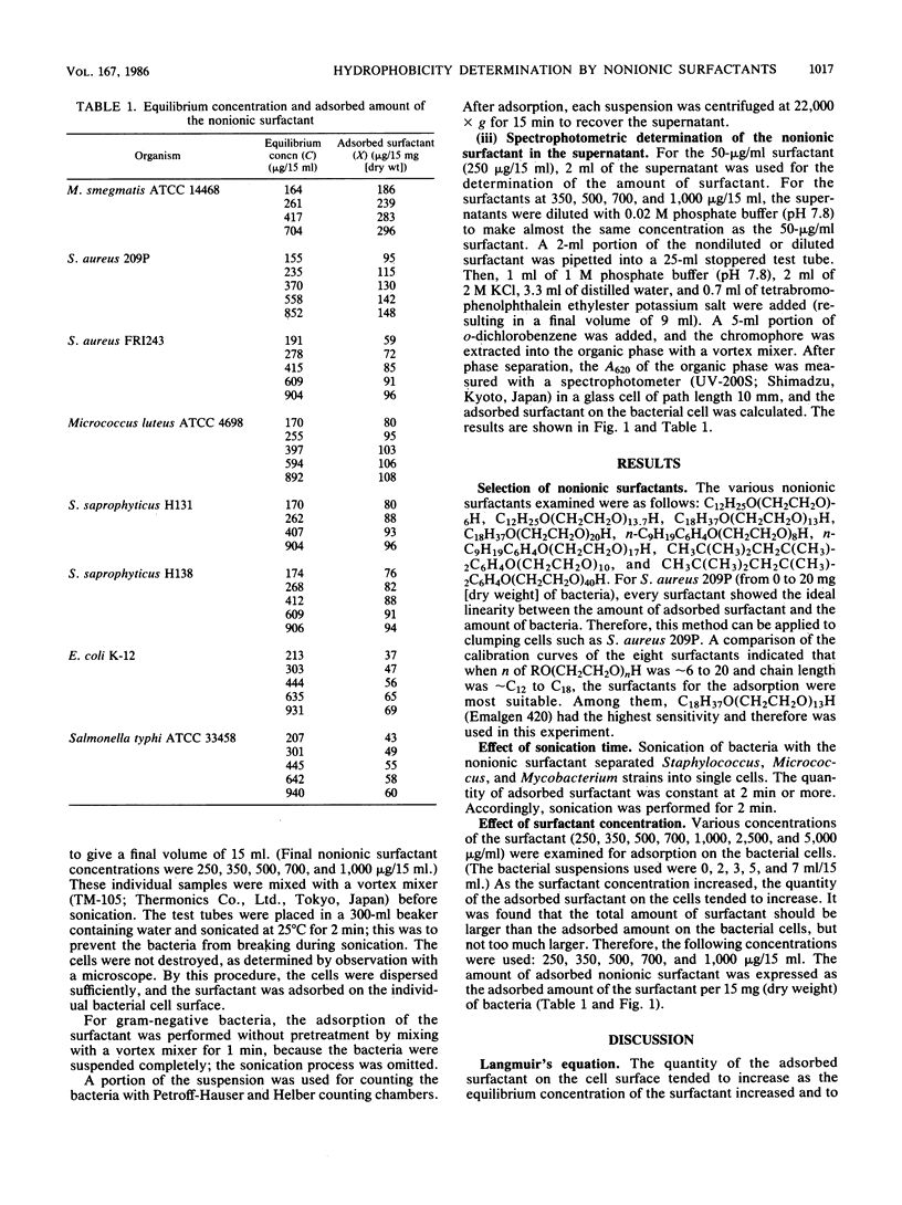
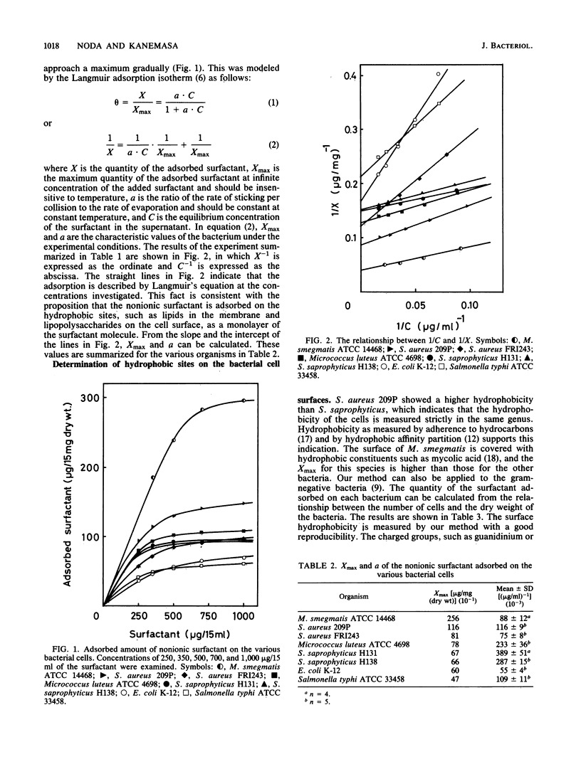
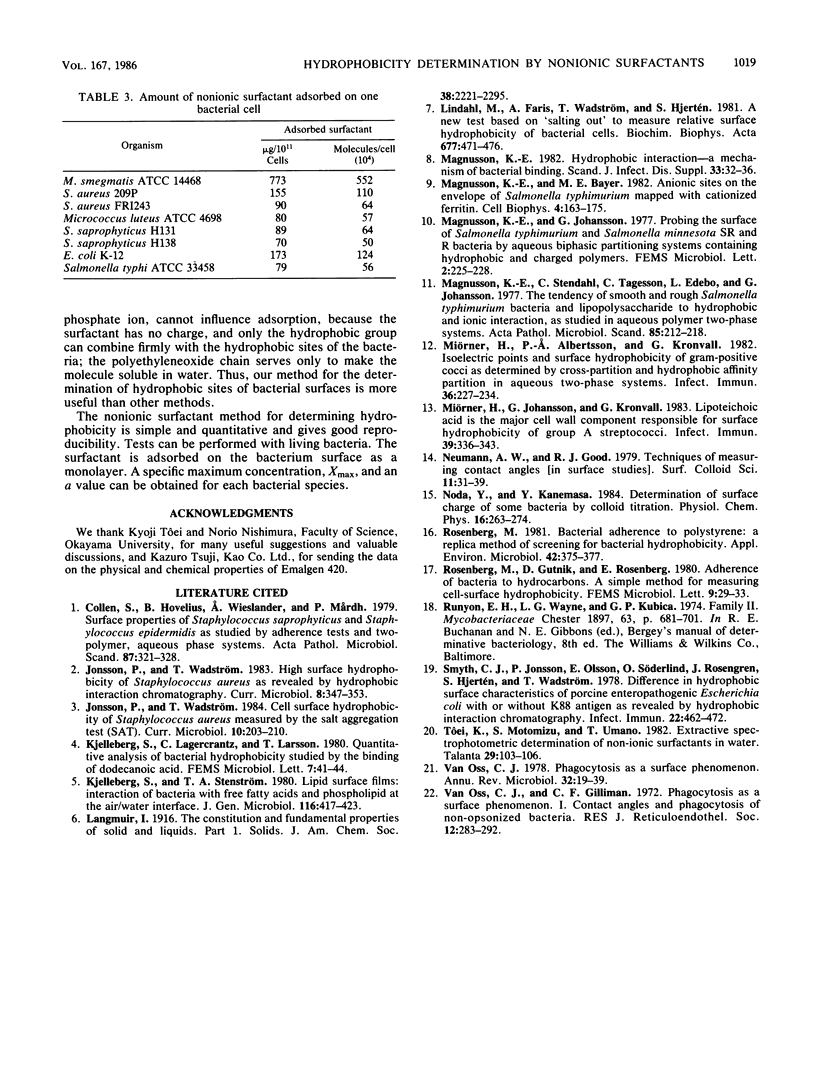
The hydrophobicity of the bacterial cell surface was determined by using nonionic surfactants. The method is based on the adsorption of nonionic surfactants at the hydrophobic sites of the cell surface. Among many nonionic surfactants, C18H37O(CH2CH2O)13H was preferred. The surfactant was added in excess to a bacterial suspension, and the suspension was mixed by sonication or mechanical stirring. The amount of surfactant remaining in the supernatant after centrifugation was determined spectrophotometrically by measuring the absorbance of tetrabromophenolphthalein ethylester. Effective dispersion of bacterial cells such as Staphylococcus aureus and Mycobacterium smegmatis was achieved by sonication in the presence of the nonionic surfactant. Adsorption measurements coincided with Langmuir's equation, indicative of monolayer adsorption. The method is useful for the determination of the hydrophobicity of various bacterial cell surfaces.
Full text
PDF
Selected References
These references are in PubMed. This may not be the complete list of references from this article.
- CHESTER S. T., BELL H. G. The management of the duodenal stump in gastric resection for technically difficult duodenal ulcer. West J Surg Obstet Gynecol. 1955 Nov;63(11):701–705. [PubMed] [Google Scholar]
- Colleen S., Hovelius B., Wieslander A., Mårdh P. A. Surface properties of Staphylococcus saprophyticus and Staphylococcus epidermidis as studied by adherence tests and two-polymer, aqueous phase systems. Acta Pathol Microbiol Scand B. 1979 Dec;87(6):321–328. doi: 10.1111/j.1699-0463.1979.tb02446.x. [DOI] [PubMed] [Google Scholar]
- Lindahl M., Faris A., Wadström T., Hjertén S. A new test based on 'salting out' to measure relative surface hydrophobicity of bacterial cells. Biochim Biophys Acta. 1981 Nov 5;677(3-4):471–476. doi: 10.1016/0304-4165(81)90261-0. [DOI] [PubMed] [Google Scholar]
- Magnusson K. E., Bayer M. E. Anionic sites on the envelope of Salmonella typhimurium mapped with cationized ferritin. Cell Biophys. 1982 Jun-Sep;4(2-3):163–175. doi: 10.1007/BF02918311. [DOI] [PubMed] [Google Scholar]
- Magnusson K. E. Hydrophobic interaction--a mechanism of bacterial binding. Scand J Infect Dis Suppl. 1982;33:32–36. [PubMed] [Google Scholar]
- Magnusson K. E., Stendahl O., Tagesson C., Edebo L., Johansson G. The tendency of smooth and rough Salmonella typhimurium bacteria and lipopolysaccharide to hydrophobic and ionic interaction, as studied in aqueous polymer two-phase systems. Acta Pathol Microbiol Scand B. 1977 Jun;85(3):212–218. doi: 10.1111/j.1699-0463.1977.tb01698.x. [DOI] [PubMed] [Google Scholar]
- Miörner H., Albertsson P. A., Kronvall G. Isoelectric points and surface hydrophobicity of Gram-positive cocci as determined by cross-partition and hydrophobic affinity partition in aqueous two-phase systems. Infect Immun. 1982 Apr;36(1):227–234. doi: 10.1128/iai.36.1.227-234.1982. [DOI] [PMC free article] [PubMed] [Google Scholar]
- Miörner H., Johansson G., Kronvall G. Lipoteichoic acid is the major cell wall component responsible for surface hydrophobicity of group A streptococci. Infect Immun. 1983 Jan;39(1):336–343. doi: 10.1128/iai.39.1.336-343.1983. [DOI] [PMC free article] [PubMed] [Google Scholar]
- Noda Y., Kanemasa Y. Determination of surface charge of some bacteria by colloid titration. Physiol Chem Phys Med NMR. 1984;16(4):263–274. [PubMed] [Google Scholar]
- Rosenberg M. Bacterial adherence to polystyrene: a replica method of screening for bacterial hydrophobicity. Appl Environ Microbiol. 1981 Aug;42(2):375–377. doi: 10.1128/aem.42.2.375-377.1981. [DOI] [PMC free article] [PubMed] [Google Scholar]
- Smyth C. J., Jonsson P., Olsson E., Soderlind O., Rosengren J., Hjertén S., Wadström T. Differences in hydrophobic surface characteristics of porcine enteropathogenic Escherichia coli with or without K88 antigen as revealed by hydrophobic interaction chromatography. Infect Immun. 1978 Nov;22(2):462–472. doi: 10.1128/iai.22.2.462-472.1978. [DOI] [PMC free article] [PubMed] [Google Scholar]
- Van Oss C. J., Gillman C. F. Phagocytosis as a surface phenomenon. Contact angles and phagocytosis of non-opsonized bacteria. J Reticuloendothel Soc. 1972 Sep;12(3):283–292. [PubMed] [Google Scholar]
- van Oss C. J. Phagocytosis as a surface phenomenon. Annu Rev Microbiol. 1978;32:19–39. doi: 10.1146/annurev.mi.32.100178.000315. [DOI] [PubMed] [Google Scholar]