

Abstract
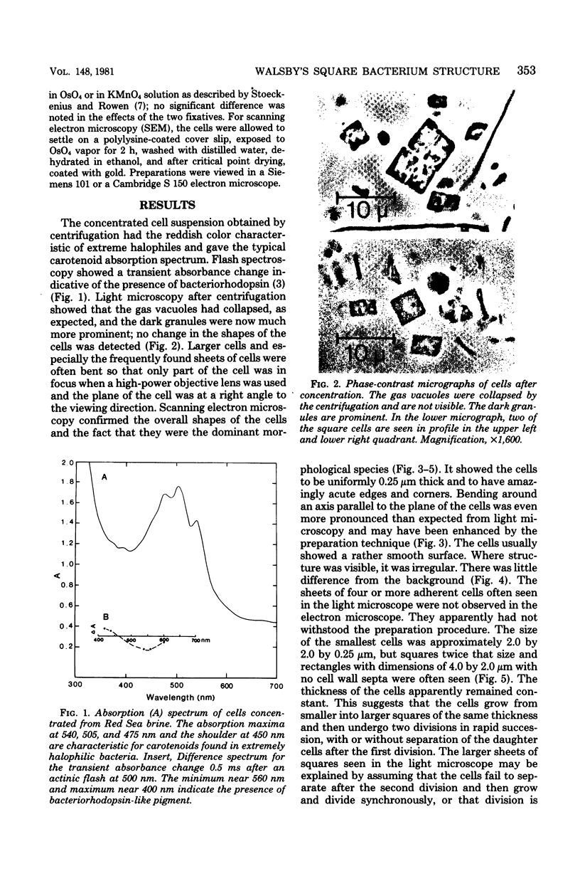
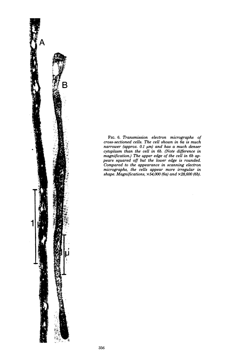
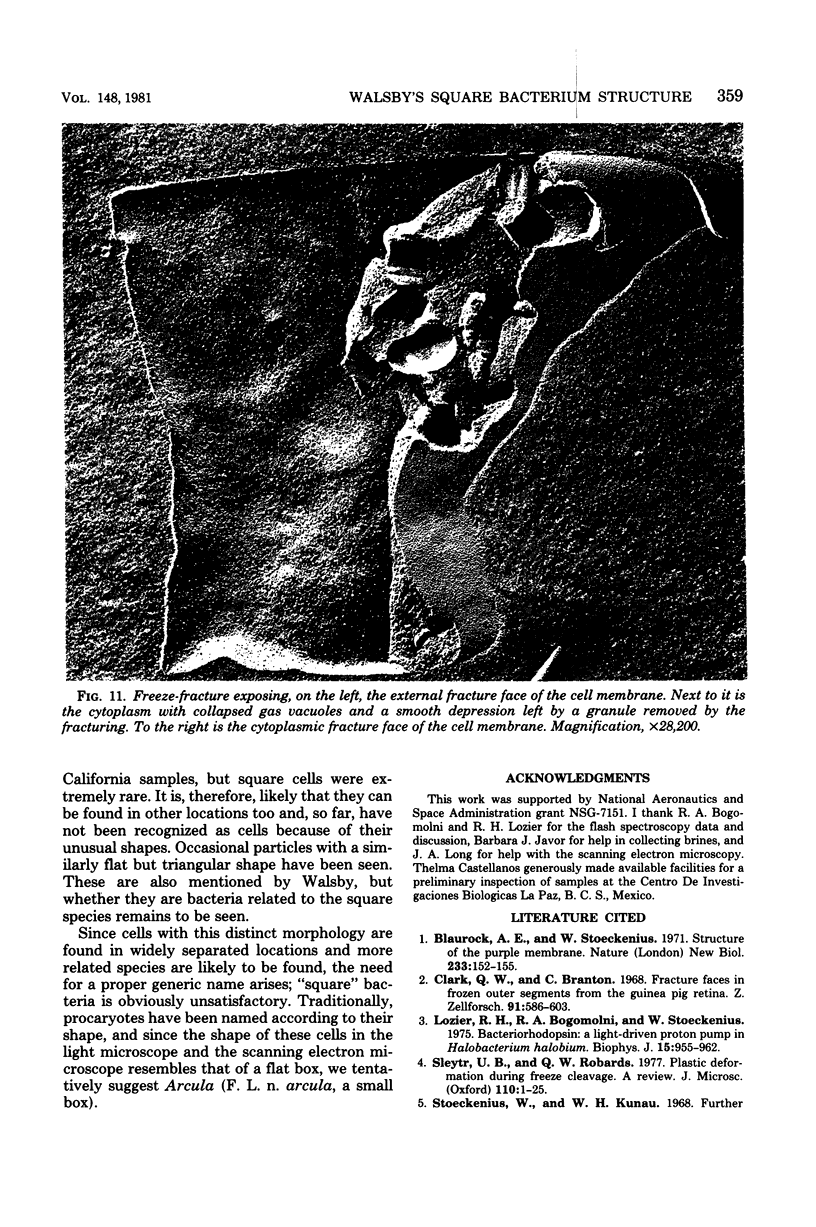
The "square" bacterium, first described by Walsby from brine collected at the Red Sea shore [A. E. Walsby, Nature (London) 283:69-71, 1980] was examined by electron microscopy. The cells appeared as flat rectangular boxes in scanning electron micrographs. In sections and freeze-fracture preparation, the edges looked more rounded. The thickness apparently remains constant as the cells grow and divide. Their sides were a few micrometers long, but the cells were only 0.25 micrometers thick. They showed typical procaryote structure, with a regular cell wall and a gas vacuole fine structure similar to that of other halophilic procaryotes. The inner fracture faces of the cell membrane showed a much denser population of intramembrane particles than the outer fracture faces, but no patches of purple membrane, despite the presence of bacteriorhodospin-like pigment in the cell suspension. Morphologically identical cells have been found in brine from Baja California, Mexico.
Full text
PDF


Images in this article


Selected References
These references are in PubMed. This may not be the complete list of references from this article.
- Blaurock A. E., Stoeckenius W. Structure of the purple membrane. Nat New Biol. 1971 Sep 29;233(39):152–155. doi: 10.1038/newbio233152a0. [DOI] [PubMed] [Google Scholar]
- Clark A. W., Branton D. Fracture faces in frozen outer segments from the guinea pig retina. Z Zellforsch Mikrosk Anat. 1968;91(4):586–603. doi: 10.1007/BF00455276. [DOI] [PubMed] [Google Scholar]
- Lozier R. H., Bogomolni R. A., Stoeckenius W. Bacteriorhodopsin: a light-driven proton pump in Halobacterium Halobium. Biophys J. 1975 Sep;15(9):955–962. doi: 10.1016/S0006-3495(75)85875-9. [DOI] [PMC free article] [PubMed] [Google Scholar]
- Sleytr U. B., Robards A. W. Plastic deformation during freeze-cleavage: a review. J Microsc. 1977 May;110(1):1–25. doi: 10.1111/j.1365-2818.1977.tb00009.x. [DOI] [PubMed] [Google Scholar]
- Stoeckenius W., Lozier R. H., Bogomolni R. A. Bacteriorhodopsin and the purple membrane of halobacteria. Biochim Biophys Acta. 1979 Mar 14;505(3-4):215–278. doi: 10.1016/0304-4173(79)90006-5. [DOI] [PubMed] [Google Scholar]
- Stoeckenius W., Rowen R. A morphological study of Halobacterium halobium and its lysis in media of low salt concentration. J Cell Biol. 1967 Jul;34(1):365–393. doi: 10.1083/jcb.34.1.365. [DOI] [PMC free article] [PubMed] [Google Scholar]
- Toeckenius W., Kunau W. H. Further characterization of particulate fractions from lysed cell envelopes of Halobacterium halobium and isolation of gas vacuole membranes. J Cell Biol. 1968 Aug;38(2):337–357. doi: 10.1083/jcb.38.2.337. [DOI] [PMC free article] [PubMed] [Google Scholar]
- Woese C. R., Fox G. E. Phylogenetic structure of the prokaryotic domain: the primary kingdoms. Proc Natl Acad Sci U S A. 1977 Nov;74(11):5088–5090. doi: 10.1073/pnas.74.11.5088. [DOI] [PMC free article] [PubMed] [Google Scholar]