Abstract
The heat resistance of foot-and-mouth disease virus (FMDV) strains isolated from outbreaks in Thailand was investigated in phosphate-buffered saline (PBS) at 50, 60, 70, 80, 90, and 100°C. The first-order kinetic model fitted most of the observed linear inactivation curves. The ranges of decimal-reduction time (D value) of FMDV strains at 50, 60, 70, 80, 90, and 100°C were 732 to 1,275 s, 16.37 to 42.00 s, 6.06 to 10.87 s, 2.84 to 5.99 s, 1.65 to 3.18 s, and 1.90 to 2.94 s, respectively. The heat resistances of FMDV strains at lower temperature (50°C) were not serotype specific. The effective inactivating temperature is approximately 60°C. Heat resistances of FMDV strains at 90 and 100°C were not statistically different (P > 0.05), while the FMDV serotype O (OPN) appeared to be the most heat resistant at 60 to 80°C. The other observed inactivation curves were linear with shoulder or tailing (biphasic curves). The shoulder effect was mostly observed at 90 and 100°C, while the tailing effect was mostly observed at 50 to 80°C. The adjusted D values in the case of shoulder and tailing effects did not affect the overall estimated heat resistance of these FMDV strains, so even unadjusted D values of deviant inactivation curves were legitimate. The z values of FMDV serotypes O, A, and Asia 1 were 21.78 to 23.26, 20.75 to 22.79, and 19.87°C, respectively. The z values of FMDV strains studied were not statistically significantly different (P > 0.05). The results of this study indicated that the heat resistance in PBS of FMDV strains from Thailand was much less than had been reported for foreign epidemic FMDV strains.
Foot-and-mouth disease virus (FMDV) is classified within the genus Aphthovirus, family Picornaviridae. FMDV is a nonenveloped, icosahedral symmetric, and single-stranded RNA virus; therefore, it is somewhat resistant to harsh environments, e.g., UV radiation, low water activities, and heat (3). FMDV causes foot-and-mouth disease (FMD) and is highly contagious to cloven-hoofed animals. The common mode of transmission of FMDV is direct contact; however, oral-route transmission via indirect contact has frequently caused epidemics in pigs (2). FMD is the first disease on the OIE List A and also was the first disease for which the OIE established an official list of free countries and zones. FMD has a great potential for causing severe economic loss for many countries (2). In order to export meat from an FMD-infected country to an FMD-free country, meat is subjected to heat treatment to reach an internal core temperature of at least 70°C for a minimum of 30 min or to any equivalent treatment which has been demonstrated to inactivate the FMD virus (38).
The efficiency of thermal inactivation is a function of time and temperature. Thermal destruction hypothetically follows first order kinetics. The decimal reduction time (D value, DRT, or DT) is the time at a specified temperature required to reduce the number of microorganisms by a factor of 10 (23, 24, 44, 45). The D value is the negative reciprocal of the slope of the inactivation curve, where the inactivation of microorganisms is a logarithmic function of time. The z value is defined as the temperature change required to reduce D value by a factor of 10 (23, 24, 44, 45). Likewise, the z value can be calculated by the negative reciprocal of the slope of the decimal reduction curve where D values of different temperatures are plotted on a semilogarithmic scale against temperatures. The z value is specific for a given strain of microorganism in a given medium or product, but the z value does not differ across media as widely as does the D value. This association of D value and temperature in a DRT curve is helpful to calculate the D value of temperatures that were not included in the experiment. Many studies have demonstrated the heat resistance of FMDV (10, 12, 14-16, 30-32, 34). However, these studies involved only a few temperatures, different media, and strains that were not epidemic in Thailand. Therefore, these data are not appropriate bases for bilateral trade negotiations involving Thai animal products. The objective of the present study was to determine the heat resistance of FMDV strains isolated from outbreaks in Thailand, in terms of D value and z value in phosphate-buffered saline (PBS) at 50, 60, 70, 80, 90, and 100°C.
MATERIALS AND METHODS
Virus and cell culture.
FMDV serotypes O (strains O189 and OPN), A (strains A118, A-Sakol, and A132), and Asia 1 (strain AS1) were obtained from the Regional Reference Laboratory for FMD in Southeast Asia, Pakchong, Thailand. All strains of these serotypes have been isolated from epidemics in Thailand and then been used as the vaccine strains produced and used domestically.
The maintenance of the baby hamster kidney (BHK-21) cell line was modified from a previous publication (11). Briefly, BHK-21 was grown in a medium composed of Eagle minimum essential medium Nissui No.1, supplemented with 5% normal bovine serum, 0.07% sodium bicarbonate, 0.25 mg of amphotericin B (Fungizone; Bristol-Myers Squibb, Ltd., Thailand)/ml, 0.1 mg of kanamycin/ml, 0.117 mg of penicillin/ml, and 0.1 mg of streptomycin/ml. The maintenance medium was like the growth medium but contained only 2% fetal bovine serum and 0.21% sodium bicarbonate. Cells for virus propagation and assay were grown in 96-well tissue culture plates (Corning).
Virus isolation.
Several serotypes of FMDV were isolated from FMD epidemics in Thailand. The epithelial tissue in the oral cavity of animals infected with FMD was prepared in 10% (wt/vol) suspension. Monolayer primary cultures of lamb kidney cells were inoculated with epithelial suspension and kept at 37°C for 60 min (21). The maintenance medium (Eagle minimum essential medium Nissui no. 1) was added on the monolayer and kept at 37°C. After observation for cytopathic effect (CPE), the harvested fluids were centrifuged at 2,000 × g for 10 min to separate the cell sediment from the fluid medium (21). The serotyping of FMDV was done by enzyme-linked immunosorbent assay as previously described (5). If no CPE was observed in the first passage within 48 h, the cells were frozen and thawed, used to inoculate fresh culture, and examined for CPE for another 48 h. The second and third blind passages used the same procedure as the first blind passage, except that the monolayer culture was BHK-21 cells (21). The other application of virus isolation was to determine any residual virus after thermal inactivation.
Virus preparation.
The viruses were prepared as previously described (35). Briefly, the FMDV was propagated in BHK-21 cells. After CPE was observed, harvested fluids were centrifuged to separate the cell sediment from the fluid medium. The sediments were mixed with the cell monolayer, which had been treated with sodium dodecyl sulfate. The supernatant and treated cell monolayer were pooled, filtered (0.2-μm pore size), and kept at −70°C until used.
Virus assay.
Tenfold virus dilutions were inoculated in equal volumes (50 μl) with BHK-21 cell suspension in minimal essential growth medium into 96-well plates. The control wells were inoculated with 50 μl of viral diluent. The viral diluent was PBS with 137 mmol of NaCl, 2.7 mmol of KCl, and 10 mmol of phosphate buffer. Plates were incubated at 37°C for 48 h to observe CPE. Virus titer was recorded as the 50% tissue culture infective dose according to the method of Reed and Muench (40).
Thermal inactivation.
The FMDV suspensions (5 ml) in borosilicate glass tubes were diluted with PBS (45 ml). The PBS was preheated before the FMDV stock was mixed and heated at 50, 60, 70, 80, 90, and 100°C in a digitally controlled water bath. The final pH of the FMDV suspension was approximately 7.0. The metal probe from digital thermocouple was submerged into another borosilicate glass tube, with PBS acting as a reference temperature tube, while this reference temperature tube was identical to all FMDV inactivation tubes. The inactivation tubes were being stirred in the shaking water bath during the thermal inactivation. The time and temperature readings were automatically recorded and printed out from the thermocouple. At 90 and 100°C, thermocouple recordings indicated that temperature increases, particularly near the target temperatures, were slightly different. FMDV suspension tubes were transferred at sampling time intervals and immersed in the ice bath to stop the reaction (17, 46). The sampling time was dependent on the temperature, i.e., the higher the temperature, the shorter the time intervals. The actual inactivating times were determined by the integration of area under temperature curve plotted against sampling time (44, 45). The randomized complete block design was used to differentiate the effect of temperatures across FMDV strains and vice versa. There were six treatments as the FMDV strains and six blocks as the inactivation temperatures. It should be emphasized that each FMDV strain was inactivated at every temperature and each temperature was assigned to inactivate every FMDV strain (13, 25). The experiments were repeated three times to decrease the error of the experiment.
Rate of viral inactivation (heat resistance).
Since the main objective was to determine that the number of virus units were activated at various inactivation times, so it was understood that some virus suspensions still had infectious virus left to be assayed. The thermal inactivation of a microorganism is dependent on time and temperature. At a constant temperature, the decrease in the number of microorganisms often follows first-order kinetics and can be expressed as:
 |
(1) |
where Nt and N0 are the concentrations of FMDV at times t and zero, respectively. The DRT is the time at specified temperature required to reduce the number of microorganisms by a factor of 10 or 90%. Therefore, the D value represents the heat resistance or rate of microbial inactivation and can be calculated by the negative reciprocal of the slope of the inactivation curve where log [Nt/N0] against inactivation time t is linear on a semilogarithmic plot. The z value is defined as the temperature change required to change the D value by a factor of 10 or 90% and can be written in the form of D values as:
 |
(2) |
where D2 and D1 are the D values of FMDV at temperatures T2 and T1, respectively. Likewise, the z value can also be calculated by the negative reciprocal of the slope of the DRT curve, where D values for different temperatures are plotted on a semilogarithmic scale against temperatures. Consequently, the z value represents the temperature dependence of the inactivation rate. The z value is specific for a given strain of microorganism in a given medium or product, but the z value does not differ across media as widely as does D.
Mathematical models for inactivation curve.
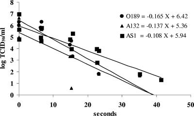
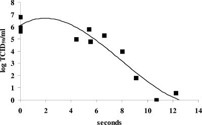
The thermal inactivation follows first-order kinetics, which yields a linear logarithmic plot of the number of microorganisms and inactivation time as shown in equation 1 (Fig. 1). However, a number of inactivation curves do not comply with this rule. If the microorganisms clump or there are multiple target sites for thermal inactivation (e.g., cell attachment site especially RGD sequence) (18), then the inactivation curve will start with a lag phase, the so-called “shoulder effect” (1, 48) (Fig. 2). Note that if microbial clumping is to cause a shoulder, virtually all of the microorganisms must be in clumps. If a substantial portion of the microbes are monodispersed, then the clumped fraction will produce a tail rather than a shoulder. Essentially for the linear inactivation curve with a shoulder during the lag phase, the microorganisms are virtually not inactivated and the actual inactivation of microorganisms follows first-order kinetics in the linear phase, which is modified from previous models (45, 48) as:
 |
(3) |
where Nt and N0 are the concentrations of FMDV at time t and time zero, respectively, and tL is the lag time, defined as the mean time required for initial inactivation of microorganisms. L is the partial log cycle of inactivation during lag time and in most cases does not exceed one log cycle.
FIG. 1.
Thermal inactivation of FMDV serotypes O, A, and Asia 1 in PBS at 70°C. The log-linear inactivation equations of FMDV serotypes are shown in the legend, where FMDV serotypes represent the number of FMDV and X is the inactivation time. D values are the reciprocal of the slope of the inactivation curve.
FIG. 2.
Thermal inactivation of FMDV strain A118 in PBS at 90°C. The inactivation curve shows the shoulder effect.
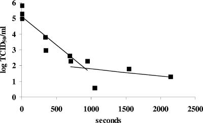
If a subpopulation of microorganisms are more resistant than others or shielded by some organic matter, e.g., soil and cell debris, or clumped, then the inactivation curve will show a “tailing effect” (Fig. 3). Thus, the plot of linear inactivation with a tail has two slopes and is called a “biphasic curve.” The first phase of the curve represents the destruction of the less resistant subpopulation and the second phase portrays the destruction of the more resistant one (8). The mathematical model that fits to this biphasic curve is modified from previous models (45, 48) as:
 |
(4) |
 |
 |
(5) |
 |
(6) |
 |
(7) |
where Nt and N0 are the concentrations of FMDV at time t and time zero, respectively. N01 and N02 are the concentrations of FMDV inactivated during the first phase and second phases, respectively, and f1 and f2 are the proportions of the FMDV subpopulation and the entire FMDV population in the first phase and second phases, respectively. D1 and D2 are the D values for N1(t) and N2(t), respectively.
FIG. 3.
Thermal inactivation of FMDV strain OPN in PBS at 50°C. The inactivation curve shows the tailing effect (biphasic curve).
Statistical analyses.
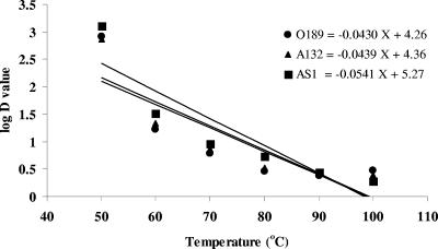
For an inactivation curve (Fig. 1), regression analysis was used to determine whether the regression coefficient (in the form of the negative reciprocal of slope or D value) is significantly different from zero, i.e., heat is capable of inactivating FMDV. Likewise, for the DRT curve (Fig. 4), regression analysis was used to determine whether the regression coefficient (in the form of the negative reciprocal of slope or z value) was significantly different from zero, i.e., the D value is temperature dependent. The correlation coefficient (r2) is calculated to determine the correlation of inactivation (time) and virus concentration. Root mean square error (RMSE) was used to determine goodness of fit of the regression equations to the observed data. The analysis of variance (ANOVA) was used to determine the statistical significance of differences in heat resistance of FMDV strains across temperatures and vice versa. In addition, ANOVA was used to determine the statistical significance of differences among z values of FMDV strains in PBS. When ANOVA indicated a statistically significant difference, a multiple comparison was used to determine specific differences between pairs of FMDV strains or temperatures (25).
FIG. 4.
DRT curve (z value) for FMDV strains in PBS. The log-linear DRT equation for FMDV serotype is shown in the legend where FMDV serotypes symbolize the log D value and X is the temperature. z values are the negative reciprocal of the slope of the DRT curve.
RESULTS
D values (rate of FMDV inactivation).
D values of FMDV at 50°C (D50) ranged from 732 to 1,275 s (Table 1). Two groups of FMDV strains were classified according to the heat resistance; FMDV strains O189, OPN, A118, and A132 were not statistically less resistant than FMDV strains A-Sakol and AS1 (P > 0.05). FMDV strain AS1 was the most heat resistant at 50°C, but its heat resistance was not statistically different from that of other FMDV strains (P > 0.05).
TABLE 1.
D values of FMDV strains in PBS at various temperatures
| Temp (°C) | FMDV | Mean D value ± SD (s)a | r2 | RMSE | Pb | Model |
|---|---|---|---|---|---|---|
| 50 | O189 | 806 ± 144A | 0.69 | 0.68 | 0.0001 | Linear |
| OPN | 890 ± 845A | 0.63 | 0.59 | NA | Tailing | |
| A118 | 851 ± 311A | 0.60 | 0.59 | 0.04 | Linear | |
| A-Sakol | 1,200 ± 406A | 0.60 | 0.47 | 0.06 | Linear | |
| A132 | 732 ± 149A | 0.88 | 0.42 | NA | Tailing | |
| AS1 | 1,275 ± 524A | 0.75 | 0.37 | 0.14 | Linear | |
| 60 | O189 | 16.37 ± 1.96A | 0.83 | 0.85 | 8 × 10⁻⁷ | Linear |
| OPN | 42.00 ± 9.11A | 0.74 | 0.50 | NA | Tailing | |
| A118 | 18.36 ± 2.79A | 0.81 | 0.79 | 0.0001 | Linear | |
| A-Sakol | 21.20 ± 3.62A | 0.77 | 0.77 | 0.0002 | Linear | |
| A132 | 21.73 ± 3.98A | 0.84 | 0.68 | 0.0002 | Linear | |
| AS1 | 32.83 ± 6.95A | 0.61 | 0.71 | 0.0003 | Linear | |
| 70 | O189 | 6.06 ± 1.02A | 0.81 | 0.67 | 0.0003 | Linear |
| OPN | 10.87 ± 7.58A | 0.59 | 0.68 | NA | Tailing | |
| A118 | 8.87 ± 1.13A | 0.86 | 0.65 | 1 × 10⁻⁵ | Linear | |
| A-Sakol | 8.68 ± 1.11A | 0.86 | 0.66 | 1 × 10⁻⁵ | Linear | |
| A132 | 7.28 ± 1.76A | 0.66 | 1.16 | 0.003 | Linear | |
| AS1 | 9.24 ± 1.25A | 0.82 | 0.69 | 9 × 10⁻⁶ | Linear | |
| 80 | O189 | 2.84 ± 0.31A | 0.89 | 0.91 | 4 × 10⁻⁶ | Linear |
| OPN | 5.99 ± 0.62B | 0.94 | 0.60 | 7 × 10⁻⁵ | Linear | |
| A118 | 3.81 ± 0.47A,B | 0.85 | 0.95 | 6 × 10⁻⁶ | Shoulder | |
| A-Sakol | 5.16 ± 0.52B | 0.91 | 0.70 | 2 × 10⁻⁶ | Shoulder | |
| A132 | 3.38 ± 0.30A | 0.94 | 0.61 | 1 × 10⁻⁶ | Linear | |
| AS1 | 5.42 ± 0.34B | 0.95 | 0.48 | 3 × 10⁻¹⁰ | Shoulder | |
| 90 | O189 | 2.53 ± 0.37A | 0.80 | 1.30 | 0.0001 | Linear |
| OPN | 3.18 ± 0.23A | 0.95 | 0.49 | 3 × 10⁻⁷ | Linear | |
| A118 | 1.65 ± 0.34A | 0.83 | 1.05 | 0.0006 | Linear | |
| A-Sakol | 1.75 ± 0.42A | 0.82 | 0.94 | 0.0009 | Linear | |
| A132 | 2.77 ± 0.38A | 0.87 | 0.87 | 0.0001 | Linear | |
| AS1 | 3.16 ± 0.32A | 0.90 | 0.67 | 2 × 10⁻⁶ | Shoulder | |
| 100 | O189 | 2.94 ± 0.35A | 0.90 | 0.65 | 3 × 10⁻⁵ | Linear |
| OPN | 2.88 ± 0.36A | 0.89 | 0.55 | 4 × 10⁻⁵ | Linear | |
| A118 | 2.64 ± 0.53A | 0.76 | 0.64 | 0.001 | Linear | |
| A-Sakol | 2.52 ± 0.38A | 0.78 | 1.04 | 3 × 10⁻⁵ | Linear | |
| A132 | 2.49 ± 0.31A | 0.84 | 0.87 | 4 × 10⁻⁶ | Linear | |
| AS1 | 1.90 ± 0.31A | 0.83 | 0.88 | 3 × 10⁻⁵ | Linear |
Values are means of three replications. Values followed by the same capital superscript letters within each temperature group are not statistically different (P > 0.05).
H0: regression coefficient = 0. NA, not applicable.
Although ranges of D60 and D70 of FMDV strains were 16.37 to 42.00 s and 6.06-10.87 s (Table 1), respectively, D60 and D70 were not statistically significantly different across FMDV strains (P > 0.05). D60 and D70 were increased exponentially compared to D50. For FMDV strains A-Sakol and A118, 60 and 70°C render FMDV much less heat resistant than 50°C. The r2 of the fitted inactivation curves at 60 and 70°C was greatly enhanced from 0.10 to 0.13 up to 0.86, and r2 was significantly different from zero (P < 0.0002), whereas the RMSEs of both fitted inactivation curves were essentially constant. FMDV strain A132 showed a tailing effect for inactivation only at 50°C. On the other hand, FMDV strain OPN showed biphasic inactivation at 50, 60, and 70°C, whereas the RMSEs of linear inactivation curves are higher than those of linear curves with tailing effect. In the case of tailing effect, r2 and RMSEs of inactivation curves were calculated corresponding to the proportions of more and less heat-resistant subpopulations. Therefore, there no P values for FMDV strains OPN and A132 are shown. The inactivation curve of FMDV strain O189 at 60°C and 70°C showed a lag time (tL) during initial inactivation. However, the inactivations at 60 and 70°C did not have a shoulder effect, since tL of inactivations at 60 and 70°C were not significantly different from D60 and D70, respectively (45).
Like 50°C inactivation, the D80 of FMDV strains can be categorized into two heat resistance groups. FMDV strains O189 and A132 are statistically significantly less heat resistant than FMDV strains OPN, A-Sakol, and AS1 (P < 0.05), while A118 had moderate heat resistance (Table 1). There was no shoulder effect or tailing effect for thermal inactivations of FMDV strains at 80°C. Ranges of D90 and D100 were 1.65 to 3.18 s and 1.90 to 2.94 s (Table 1), respectively; but these ranges of D90 and D100 were somewhat narrow and did not vary as much as those of D values at 50 to 80°C. For all FMDV strains except OPN inactivated at 90°C, the inactivation curves deviated slightly from linear and revealed an initial lag phase; however, only FMDV strains A118 and A-Sakol displayed a shoulder effect. Whereas D90 and D100 were not statistically significantly different across FMDV strains (P > 0.05); only FMDV strain AS1 had a shoulder effect at 100°C inactivation. The statistical significant differences in heat resistance of FMDV strains across temperatures and the statistical significant differences of DT values across FMDV strains are reported in Table 2.
TABLE 2.
D values of FMDV strains in PBS at 50 to 100°C
| Temp (°C) |
D value (s)a in FMDV strain:
|
|||||
|---|---|---|---|---|---|---|
| O189 | OPN | A118 | A-Sakol | A132 | AS1 | |
| 50 | 806A,a | 890A,a | 851A,a | 1,200A,a | 732A,a | 1,275A,a |
| 60 | 16.37B,a | 42.00B,a | 18.36B,a | 21.20B,a | 21.73B,a | 32.83B,a |
| 70 | 6.06C,a | 10.87C,a | 8.87C,a | 8.68C,a | 7.28B,a | 9.24C,a |
| 80 | 2.84D,a | 5.99C,b | 3.81C,D,a,b | 5.16D,b | 3.38C,a | 5.42D,b |
| 90 | 2.53D,a | 3.18D,a | 1.65E,a | 1.75E,a | 2.77C,a | 3.16E,a |
| 100 | 2.94D,a | 2.88D,a | 2.64D,E,a | 2.52E,a | 2.49C,a | 1.90E,a |
Values are means of three replications. In the same column, mean values with different letters imply that there are statistically significant differences (P < 0.05) among the different temperatures for the same FMDV strain (letters A through E). In the same row, mean values with different letters imply that there are statistically significant differences (P < 0.05) among the different FMDV strains for the same temperature (letters a and b).
The D50 of FMDV strain O189 was much higher than the D60 and D70, and the D50, D60 and D70 were statistically significantly different (P < 0.05). In addition, the D50 and D60 of all FMDV strains were statistically significantly different (P < 0.05). However, the D90 and D100 of all FMDV strains were not statistically significantly different (P > 0.05). A similar situation was also evident for FMDV strains OPN and A118 in that two pairs of D values, which are D70 versus D80 and D90 versus D100, were not statistically significantly different (P > 0.05). For FMDV strain O189, D80, D90, and D100 were not statistically significantly different (P > 0.05).
z values.
Given the broad temperatures tested in the present study, fitting DRT curves to determine the z values was more difficult than with a narrow range of temperatures. The conventional DRT curve fitting included all temperatures tested to calculate z values so that other DT can be interpolated within the range of inactivation temperatures. The range of z values of FMDV strains using D50 to D100 was between 19.87 and 23.26°C (Table 3). However, because D50 of all FMDV strains were much higher than their D60, z values calculated using D values of six temperatures could be incorrectly lowered by outlier D50. Therefore, the second method of fitting DRT adopted only D60 to D100, and z values of FMDV strains using five temperatures ranged from 33.25 to 52.55°C (Table 4). Obviously, the five-temperature z values were much higher than six-temperature z values. The alternative mathematical model that could be fitted to the DRT curve is a linear curve with tailing effect. The range of z values of FMDV strains, taking into account six temperatures with a tailing effect, was between 13.94 and 20.76°C (data not shown). From Table3, z values of FMDV strains were not statistically significantly different (P > 0.05) by three means of fitting DRT curves. FMDV strain O189 had the highest z values, calculated by five temperatures and by six temperatures without tailing effect.
TABLE 3.
z values of FMDV strains in PBS at 50 to 100°C
| FMDV | Mean z value (°C) ± SDa | r2 | RMSE | Pb |
|---|---|---|---|---|
| O189 | 23.26 ± 11.78A | 0.69 | 0.60 | 0.04 |
| OPN | 21.78 ± 6.54A | 0.82 | 0.44 | 0.01 |
| A118 | 20.81 ± 8.81A | 0.75 | 0.55 | 0.03 |
| A-Sakol | 20.75 ± 8.19A | 0.76 | 0.57 | 0.02 |
| A132 | 22.79 ± 9.15A | 0.75 | 0.53 | 0.03 |
| AS1 | 19.87 ± 6.20A | 0.82 | 0.50 | 0.01 |
Values are means of three replications. Values followed by the same superscript letters are not statistically different (P > 0.05).
H0: regression coefficient = 0.
TABLE 4.
z values of FMDV strains in PBS at 60 to 100°C
| FMDV | Mean z value (°C) ± SDa | r2 | RMSE | Pb |
|---|---|---|---|---|
| O189 | 52.55 ± 26.62A | 0.75 | 0.20 | 0.06 |
| OPN | 34.95 ± 8.16A | 0.90 | 0.17 | 0.01 |
| A118 | 41.41 ± 14.67A | 0.83 | 0.20 | 0.03 |
| A-Sakol | 39.29 ± 11.26A | 0.87 | 0.18 | 0.02 |
| A132 | 43.45 ± 13.52A | 0.86 | 0.17 | 0.02 |
| AS1 | 33.25 ± 4.95A | 0.95 | 0.12 | 0.00 |
Values are means of three replications. Values followed by the same letters are not statistically different (P > 0.05).
H0: regression coefficient = 0.
DISCUSSION
FMDV is nonenveloped RNA virus within the Picornaviridae family. This virus is notorious for its exceptional stability in a harsh environment, since FMDV is resistant to UV, chemicals, and heat (14, 27, 38). The essential heat resistance of many food-borne and waterborne picornaviruses, such as hepatitis A virus and poliovirus (PV) (36), are derived from the capsid. One primary function of viral capsid is attachment to the host cell receptor during the first step of viral replication (33). Engulfment, or endocytosis, follows in PV and is believed to happen similarly in FMDV, given their similarities in capsid symmetry and their phylogeny (20). If infectious RNA can get inside an otherwise-insusceptible host cell, the replicative cycle can be completed without a capsid, so the capsid integrity essentially mediates virus and tissue interactions (37). The capsid integrity and immunological properties of 37°C-inactivated human picornaviruses and FMDV serotype O (7) were intact, while the capsid protein of human picornaviruses was unfolded at 72°C and readily hydrolyzed by protease (36). Heat inactivation of FMDV in suspension was temperature-dependent (Tables 3 and 4). The effective inactivating temperature is approximately at 60°C (36) since the D value dropped dramatically at 60°C. This result agreed with previous studies, indicating that hepatitis A virus and PV were heat resistant up to 60°C (39, 43).
Theoretically, the D value was temperature dependent or negatively correlated with temperature (Tables 3 and 4), i.e., the higher the temperature, the lower the D value (23). At the low temperature the D value for 50°C inactivation was large, since the slope of the inactivation curve was low. This is especially the case for heat inactivation of FMDV strains A-Sakol and AS1, where the D values were as high as 1,200 and 1,275 s (Table 1).
It was evident that the D values at temperatures higher than 80°C were not statistically different. This is more favorable to the food processors because the lower temperatures are easier to achieve, while the texture of food, especially pork, will be more appetizing, with a lower cost of processing.
From Table 3, z values of FMDV strains were not statistically significantly different (P > 0.05) by three means of fitting DRT curves. In terms of temperatures, FMDV strains had statistically equivalent heat resistance. Taking into account both correlation coefficient and goodness of fit, it was evident that z value using six temperatures without a tailing effect was the most appropriate. FMDV strain O189 had highest z values, calculated by five temperatures and by six temperatures without a tailing effect. Therefore, it was tempting to speculate that FMDV strain O189 was the most heat resistant at all temperatures. Surprisingly, from Table 2, it was evident that FMDV strain OPN had higher DT between 60 and 90°C than FMDV strain O189, although some of the DT values were not statistically significantly different. The discrepancy might be explained by the fact that the D50 of FMDV strain O189 is somewhat less than that of FMDV strain OPN, although the D50 values of both FMDV strains were not statistically significantly different (P > 0.05).
A serotype is the antigenic property that is determined by the ability of an antiserum, which was raised in an animal not previously exposed to the related virus, to neutralize the infectivity of a virus (6). Human echoviruses and PVs are enterovirus genera in the Picornaviridae family (20). Both genera have comparable structures, but human echoviruses and PVs have 33 and 3 serotypes, respectively (4, 20, 41, 47). Probably, the serotypes vary by the receptor attachment or serological reactions rather than virus structures (19, 42). The heat resistances of FMDV strains in the present study were not different at 60, 70, 90, and 100°C. While the pattern of inactivation of FMDV at 50°C revealed two heat resistance groups, it was noteworthy that the same FMDV serotypes (serotypes O and A) were classified into different heat resistance groups. Perhaps heat inactivation randomly attacked the target inactivating sites on the viral capsid protein (29). Therefore, at a low temperature (50°C), heat resistance of FMDV is independent of the surface antigen of the virus structure or not serotype specific (36).
Even though a sigmoidal curve is also commonly found in thermal and nonthermal inactivation (48), heat inactivation of FMDV strains in the present study did not show this inactivation shape. In the present study, there were three types of inactivation curves: linear curves, linear curves with a shoulder, and linear curves with a tailing. Based on the first-order chemical kinetics to describe the thermal inactivation, the log-linear model was applied for microbial heat inactivation (9). The primary concept of microbial thermal inactivation, which is analogous to chemical reaction, is the single lethal hit or one-hit kinetics, where the virus particle is completely inactivated by only a single hit (26). Arguably, some thermal inactivation curves of FMDV showed deviation from the log-linear law, since in fact thermal inactivation of the virus was not exactly the same as the chemical reaction. The tailing effect was found only at 50 to 70°C, and FMDV strain OPN showed this effect at temperatures within this range (Table 1). It is arguable to assume that OPN suspension in fact had a subpopulation, since the tailing effect did not happen at the higher temperatures. One possible explanation is that some organic matter that was derived from the cell culture debris during virus propagation could more effectively shield the virus particle from inactivation at lower temperatures than at the higher temperatures (48). Supposedly, the protective organic matters are proteinaceous substances, which are prone to denature or degrade more easily at the higher temperatures (22, 36). The shoulder effect was observed only at 90 to 100°C (Table 1), so it was more prevalent at the higher temperatures, perhaps because the higher temperatures had very low DT values, and special procedures were needed to catch the fast drop of FMDV concentration in the suspension. This is why the first sampling time is so close to the time zero. The first sampling time was arbitrarily set on the basis of a preliminary study (data not shown), so it is possible that the sample was taken prematurely while the heat was still being conducted through the glass tube and throughout the viral suspension.
In the present study, the deviations of the inactivation curves only slightly change the D values of FMDV inactivation, and the multiple comparisons remained the same. If the 95% confidence intervals of the original D value and adjusted D value were overlapped, the deviated inactivation curve did not have shoulder effect (Table 1). It was noted that the shoulder effect sometimes changed the D value but did not change overall heat resistances among FMDV strains at 90 and 100°C. On the other hand, the choice of D values from the original linear regression and adjusted D values from the tailing-effect inactivation curve was dependent on the RMSE, i.e., the smaller the RMSE, the better the fit (Table 1). The results of multiple comparisons of D50, D60, and D70 did not change, regardless of taking the tailing effect into consideration. These observations indicated that the unadjusted D values, even from inactivation curves with deviations, were statistically and essentially legitimate.
The heat resistances of FMDV strains at lower temperature varied dependent upon the temperature; however, those of FMDV strains at 90 and 100°C were opposite. One possible reason is that heat resistance (or in turn sensitivity) has a threshold or plateau temperature (28). The heat resistance of the virus is linearly correlated within a certain range of temperatures, and in the case of FMDV the range of temperature is 60 to 90°C (Table 3). Care must be taken to avoid extrapolating the data out of the range of experimental temperatures, so it is not recommended to apply the D value from the DRT curve or the z value unless the D value is in the range of experimental temperatures. Nevertheless, if one proved that viral suspension and pork demonstrated the same thermal conductivity, doing thermal inactivation of FMDV strains in pork at 90°C could also sufficiently represent that of FMDV strains in pork at 100°C.
The heat resistance of FMDV strains in Thailand is apparently less than those reported for FMDV strains elsewhere. A valid comparison of FMDV strains in Thailand and other countries must be based on suspensions in the same medium, which was PBS (34). The heat resistance of FMDV serotypes O and A at 50 and 54°C is shown in Tables 5 and 6 (34). The D50 of FMDV strains O189 and OPN in Thailand is greater than that of FMDV strain OiBFS 1860. When calculated based on z values of FMDV strains O189 and OPN, which were 23.26 and 21.78, respectively, the D54 of FMDV strains O189 and OPN in Thailand is at least 6- to 10-fold lower than that of FMDV strain OiBFS 1860 and all other FMDV serotypes O. Likewise D54 of FMDV strains A118, A-Sakol, and A132 were at least fivefold lower than other FMDV serotype A. Even though there were very limited data for comparisons, it was apparent that FMDV strains in Thailand show much less heat resistance, in PBS, than those in other countries. Therefore, these data indicate that the heat resistance of FMDV strains in pork will follow the same trend as that of FMDV in PBS. From the practical point of view, the industry needs to know the D value of FMDV in the pork, since the medium in the thermal inactivation plays a vital role. The state of the medium (liquid versus solid) or the composition of the medium (e.g., fat, protein, pH, etc.) always influences the heat resistance, aside from the inherent heat resistance of the microorganisms (23). Further experiments will evaluate the thermal inactivation of FMDV serotypes in pork.
TABLE 5.
D values of FMDV serotype O in PBS from various countries
| FMDV straina |
D value (min) at:
|
Reference(s) | |
|---|---|---|---|
| 50°C | 54°C | ||
| O189 | 13.4 | 1.44 | 24,45 |
| OPN | 14.8 | 2.56 | 24,45 |
| OiBFS 1860 | 4.17 | 25.21 | 34 |
| O Lausanne | NAb | 26.20 | 34 |
| O Pacheco | NA | 32.96 | 34 |
| O Campos | NA | 25.97 | 34 |
| O Colombia 7250 | NA | 18.07 | 34 |
| O Susan 5/75 | NA | 23.81 | 34 |
| O Susan 6/75 | NA | 20.07 | 34 |
| O Susan 1/76 | NA | 18.07 | 34 |
| OK 120/64 | NA | 16.43 | 34 |
Strains O189 and OPN were from the present study; the remaining strains were from the World Reference Laboratory for FMD, Animal Virus Research Institure, Pirbright, United Kingdom. The z value of O189 is 23.26°C; the z value of OPN is 21.78°C.
NA, not applicable.
TABLE 6.
D values of FMDV serotype A in PBS
| FMDV straina | D (min) value at 54°C | Reference |
|---|---|---|
| A118 | 1.76 | 24,45 |
| A-Sakol | 2.24 | 24,45 |
| A132 | 1.61 | 24,45 |
| A5 France 1/68 | 26.79 | 34 |
| A22 Mahmatli | 33.90 | 34 |
| A22 Iraq 24/64 | 36.81 | 34 |
| A Pando (1970) | 37.97 | 34 |
| A24 Cruzeiro | 33.15 | 34 |
| A Colombia 8046 | 15.35 | 34 |
| A Bage | 14.93 | 34 |
| A Venceslau (Bra 1/77) | 25.32 | 34 |
| AK 18/66 | 18.34 | 34 |
| A Sudan 2/75 | 15.23 | 34 |
| A Morocco 5/77 | 18.58 | 34 |
| A Morocco 8/77 | 12.34 | 34 |
| A Philippine 10/75 | 12.07 | 34 |
| A Philippine 14/75 | 13.95 | 34 |
Strains A118 (z value = 21.81°C), A-Sakol (z value = 20.75°C), and A132 (z value = 22.79°C) were from the present study; all remaining strains were from the World Reference Laboratory for FMD, Animal Virus Research Institute, Pirbright, United Kingdom.
Acknowledgments
This study was supported by Thailand Research Fund grant RDG 4720024.
We thank Wantanee Kalpravidh, Wilai Linjongsubongkoch, and Dean O. Cliver for critical assistance.
Footnotes
Published ahead of print on 27 July 2007.
REFERENCES
- 1.Adams, M. R., M. O. Moss, et al. 2000. Food microbiology, 2nd ed. Royal Society of Chemistry, Cambridge, United Kingdom.
- 2.Alexandersen, S., Z. Zhang, A. I. Donaldson, and A. J. Garland. 2003. The pathogenesis and diagnosis of foot-and-mouth disease. J. Comp. Pathol. 129:1-36. [DOI] [PubMed] [Google Scholar]
- 3.Bartley, L. M., C. A. Donnelly, and R. M. Anderson. 2002. Reviews of foot-and-mouth disease virus survival in animal excretions and on formites. Vet. Rec. 151:667-669. [DOI] [PubMed] [Google Scholar]
- 4.Beck, E., S. Forss, K. Strebel, R. Cattaneo, and G. Feil. 1983. Structure of the FMDV translation initiation site and of the structural proteins. Nucleic Acids Res. 11:7873-7885. [DOI] [PMC free article] [PubMed] [Google Scholar]
- 5.Blacksell, S. D., R. A. Lunt, C. Chamnanpood, W. Linchongsubongkoch, N. Nakarungkul, L. J. Gleeson, and C. Megkamol. 1994. Establishment of a typing enzyme-linked immunosorbent assay for foot and mouth disease antigen, using reagents against viruses endemic in Thailand. Rev. Sci. Technol. 13:701-709. [DOI] [PubMed] [Google Scholar]
- 6.Bolwell, C., A. L. Brown, P. V. Barnett, R. O. Campbell, B. E. Clarke, N. R. Parry, E. J. Ouldridge, F. Brown, and D. J. Rowlands. 1989. Host cell selection of antigenic variants of foot-and-mouth disease virus. J. Gen. Virol. 70(Pt. 1):45-57. [DOI] [PubMed] [Google Scholar]
- 7.Brown, F., and T. F. Wild. 1966. The effect of heat on the structure of foot-and-mouth disease virus and the viral ribonucleic acid. Biochim. Biophys. Acta 119:301-308. [DOI] [PubMed] [Google Scholar]
- 8.Cerf, O. 1977. Tailing of survival curves of bacterial spores. J. Appl. Bacteriol. 42:1-19. [DOI] [PubMed] [Google Scholar]
- 9.Chick, H. 1908. An investigation of the laws of disinfection. J. Hyg. 8:92-158. [DOI] [PMC free article] [PubMed] [Google Scholar]
- 10.Chou, C. C., and S. E. Yang. 2004. Inactivation and degradation of O Taiwan 97 foot-and mouth-disease virus in pork sausage processing. Food Microbiol. 21:737-742. [Google Scholar]
- 11.Clarke, J. B., and R. E. Spier. 1980. Variation in the susceptibility of BHK populations and cloned cell lines to three strains of foot-and-mouth disease virus. Arch. Virol. 63:1-9. [DOI] [PubMed] [Google Scholar]
- 12.Cottral, G. E., B. F. Cox, and D. E. Baldwin. 1960. The survival of foot-and-mouth disease virus in cured and uncured meat. Am. J. Vet. Res. 21:288-297. [PubMed] [Google Scholar]
- 13.Daniel, W. W. 1995. Biostatistics: a foundation for analysis in the health sciences, 6th ed. Wiley, New York, NY.
- 14.Dekker, A. 1998. Inactivation of foot-and-mouth disease virus by heat, formaldehyde, ethylene oxide, and gamma radiation. Vet. Rec. 143:168-169. [DOI] [PubMed] [Google Scholar]
- 15.de Leeuw, P. W., J. W. Tiessink, and J. G. van Bekkum. 1980. Aspects of heat inactivation of foot-and-mouth disease virus in milk from intramammarily infected susceptible cows. J. Hyg. 84:159-172. [DOI] [PMC free article] [PubMed] [Google Scholar]
- 16.Doel, T. R., and P. J. Baccarini. 1981. Thermal stability of foot-and-mouth disease virus. Arch. Virol. 70:21-32. [DOI] [PubMed] [Google Scholar]
- 17.Forbes, L. S., and G. E. Cottral. 1969. Heat inactivation of foot-and-mouth disease virus in blood products. Res. Vet. Sci. 10:98-100. [PubMed] [Google Scholar]
- 18.Fox, G., N. R. Parry, P. V. Barnett, B. McGinn, D. J. Rowlands, and F. Brown. 1989. The cell attachment site on foot-and-mouth disease virus includes the amino acid sequence RGD (arginine-glycine-aspartic acid). J. Gen. Virol. 70(Pt. 3):625-637. [DOI] [PubMed] [Google Scholar]
- 19.Fry, E. E., J. W. Newman, S. Curry, S. Najjam, T. Jackson, W. Blakemore, S. M. Lea, L. Miller, A. Burman, A. M. King, and D. I. Stuart. 2005. Structure of foot-and-mouth disease virus serotype A10 61 alone and complexed with oligosaccharide receptor: receptor conservation in the face of antigenic variation. J. Gen. Virol. 86:1909-1920. [DOI] [PubMed] [Google Scholar]
- 20.International Committee on Taxonomy of Viruses, M. H. V. Van Regenmortel, and International Union of Microbiological Societies. Virology Division. 2000. Family: Picornaviridae, p. 657-678. In Virus taxonomy: classification and nomenclature of viruses: seventh report of the International Committee on Taxonomy of Viruses. Academic Press, Inc., San Diego, CA.
- 21.International Office of Epizootics, Standards Commission, and International Office of Epizootics. 2000. Manual of standards for diagnostic tests and vaccines: lists A and B diseases of mammals, birds and bees, 4th ed. Office international des Pizooties, Paris, France.
- 22.Jordan, L., and H. D. Mayor. 1974. Studies on the degradation of poliovirus by heat. Microbios 9:51-60. [PubMed] [Google Scholar]
- 23.Juneja, V. K., B. S. Eblen, and G. M. Ransom. 2001. Thermal inactivation of Salmonella spp. in chicken broth, beef, pork, turkey, and chicken: determination of D and z values. J. Food Sci. 66:146-152. [Google Scholar]
- 24.Juneja, V. K., O. P. Snyder, Jr., and B. S. Marmer. 1997. Thermal destruction of Escherichia coli O157:H7 in beef and chicken: determination of D and z values. Int. J. Food Microbiol. 35:231-237. [DOI] [PubMed] [Google Scholar]
- 25.Kleinbaum, D. G., L. L. Kupper, and K. E. Muller. 1988. Applied regression analysis and other multivariable methods, 2nd ed. PWS-Kent Publishing Co., Boston, MA.
- 26.Lambert, R. J. 2003. A model for the thermal inactivation of micro-organisms. J. Appl. Microbiol. 95:500-507. [DOI] [PubMed] [Google Scholar]
- 27.Lasta, J., J. H. Blackwell, A. Sadir, M. Gallinger, F. Marcoveccio, M. zamorano, B. Ludden, and R. Rodriguez. 1992. Combined treatments of heat, irradiation, and pH effects on infectivity of foot-and-mouth disease virus in bovine tissues. J. Food Sci. 57:36-39. [Google Scholar]
- 28.Laurent, Y., S. Arino, and L. Rosso. 1999. A quantitative approach for studying the effect of heat treatment conditions on resistance and recovery of Bacillus cereus spores. Int. J. Food Microbiol. 48:149-157. [DOI] [PubMed] [Google Scholar]
- 29.Mansfeld, J., and R. Ulbrich-Hofmann. 2000. Site-specific and random immobilization of thermolysin-like proteases reflected in the thermal inactivation kinetics. Biotechnol. Appl. Biochem. 32(Pt. 3):189-195. [DOI] [PubMed] [Google Scholar]
- 30.Masana, M. O., N. A. Fondevila, M. M. Gallinger, J. A. Lasta, H. R. Rodriguez, and B. Gonzalez. 1995. Effect of low-temperature long-time thermal processing of beef-cuts on the survival of foot-and-mouth disease virus. J. Food Prot. 58:165-169. [DOI] [PubMed] [Google Scholar]
- 31.McColl, K. A., H. A. Westbury, R. P. Kitching, and V. M. Lewis. 1995. The persistence of foot-and-mouth disease virus on wool. Aust. Vet. J. 72:286-292. [DOI] [PubMed] [Google Scholar]
- 32.Mebus, C. A., C. House, F. R. Gonzalvo, J. M. Pineda, J. Tapiador, J. J. Pire, J. Bergada, R. J. Yedloutschning, S. Sahu, V. Becerra, and J. M. Sanchez-Vizcaino. 1993. Survival of foot-and-mouth disease, African swine fever and hog cholera viruses in Spanish Serrano cured hams and Iberian cured hams, shoulders and loins. Food Microbiol. 10:133-143. [Google Scholar]
- 33.Minor, P. D., G. C. Schild, J. Bootman, D. M. Evans, M. Ferguson, P. Reeve, M. Spitz, G. Stanway, A. J. Cann, R. Hauptmann, L. D. Clarke, R. C. Mountford, and J. W. Almond. 1983. Location and primary structure of a major antigenic site for poliovirus neutralization. Nature 301:674-679. [DOI] [PubMed] [Google Scholar]
- 34.Nettleton, P. F., M. J. Davies, and M. M. Rweyemamu. 1982. Guanidine and heat sensitivity of foot-and-mouth disease virus strains. J. Hyg. 89:129-138. [DOI] [PMC free article] [PubMed] [Google Scholar]
- 35.Nuanualsuwan, S., and D. O. Cliver. 2002. Pretreatment to avoid positive RT-PCR results with inactivated viruses. J. Virol. Methods 104:217-225. [DOI] [PubMed] [Google Scholar]
- 36.Nuanualsuwan, S., and D. O. Cliver. 2003. Capsid functions of inactivated human picornaviruses and feline calicivirus. Appl. Environ. Microbiol. 69:350-357. [DOI] [PMC free article] [PubMed] [Google Scholar]
- 37.Nuanualsuwan, S., and D. O. Cliver. 2003. Infectivity of RNA from inactivated poliovirus. Appl. Environ. Microbiol. 69:1629-1632. [DOI] [PMC free article] [PubMed] [Google Scholar]
- 38.OIE. 2003. Food and mouth disease virus inactivation procedures: appendix 3.6.2, article 3.6.2.1. In Terrestrial animal health code, 11th ed. OIE, Paris, France.
- 39.Parry, J. V. 1984. Diagnosis of hepatitis A infection: comparative specificity of IgM capture assays using antigens derived from tissue cultures and marmoset feces. J. Virol. Methods 9:35-44. [DOI] [PubMed] [Google Scholar]
- 40.Reed, L., and H. Muench. 1938. A simple method for estimating fifty percent endpoints. Am. J. Hyg. 27:493. [Google Scholar]
- 41.Rosenwirth, B., and H. J. Eggers. 1978. Structure and replication of echovirus type 12. 2. Viral polypeptides synthesized in the infected cell. Eur. J. Biochem. 92:61-67. [DOI] [PubMed] [Google Scholar]
- 42.Short, J. J., A. V. Pereboev, Y. Kawakami, C. Vasu, M. J. Holterman, and D. T. Curiel. 2004. Adenovirus serotype 3 utilizes CD80 (B7.1) and CD86 (B7.2) as cellular attachment receptors. Virology 322:349-359. [DOI] [PubMed] [Google Scholar]
- 43.Siegl, G., M. Weitz, and G. Kronauer. 1984. Stability of hepatitis A virus. Intervirology 22:218-226. [DOI] [PubMed] [Google Scholar]
- 44.Singh, R. P., and D. R. Heldman. 2001. Introduction to food engineering, 3rd ed. Academic Press, London, United Kingdom.
- 45.Toledo, R. T. 1991. Fundamentals of food processing engineering, 2nd ed. Van Nostrand Reinhold, New York, NY.
- 46.Turner, C., S. M. Williams, and T. R. Cumby. 2000. The inactivation of foot and mouth disease, Aujeszky's disease and classical swine fever viruses in pig slurry. J. Appl. Microbiol. 89:760-767. [DOI] [PubMed] [Google Scholar]
- 47.Wilson, V., P. Taylor, and U. Desselberger. 1988. Crossover regions in foot-and-mouth disease virus recombinants correspond to regions of high local secondary structure. Arch. Virol. 102:131-139. [DOI] [PMC free article] [PubMed] [Google Scholar]
- 48.Xiong, R., G. Xie, A. E. Edmondson, and M. A. Sheard. 1999. A mathematical model for bacterial inactivation. Int. J. Food Microbiol. 46:45-55. [DOI] [PubMed] [Google Scholar]


