Abstract
Bacillus collagen-like protein of anthracis (BclA) is the immunodominant glycoprotein on the exosporium of Bacillus anthracis spores. Here, we sought to assess the impact of BclA on spore germination in vitro and in vivo, surface charge, and interaction with host matrix proteins. For that purpose, we constructed a markerless bclA null mutant in B. anthracis Sterne strain 34F2. The growth and sporulation rates of the ΔbclA and parent strains were nearly indistinguishable, but germination of mutant spores occurred more rapidly than that of wild-type spores in vitro and was more complete by 60 min. Additionally, the mean time to death of A/J mice inoculated subcutaneously or intranasally with mutant spores was lower than that for the wild-type spores even though the 50% lethal doses of the two strains were similar. We speculated that these in vitro and in vivo differences between mutant and wild-type spores might reflect the ease of access of germinants to their receptors in the absence of BclA. We also compared the hydrophobic and adhesive properties of ΔbclA and wild-type spores. The ΔbclA spores were markedly less water repellent than wild-type spores, and, probably as a consequence, the extracellular matrix proteins laminin and fibronectin bound significantly better to mutant than to wild-type spores. These studies suggest that BclA acts as a shield to not only reduce the ease with which spores germinate but also change the surface properties of the spore, which, in turn, may impede the interaction of the spore with host matrix substances.
Bacillus anthracis is a gram-positive, spore-forming bacillus that can cause anthrax (15). The spore is the form of the organism found in its natural habitat, the soil, and is also the infectious form for herbivores, the typical vertebrate host for the bacterium, and humans (15). The B. anthracis spore is covered by a loose balloon-like membranous structure called the exosporium (8). BclA (for bacillus collagen-like protein of anthracis) was first described by Sylvestre et al. (23), who constructed an insertional bclA mutant and compared it to its wild-type parent. These investigators and, subsequently, others found that BclA is a glycoprotein and a major component of the hair-like projections that cover the exosporium (16, 22, 23, 25). BclA is also an immunodominant marker on the outside of the spore (22). The finding that BclA does not play a significant role in the virulence of a Sterne-like strain for mice was first reported by Sylvestre et al. (23). Sterne strains contain pXO1 but not pX02 and are attenuated in humans and many other animals except certain mouse strains (26). In support of the findings of Sylvestre and colleagues, Bozue and coworkers recently constructed a bclA mutant of the fully virulent B. anthracis Ames strain and showed that the absence of BclA had no impact on the lethality of that strain for guinea pigs or mice (5). Whether BclA, the substance on the spore with which the host cells probably first interact, plays a more subtle role in B. anthracis pathogenicity remains to be determined, as does the function for this highly expressed glycoprotein in the spore stage of the B. anthracis life cycle. Here, we report that BclA-positive spores, compared to spores generated from an isogenic markerless bclA mutant of Sterne, display a reduced extent of spore germination by 60 min of incubation in vitro and a dramatic decrease in the extent of association with the human extracellular matrix proteins laminin and fibronectin. We also show that BclA contributes to spore surface hydrophobicity.
MATERIALS AND METHODS
Construction of a ΔbclA B. anthracis strain.
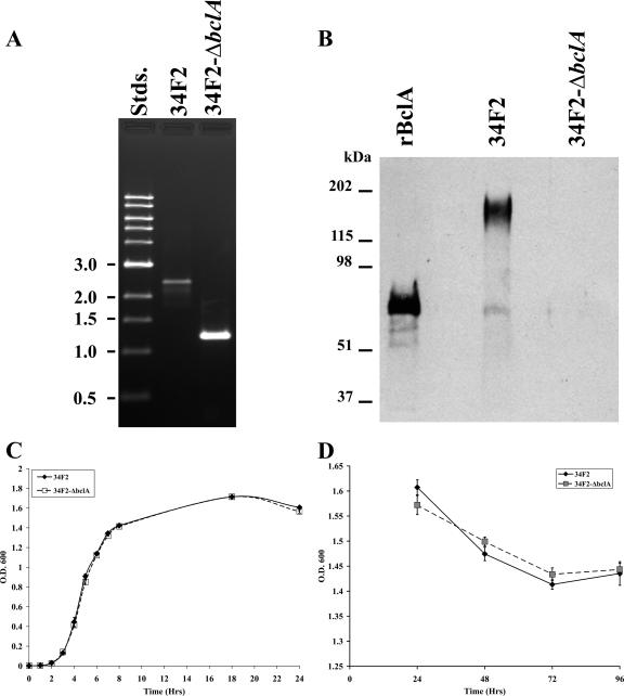
A deletion construct of the bclA gene suitable for allelic exchange was created by cloning two NotI-XmaI DNA fragments containing bclA flanking regions into the NotI site of pBKJ236 (11). These fragments were created by PCR with primers cgcGCGGCCGCagttaggtgcggacagaccttcgaa and tttCCCGGGttttttacttagcagtaaaactgat to one side and aaaCCCGGGaaattcacctccataaagcgttca and gcgGCGGCCGCacgctttacaagtcgatttctttttg to the other (capitalized bases denote NotI and XmaI sites). Ligation of the two fragments created a precise deletion of the bclA open reading frame from the start to the stop codon, inclusive, replacing it with an XmaI site. The resulting construct was used to perform allelic exchange in B. anthracis Sterne strain 34F2 (obtained from Robert L. Bull, NMRC) to create a bclA null mutant by a procedure described previously (11). In brief, integrants of the bclA-pBKJ236 plasmid construct were isolated by a shift to the replication-nonpermissive temperature after conjugative transfer and growth at the permissive temperature while maintaining selection for erythromycin resistance. A second plasmid, pBKJ223, was then introduced by electroporation and selection for tetracycline resistance. This plasmid mediates cleavage within the vector sequences, thus stimulating recombination and the loss of the integrated plasmid, resulting in gene replacement in a portion of the erythromycin-sensitive candidates. This strain was named 34F2-ΔbclA. As shown in Fig. 1A, the absence of the bclA locus in 34F2-ΔbclA was demonstrated by PCR using primers 5′-AATCACCGTAATCTTTTGGGTATTG-3′ (upstream) and 5′-CATGGACCTTTCCAAACCATTGAAA-3′ (downstream). These primers were designed to bind to sequences flanking the region included in the deletion construct described above.
FIG. 1.
Characterization of 34F2-ΔbclA. (A) Comparison of mutant and wild-type strains by PCR using primers that flank the bclA gene as described in Materials and Methods. The predicted sizes of the diagnostic fragments are 2,380 bp and 1,237 bp for the wild type or deleted bclA locus, respectively. Stds., standards. (B) Western blot of purified rBclA expressed from E. coli and SSPE from wild-type 34F2 and 34F2-ΔbclA probed with rabbit anti-rBclA (V683) antiserum. Note that the difference in mobility between rBclA (lane 1) and BclA contained in Sterne strain spore extracts (lane 2) is likely due to the glycosylation of BclA in wild-type spores. Sporulation is shown as a measure of the OD600. The growth rate and sporulation of the mutant and wild-type strains are depicted in C and D, respectively. Time zero is defined as the time of inoculation of the broth culture with bacteria. The change in size as the vegetative rod (large particle) becomes a spore (small particle) corresponds to a reduction in the optical density as sporulation occurs.
Cloning, expression, and purification of rBclA.
Sequence data for the B. anthracis (Ames strain) genome were obtained from the National Center for Biotechnology Information (NCBI) website at http://www.ncbi.nlm.nih.gov/entrez/viewer.fcgi?db=nucleotide&val=30260195. Based on this sequence, DNA primers were designed to amplify the entire bclA open reading frame of the Sterne strain by the PCR. The sequences of primers were 5′-CACACACATATGTCAAATAATAATTATTCAAATGG-3′ and 5′-CACACAGGATCCTATTAAGCAACTTTTTCAATAATAATGGATGC-3′. Genomic DNA was extracted and purified from a 3-ml culture grown overnight with the Easy DNA kit (Invitrogen, Carlsbad, CA). A 10-ng sample of genomic DNA was added to a PCR mixture as a source of template DNA. PCR amplification was done with High Fidelity DNA polymerase (Roche, Indianapolis, IN). The PCR conditions were 94°C for 5 min; 94°C for 30 s, 50°C for 30 s, and 68°C for 1.5 min for 45 cycles; and, finally, 68°C for 10 min. The PCR product was purified by agarose gel electrophoresis with Geneclean (Qbiogene, Irvine, CA), digested with NdeI and BamHI restriction enzymes (Roche, Indianapolis, IN), and inserted into the corresponding cloning sites of the expression vector pET15b (Novagen, San Diego, CA). The inserted DNA regions in the resulting recombinant plasmids were sequenced to confirm that no changes had occurred during the PCR process. A bclA-containing plasmid was transformed into Escherichia coli BL21(DE3) pLysS to express the cloned gene according to the pET system manual (Novagen, San Diego, CA). The expressed BclA protein, with a six-His tag, was purified by His-Trap nickel affinity column chromatography using the fast protein liquid chromatography system from GE Healthcare (Piscataway, NJ).
Immune sera and antibody preparation.
Immune sera from rabbits were obtained from animals vaccinated multiple times at monthly intervals with 50 ng of purified rBclA in Freund's complete adjuvant for the first inoculation and Freund's incomplete adjuvant for all subsequent immunizations. Immunoglobulin G was purified using a protein G column. Protein concentrations were determined by a microtiter BCA assay (Pierce, Rockford, IL).
Spore preparation.
B. anthracis wild-type and 34F2-ΔbclA vegetative cells were induced to sporulate by growth at 30°C for 3 to 4 days on modified G medium (11) that had been rendered solid by the addition of 0.5% Difco agar. Spores were scraped from the agar plates and then purified through a Hypaque-76 gradient (Nycomed, Inc., Princeton, NJ).
Sporulation assays.
Two approaches were used to compare the degrees of sporulation of B. anthracis wild-type and 34F2-ΔbclA vegetative cells over time. For each of these methods, vegetative cells were cultured in Luria Bertani broth for 5 days at 37°C with agitation at 100 rpm. For the first protocol, the optical density at 600 nm (OD600) of the culture was monitored at 24, 48, 72, and 96 h, a measurement that reflects the density of the particles in suspension. Since spores are less dense than vegetative cells, the OD600 should decrease as the bacilli in the culture become spores (20, 21). For the second technique, samples were obtained at the time points listed above and assessed by phase microscopy for sporulation.
Spore surface protein extraction and Western blot analysis.
Spore surface protein extracts (SSPEs) were prepared from wild-type Sterne 34F2 and 34F2-ΔbclA spores as outlined previously by Aronson and Fitz-James (3). Briefly, 109 spores were incubated with 1 ml buffer that contained 0.1 M dithiothreitol, 0.5% sodium dodecyl sulfate, and 0.1 M NaCl (pH 10.0) at 37°C for 2 1/2 h. The spores were then centrifuged at 16,000 × g for 10 min. The supernatant was removed and stored at 4°C. SSPEs were diluted in sample loading buffer, loaded onto 8% Tris-glycine sodium dodecyl sulfate-polyacrylamide gels, electrophoresed, and transferred onto nitrocellulose. The resulting blots were probed with rabbit anti-BclA (V683) antiserum at a 1:5,000 dilution. Blots were developed using the ECL Western blot analysis system (GE Healthcare) with the secondary antibody at 1:10,000 (Bio-Rad). The primary antiserum (anti-rBclA V683) used for this Western analysis was generated by immunizing rabbits with rBclA as outlined above.
Lethal dose assays.
To study the effect of our bclA null mutation on virulence of spores, we used the sensitive A/J mouse model (26). Six- to eight-week-old A/J mice were obtained from Jackson Laboratories (Bar Harbor, ME) and quarantined for 1 week before use. The mice were then inoculated via intranasal and subcutaneous routes with heat-treated spores of either 34F2 or 34F2-ΔbclA. Mice were monitored for 14 days for survival. The 50% lethal dose (LD50) of each B. anthracis strain in the A/J mouse was calculated by use of methods described previously by Reed and Muench (17).
Spore germination procedure.
Spores were heated at 65°C for 30 min, inoculated into brain heart infusion broth, and incubated at 37°C with slow agitation. Samples were taken at 0, 15, 30, 45, and 60 min and heat-treated at 65°C for 30 min, and dilutions were plated on Trypticase soy agar plates. After incubation of the plates at 37°C for approximately 8 h, the colonies were counted.
Assay to assess host matrix protein binding to spores.
To evaluate the relative degree of interaction of wild-type versus bclA mutant spores with fibronectin and laminin, we developed a binding assay based on a method described previously by Kuusela et al. (14). Those researchers examined the attachment of staphylococci and streptococci to fibrinogen bound to microtiter wells. We made two major modifications to the format of their protocol: we used an enzyme-linked immunosorbent assay rather than a direct radioimmunoassay, and we first allowed spores to coat the wells of a microtiter plate and then added the matrix proteins rather than vice versa. The reason for the latter configuration was that such a design gave the most consistent results in preliminary studies. Therefore, in our assay, we coated wells with spores at 37°C for 1 h, washed the wells with phosphate-buffered saline with 0.1% Tween-20 (PBS-T), and then blocked the wells with 5% nonfat dry milk in PBS-T for 1 h. After washing the wells of the plates three times with PBS-T, either fibronectin or laminin was added at various concentrations. The plates were then incubated for 1 h at 37°C, and the wells were washed again as described above. Next, rabbit anti-fibronectin or anti-laminin antibodies (Sigma-Aldrich, St. Louis, MO) at a dilution of 1:10,000 in PBS-T were added to the wells. The plates were then incubated for 1 h, and the wells were washed as described above. Horseradish peroxidase-conjugated goat anti-rabbit serum (Bio-Rad) at a 1:10,000 dilution was then added to the wells. After a 45-min incubation, the plates were then washed, and 3,3′,5,5′-tetramethylbenzidine substrate (Bio-Rad) was added to the wells. After 15 min, the reaction was stopped with 1 N H2SO4, and the plates were read at 450 nm on an ELX-800 (Bio-Tek Instruments Inc.) plate reader. The assay was done in triplicate, and all control wells reacted as expected. Specifically, wells that contained spores only did not react with anti-fibronectin or anti-laminin antibodies, but fibronectin and laminin were recognized by the specific primary antibody and not by the secondary antibody. Additionally, neither fibronectin nor laminin bound to any significant degree to microtiter wells treated with buffer alone (OD450 of 0.05 for binding of laminin and fibronectin [at the highest concentrations used in the assay] to buffer-treated wells). The latter results indicate that nonspecific adherence of the matrix proteins to the buffer-treated plastic wells did not occur as the assay was configured.
Measurement of hydrophobicity of spores.
The relative hydrophobicities of spores can be measured by a number of established techniques that include adherence to hydrocarbons (13, 18, 19, 27), hydrophobic interaction chromatography (HIC) (7, 10), salt aggregation (24), and contact angle measurements (14). To investigate the role of BclA in surface hydrophobicity, we compared spores with and without BclA (wild-type versus bclA mutant spores) in the bacterial adherence to hydrocarbon (BATH) assay as described previously by Rosenberg et al. (18). For the BATH technique, spore suspensions in sterile water were prepared at turbidities of 0.4 to 0.6 at an absorbance of 440 nm and then mixed with a nonaqueous solvent. Adherence to the hydrocarbon was measured by the loss of turbidity in the aqueous phase. A ratio of 0.133 ml hydrocarbon per ml of spore solution is reported to be sufficient for maximal adherence (7). Here, we used 0.01, 0.05, 0.1, and 0.2 ml hexadecane/ml spores or 34 to 570 μM hexadecane. The suspensions were vortexed for 30 s, and the phases were allowed to separate by standing for 15 min. The aqueous phase was carefully removed from the mixture, and the turbidity of the aqueous phase was then measured. The percent hydrophobicity was calculated by the formula 100 − [(Aafter/Abefore) × 100], where A is absorbance at 440 nm.
A second approach, HIC with Sepharose CL-4B, as described previously by Ismaeel et al. (10), was also used to examine differences between the hydrophobicities of wild-type and ΔbclA spores. In brief, columns of CL-4B Sepharose were prepared to a height of 1.7 cm in wide-bore Pasteur pipettes plugged with glass wool, and the columns were washed extensively with 4 M sodium chloride buffered to pH 7.0 with 0.01 M sodium phosphate. The spores were suspended in the same buffer, and 5 ml of this mixture was loaded onto the column. The eluate was collected, and the absorbance was measured and compared to that of the applied spore suspension.
Assessment of effect of heat on spore hydrophobicity.
To examine the impact of heat on the relative hydrophobicity of the wild-type and mutant spores, we first had to test the effect of heat treatment alone on spore survival over time. For that purpose, spores of 34F2 or 34F2-ΔbclA in sterile water were sampled for colony counts (see below) and then subjected to 75°C heat. Aliquots were drawn every hour, diluted, and plated in triplicate onto Trypticase soy agar plates. After incubation of the plates at 37°C for approximately 8 h, the colonies were counted. We then used a method described previously by Wiencek et al. (27) to measure the impact of heat on hydrophobicity. In brief, spore suspensions were heated in glass tubes at 37, 75, 85, or 100°C for 10 min. The heated spores were then rapidly cooled on ice and refrigerated overnight. The hydrophobicities of heated spores were assessed by the BATH assay as described above (with 0.1 ml hexadecane).
RESULTS AND DISCUSSION
We first constructed a bclA deletion mutant of B. anthracis Sterne strain 34F2. This deletion cleanly excised the entire bclA open reading frame, replacing it with an XmaI site (CCCGGG). Thus, this mutation was not expected to exert polarity on downstream genes. It should also be noted that although an examination of the annotated genomic sequence of B. anthracis Ames indicates the presence of additional genes downstream of, and in the same orientation as, bclA, the presence of a predicted rho-independent terminator just downstream of bclA also supports the view that this deletion does not affect the expression of downstream genes.
Next, we tested SSPEs of wild-type and 34F2-ΔbclA spores for the presence or absence of BclA by Western blot analysis (Fig. 1B). We observed a broad high-molecular-weight band in the SSPE from 34F2 spores. That band (∼180 kDa) was smaller but similar in size to that for glycosylated wild-type BclA (∼250 kDa) reported previously by Sylvestre et al. (23). Our E. coli-derived rBclA control migrated at ∼70 kDa, a size consistent with that noted previously by Steichen et al. (22) for rBclA. As anticipated from the manner in which the 34F2-ΔbclA mutant was derived, we detected no BclA band in the SSPE of 34F2-ΔbclA.
We then compared the growth and sporulation capacities of wild-type 34F2 with 34F2-ΔbclA. We found that the strains grew with comparable kinetics and to indistinguishable levels (Fig. 1C). To evaluate the sporulation capacity of the wild-type and mutant strains, spore formation over time in broth culture was assessed by both optical density measurement at 600 nm (Fig. 1D) and phase-contrast microscopy of samples taken at 24-, 48-, 72-, 96-h time points (data not shown). Results by both sporulation assessment methods were similar for the wild-type and mutant strains.
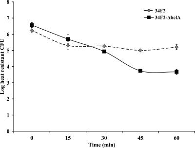
We then used 34F2-ΔbclA to address the influence of BclA on four properties of the spore: germination rate, virulence, adhesion to extracellular matrix proteins, and hydrophobicity. For the germination assay, we elected to use CFU in the presence and absence of heat as our measurement because a change from heat resistance to heat sensitivity is considered to be an irreversible step in the process of spore germination (12). We found that the mutant spores germinated at a faster rate in vitro than did wild-type spores between 30 and 60 min of incubation in germination medium and that the mutant spores germinated to a statistically significantly greater level than the wild type at 45 and 60 min of incubation (Fig. 2). Although Bozue et al. reported no statistically significant differences in germination between the Ames strain and its bclA mutant (5), that mutant appeared to germinate to a somewhat greater extent and at a slightly faster rate than the wild type, as evaluated by spectrofluorometric assays for germination.
FIG. 2.
Germination curve of 34F2 and 34F2-ΔbclA spores. Germination is measured here as a loss of heat resistance. The experiment was repeated three times. Analysis of variance showed statistically significant differences (P values of 0.001 and 0.007 for 45- and 60-min time points, respectively) in the extent of germination between wild-type and mutant spores. In the analysis of variance, three experiments were combined and then adjusted for variation.
When we compared the relative LD50s of mutant and wild-type spores by intranasal or subcutaneous routes of administration, no statistically significant differences between 34F2 and 34F2-ΔbclA were noted (Table 1). This finding is in keeping with reports described previously by Sylvestre et al. (23), who used a Sterne-like strain, and Bozue and colleagues, who used Ames (5). However, the LD50 values that we obtained were always lower for the mutant strain (Table 1). Furthermore, the mean time to death (MTD) of mice given a 106 dose of wild-type 34F2 spores was 4.2 days for the intranasal route and 4.4 days for the subcutaneous route compared to 3 days for mice that received 34F2-ΔbclA spores by both the intranasal and subcutaneous routes. Thus, the MTD was shorter for the 106 dose by both the subcutaneous and intranasal routes for the mutant strain (P = 0.006). These two findings taken together indicate that 34F2-ΔbclA spores germinate slightly better or faster in vivo (as they appear to do in vitro) or perhaps associate with host cells more avidly than do wild-type spores. Indeed, Bozue et al. reported less efficient recovery of ΔbclA spores of the Ames strain from lungs of mice upon bronchoalveolar lavage at days 2 and 4 in an aerosolized spore challenge (5). Those authors speculated that this reduced clearance of the mutant spores from the lungs of these animals might reflect the better binding of mutant spores than wild-type spores to host cells lining the lungs or airways. In fact, very recently, Bozue et al. (6) demonstrated that bclA mutant spores adhered to epithelial cell lines much better than did wild-type Ames spores. However, those investigators noted that such adhesion differences between their BclA Ames mutant strain and Ames did not extend to macrophages (6).
TABLE 1.
Comparison of LD50 values of spores of B. anthracis Sterne strain 34F2 and 34F2-ΔbclA
| Spore source | Route of infection | Expt | LD50 |
|---|---|---|---|
| 34F2 | Subcutaneous | 1 | 8.4 × 10² |
| 2 | 6.3 × 10² | ||
| 34F2-ΔbclA | Subcutaneous | 1 | 3.0 × 10² |
| 2 | 1.3 × 10² | ||
| 34F2 | Intranasal | 1 | 6.8 × 10⁴ |
| 2 | 7.9 × 10⁴ | ||
| 34F2-ΔbclA | Intranasal | 1 | 3.5 × 10⁴ |
| 2 | 4.0 × 10⁴ |
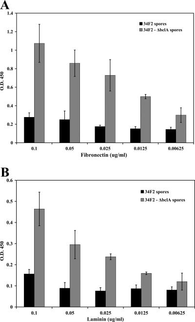
To evaluate the possibility that BclA might modulate the surface of the spore in a manner that influences the degree of association of spores with host matrix proteins, we compared how well fibronectin and laminin bound to wild-type and bclA mutant spores We found that the interaction of both fibronectin and laminin with 34F2-ΔbclA spores was more extensive than that with 34F2 spores (Fig. 3A and B). These results suggest that when BclA is removed from the surface of the spores, another protein(s) may become exposed and adhere to the two extracellular matrix proteins tested. Moreover, our data on the greater stickiness of the extracellular matrix components laminin and fibronectin to BclA-negative spores is consistent with the aforementioned report by Bozue et al. (6) on the enhanced adhesion of bclA mutant Ames spores to epithelial cells compared to the parental strain.
FIG. 3.
Comparison of adherence of matrix proteins to 34F2 and 34F2-ΔbclA spores. (A) Binding of fibronectin to spores. (B) Binding of laminin to spores. Data are shown as means ± standard deviations of values obtained from three experiments done in triplicate.
One possible explanation as to why matrix proteins bound less well to BclA-positive spores than to BclA-negative spores is that BclA affects the hydrophobic nature of the spore. That bacterial spores are hydrophobic has been demonstrated for several species of Bacillus and Clostridium (27), and, probably as a consequence, these types of spores adhere strongly to inanimate surfaces like microtiter plates (1). Moreover, Bacillus cereus spores bind to Caco-2 cells through surface hydrophobic interactions (2). The possibility that the degree of hydrophobicity of spores may be linked to the presence of an exosporium was first suggested by Takubo et al. (24), who showed that Bacillus megaterium QMB1551 spores with a defective or absent exosporium exhibit reduced affinity for hexadecane. Moreover, spores of Bacillus subtilis, B. licheniformis, and B. macerans do not have distinct exosporium layers and are less hydrophobic than are exosporium-containing B. cereus, B. brevis, and B. thuringiensis spores (13). Furthermore, a reduction in partitioning to hexadecane was successfully used by Bailey-Smith et al. to enrich for exosporium mutants of B. cereus (4). Thus, several studies indicate that the surface hydrophobicity of certain spores is attributable to the presence of an exosporium. However, the specific structural component(s) that contributes to the hydrophobic nature of the spore is not known.
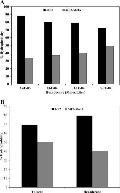
To test our theory about the impact of BclA on the hydrophobicity of spores, we evaluated the percent hydrophobicity of spores at various molarities of hexadecane. We found that the hydrophobicity of wild-type spores was greater than that of ΔbclA spores at each of the four hexadecane concentrations selected (Fig. 4A). We confirmed the findings of this BATH assay with a second hydrocarbon, toluene, at 0.1 ml per ml of spore suspension (853 μM toluene). Again, the wild-type spores showed higher hydrophobicity with toluene than did mutant spores (Fig. 4B). The 34F2 spores were more hydrophobic by HIC than were 34F2-ΔbclA spores (data not shown), although the absolute hydrophobicities of the spores as measured by the BATH and HIC techniques did not agree.
FIG. 4.
Comparison of hydrophobicity of 34F2 and 34F2-ΔbclA spores. (A) Hydrophobicity measured with various volumes of hexadecane. (B) Hydrophobicity differences between 34F2 and 34F2-ΔbclA spores compared with a second hydrocarbon, toluene. Data represent the average of data from two experiments.
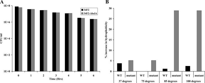
Next, we assessed whether the greater hydrophobicity of wild-type versus bclA mutant spores would still be evident in the presence of heat. We asked this question because Howell et al. (9) previously reported that increased temperatures can alter the structure of macromolecules and expose internal hydrophobic moieties. Moreover, Doyle et al. suggested that increases in the hydrophobicity of spores because of heat treatment may result from the disruption of outer coat or exosporium proteins (7). To examine the impact of heat on the relative hydrophobicity of the BclA-positive and -negative spores, we first had to test the effect of heat treatment alone on spore survival over time. No differences in the relative viabilities of wild-type and mutant spores after this heat treatment were noted (Fig. 5A). We then used a method described previously by Wiencek et al. (27) to measure the effect of heat on hydrophobicity. The values obtained were compared with the values determined for the unheated controls, and the results are represented as the percent increase in hydrophobicity. As shown in Fig. 5B, the mutant spores became strikingly more hydrophobic in the presence of heat than did wild-type spores even though heat did not differentially impact the viability of mutant and wild-type spores (Fig. 5A). One interpretation of the findings shown in Fig. 5B is that spore proteins that are normally obscured by BclA are exposed in its absence and that these now-uncovered proteins are altered by heat treatment in a manner that leads to the increased hydrophobicity of mutant spores compared to that of wild-type spores.
FIG. 5.
Evaluation of the impact of heat on hydrophobicity of 34F2 and 34F2-ΔbclA spores. (A) Effect of heat treatment on survival. (B) Percent increase in hydrophobicity compared to unheated controls. Both experiments were done in duplicate, and averages are presented. WT, wild type.
Further insight into how BclA affects the B. anthracis spore surface is important for at least two reasons. First, such information may lead to a better understanding of the initial interaction of the spore with its host. Indeed, our data on a shorter MTD of 34F2-ΔbclA versus 34F2 at a challenge dose of 106 spores, taken with a lower, albeit not statistically significantly different, LD50 of the BclA mutant than that of the wild-type strain for A/J mice, hint that BclA may slow spore germination in vivo. The fact that others have not reported any impact of BclA on virulence of B. anthracis spores (5, 23) may reflect the subtlety of a BclA effect. If that postulate is correct, variations between our group and others in bacterial or mouse strains used for these assays may explain the apparent discordance in the in vivo results. A second reason for further analyzing the influence of BclA on the B. anthracis spore surface is the possibility that the hydrophobicity of BclA may dictate how spores initially interact with inanimate surfaces. If this theory proves to be the case, then an evaluation of chemical ways to interfere with or modify BclA may lead to more efficient methods of surface sanitation in cases of an inadvertent or deliberate release of spores as well as sterilization of equipment in the laboratory environment.
Acknowledgments
This work was supported by NIH/NIAID grant AI53397, NIAID-NIH Middle Atlantic Regional Centers for Excellence grant 454 AI57168, and USUHS grant G173HS.
We thank Farhang Alem and Robert Cybulski for their technical assistance and Cara Olsen, the USUHS Biostatistician, for her expert statistical analysis.
The opinions and assertions in this paper are the private views of the authors and are not to be construed as official or as reflecting the views of the Department of the Navy or the Department of Defense. This research was conducted in compliance with the Animal Welfare Act. All animal use protocols were reviewed and approved by the Institutional Animal Care and Use Committee of the Uniformed Services University.
Editor: V. J. DiRita
Footnotes
Published ahead of print on 20 August 2007.
REFERENCES
- 1.Absolom, D. R. 1988. The role of bacterial hydrophobicity in infection: bacterial adhesion and phagocytic ingestion. Can. J. Microbiol. 34:287-298. [DOI] [PubMed] [Google Scholar]
- 2.Andersson, A., P. E. Granum, and U. Rönner. 1998. The adhesion of Bacillus cereus spores to epithelial cells might be an additional virulence mechanism. Int. J. Food Microbiol. 39:93-99. [DOI] [PubMed] [Google Scholar]
- 3.Aronson, A. I., and P. Fitz-James. 1976. Structure and morphogenesis of the bacterial spore coat. Bacteriol. Rev. 40:360-402. [DOI] [PMC free article] [PubMed] [Google Scholar]
- 4.Bailey-Smith, K., S. J. Todd, T. W. Southworth, J. Proctor, and A. Moir. 2005. The ExsA protein of Bacillus cereus is required for assembly of coat and exosporium onto the spore surface. J. Bacteriol. 187:3800-3806. [DOI] [PMC free article] [PubMed] [Google Scholar]
- 5.Bozue, J., C. K. Cote, K. L. Moody, and S. L. Welkos. 2007. Fully virulent Bacillus anthracis does not require the immunodominant protein BclA for pathogenesis. Infect. Immun. 75:508-511. [DOI] [PMC free article] [PubMed] [Google Scholar]
- 6.Bozue, J., K. L. Moody, C. K. Cote, B. G. Stiles, A. M. Friedlander, S. L. Welkos, and M. L. Hale. 2007. Bacillus anthracis spores of the bclA mutant exhibit increased adherence to epithelial, fibroblast, and endothelial cells but not macrophages. Infect. Immun. 75:4498-4505. [DOI] [PMC free article] [PubMed] [Google Scholar]
- 7.Doyle, R. J., F. Nedjat-Haiem, and J. S. Singh. 1984. Hydrophobic characteristics of Bacillus spores. Curr. Microbiol. 10:329-332. [Google Scholar]
- 8.Gerhardt, P., and E. Ribi. 1964. Ultrastructure of the exosporium enveloping spores of Bacillus cereus. J Bacteriol. 88:1774-1789. [DOI] [PMC free article] [PubMed] [Google Scholar]
- 9.Howell, N. K., G. Arteaga, S. Nakai, and E. C. Li-Chan. 1999. Raman spectral analysis in the C-H stretching region of proteins and amino acids for investigation of hydrophobic interactions. J. Agric. Food Chem. 47:924-933. [DOI] [PubMed] [Google Scholar]
- 10.Ismaeel, N., J. R. Furr, W. J. Pugh, and A. D. Russell. 1987. Hydrophobic properties of Providencia stuartii and other gram-negative bacteria measured by hydrophobic interaction chromatography. Lett. Appl. Microbiol. 5:91-95. [Google Scholar]
- 11.Janes, B. K., and S. Stibitz. 2006. Routine markerless gene replacement in Bacillus anthracis. Infect. Immun. 74:1949-1953. [DOI] [PMC free article] [PubMed] [Google Scholar]
- 12.Knaysi, G. 1959. Optical density of the endospore of Bacillus cereus and its relation to germination and resistance. J. Bacteriol. 78:206-216. [DOI] [PMC free article] [PubMed] [Google Scholar]
- 13.Koshikawa, T., M. Yamazaki, M. Yoshimi, S. Ogawa, A. Yamada, K. Watabe, and M. Torii. 1989. Surface hydrophobicity of spores of Bacillus spp. J. Gen. Microbiol. 135:2717-2722. [DOI] [PubMed] [Google Scholar]
- 14.Kuusela, P., T. Vartio, M. Vuento, and E. B. Myhre. 1985. Attachment of staphylococci and streptococci on fibronectin, fibronectin fragments, and fibrinogen bound to a solid phase. Infect. Immun. 50:77-81. [DOI] [PMC free article] [PubMed] [Google Scholar]
- 15.Mock, M., and A. Fouet. 2001. Anthrax. Annu. Rev. Microbiol. 55:647-671. [DOI] [PubMed] [Google Scholar]
- 16.Redmond, C., L. W. Baillie, S. Hibbs, A. J. Moir, and A. Moir. 2004. Identification of proteins in the exosporium of Bacillus anthracis. Microbiology 150:355-363. [DOI] [PubMed] [Google Scholar]
- 17.Reed, L. J., and H. Muench. 1938. A simple method of estimating fifty percent endpoints. Am. J. Hyg. 27:493-497. [Google Scholar]
- 18.Rosenberg, M., D. Gutnick, and E. Rosenberg. 1980. Adherence of bacteria to hydrocarbons: a simple method for measuring cell-surface hydrophobicity. FEMS Microbiol. Lett. 9:29-33. [Google Scholar]
- 19.Rosenberg, M. 2006. Microbial adhesion to hydrocarbons: twenty-five years of doing MATH. FEMS Microbiol. Lett. 262:129-134. [DOI] [PubMed] [Google Scholar]
- 20.Sarrafzadeh, M. H., L. Belloy, G. Esteban, J. M. Navarro, and C. Ghommidh. 2005. Dielectric monitoring of growth and sporulation of Bacillus thuringiensis. Biotechnol. Lett. 27:511-517. [DOI] [PubMed] [Google Scholar]
- 21.Sarrafzadeh, M. H., J. P. Guiraud, C. Lagneau, B. Gaven, A. Carron, and J. M. Navarro. 2005. Growth, sporulation, delta-endotoxins synthesis, and toxicity during culture of Bacillus thuringiensis H14. Curr. Microbiol. 51:75-81. [DOI] [PubMed] [Google Scholar]
- 22.Steichen, C., P. Chen, J. F. Kearney, and J. C. Turnbough, Jr. 2003. Identification of the immunodominant protein and other proteins of the Bacillus anthracis exosporium. J. Bacteriol. 185:1903-1910. [DOI] [PMC free article] [PubMed] [Google Scholar]
- 23.Sylvestre, P., E. Couture-Tosi, and M. Mock. 2002. A collagen-like surface glycoprotein is a structural component of the Bacillus anthracis exosporium. Mol. Microbiol. 45:169-178. [DOI] [PubMed] [Google Scholar]
- 24.Takubo, Y., M. Atarashi, T. Nishihara, and M. Kondo. 1988. Isolation and characterization of outermost layer deficient mutant spores of Bacillus megaterium. Microbiol. Immunol. 32:973-979. [DOI] [PubMed] [Google Scholar]
- 25.Todd, S. J., A. J. Moir, M. J. Johnson, and A. Moir. 2003. Genes of Bacillus cereus and Bacillus anthracis encoding proteins of the exosporium. J. Bacteriol. 185:3373-3378. [DOI] [PMC free article] [PubMed] [Google Scholar]
- 26.Welkos, S. L., T. J. Keener, and P. H. Gibbs. 1986. Differences in susceptibility of inbred mice to Bacillus anthracis. Infect. Immun. 51:795-800. [DOI] [PMC free article] [PubMed] [Google Scholar]
- 27.Wiencek, K. M., N. A. Klapes, and P. M. Foegeding. 1990. Hydrophobicity of Bacillus and Clostridium spores. Appl. Environ. Microbiol. 56:2600-2605. [DOI] [PMC free article] [PubMed] [Google Scholar]


