Abstract
The Escherichia coli hygromycin phosphotransferase (hph) gene, which confers hygromycin resistance, is commonly used as a dominant selectable marker in genetically modified bacteria, fungi, plants, insects, and mammalian cells. Expression of the hph gene has rarely been reported to induce effects other than those expected. Hygromycin B is the most common dominant selectable marker used in the molecular manipulation of Histoplasma capsulatum in the generation of knockout strains of H. capsulatum or as a marker in mutant strains. hph-expressing organisms appear to have no defect in long-term in vitro growth and survival and have been successfully used to exploit host-parasite interaction in short-term cell culture systems and animal experiments. We introduced the hph gene as a selectable marker together with the gene encoding green fluorescent protein into wild-type strains of H. capsulatum. Infection of mice with hph-expressing H. capsulatum yeast cells at sublethal doses resulted in lethality. The lethality was not attributable to the site of integration of the hph construct into the genomes or to the method of integration and was not H. capsulatum strain related. Death of mice was not caused by altered cytokine profiles or an overwhelming fungal burden. The lethality was dependent on the kinase activity of hygromycin phosphotransferase. These results should raise awareness of the potential detrimental effects of the hph gene.
Hygromycin B is commonly used as a dominant selectable marker in selection of genetically manipulated organisms. Expression of an Escherichia coli hygromycin phosphotransferase (hph) gene results in hygromycin B resistance in bacteria, fungi, plants, insects, and mammalian cells (7, 8, 10-12, 16, 21, 27, 28). Expression of the hph gene has rarely been reported to result in effects other than those expected by the expression of hygromycin B resistance, insertional mutagenesis, or the expression of the recombinant DNA carrying the hph gene (3, 19). hph gene expression is generally well tolerated and has been used in the generation of transgenic mice, viable Drosophila, and infectious bacteria and fungi.
Histoplasma capsulatum is an ascomycetous fungus with worldwide distribution and is the causative agent of histoplasmosis. Hygromycin B is the most common dominant selectable marker used in the molecular manipulation of H. capsulatum (20, 25-27, 31). Recombinant molecular genetics is limited in H. capsulatum, and few selectable markers have been characterized (18). Several studies have used hygromycin as a selectable marker in the generation of knockout strains of H. capsulatum or as a marker in mutant strains (20, 25-27, 31). hph-expressing organisms appeared to have no defect in long-term in vitro growth and survival and have been successfully used to exploit host-parasite interaction in short-term cell culture systems and animal experiments. We sought to introduce the gene encoding green fluorescent protein (GFP) into wild-type strains of H. capsulatum, and we selected the hygromycin resistance gene as our selectable marker. This work was being conducted to examine compartmentalization and reactivation of H. capsulatum. Surprisingly, infection of mice with hph-expressing H. capsulatum at normally sublethal numbers resulted in lethality. Five independent hph-expressing strains in the H. capsulatum G217B background all displayed the unexpected lethality. The lethality was not related to the site of integration of the hph construct into the genomes, the method of integration, or the strain of H. capsulatum. Lethality was dependent on kinase activity of hygromycin phosphotransferase.
MATERIALS AND METHODS
Fungal and bacterial strains.
Escherichia coli was grown in LB broth with appropriate antibiotics. H. capsulatum was grown in HMM medium with the addition of 200 μg/ml hygromycin, 200 μM uracil, or 150 μg/ml zeocin as appropriate (32). H. capsulatum strain WU 15 was obtained from William Goldman. Chemically competent E. coli strain TOP10 was used for plasmid cloning. Agrobacterium tumefaciens strains for H. capsulatum transformation were generated by electroporation of plasmid constructs generated in the pCB301 backbone into A. tumefaciens LBA1100 (15, 33). Agrobacterium-mediated transformation (AMT) of H. capsulatum was performed as previously described (20). Electroporation of H. capsulatum was performed as previously described with modifications due to the use of a square-wave electroporator (31). Electroporations were performed using a BTX 830 instrument at 375 kV for 5.5 ms using 1-mm cuvettes.
Generation of plasmid constructs.
Restriction digestions and ligations were performed under standard conditions. A BglII-SalI fragment comprising the E. coli hph gene under control of the Aspergillus nidulans GPD regulator sequences ligated to the A. nidulans TrpC terminator sequence was ligated between the BglII and XhoI sites of pUG27, resulting in a hygromycin resistance cassette flanked by loxP sequences. A SalI-SacII fragment containing the hygromycin resistance cassette flanked by loxP sequences was cloned into the polylinker of pCB301 (33) between the SacII and SalI sites to generate pCB301-HYG. To generate pCB301-BLE, the hygromycin open reading frame was replaced by the Streptoalloteichus hindustanus BLE gene. To generate pCB301-GFP-HYG, the GFP gene under control of the CBP regulatory sequence was excised from pSBB9.2 (14) and cloned downstream of the hygromycin resistance cassette within the pCB301 multiple cloning region. The vector pCB301-BLE-HYG was generated by insertion of the GPD promoter-hygromycin phosphotransferase-TrpC terminator cassette into pCB301-BLE between the ApaI and the KpnI sites. Site-directed mutagenesis was performed using a QuikChange mutagenesis kit according to the manufacturer's instructions to generate pCB301-BLE-mutHYG, resulting in a D196A substitution within the hph gene product (Stratagene, La Jolla, CA). The plasmid pCR186 was obtained as a kind gift from Chad Rappleye.
Animal studies.
C57BL/6 mice were purchased from The Jackson Laboratory. Animals were housed in microisolator cages and were maintained by the Department of Laboratory Animal Medicine (University of Cincinnati), which is accredited by the Association for Assessment and Accreditation of Laboratory Animal Care. All animal experiments were performed in accordance with the Animal Welfare Act guidelines of the National Institutes of Health, and all protocols were approved by the Institutional Animal Care and Use Committee of the University of Cincinnati. H. capsulatum was harvested in mid-log phase by centrifugation from a liquid culture in HMM medium. To produce infection, animals were inoculated intranasally with between 2 × 10⁴ and 2 × 10⁷ H. capsulatum yeast cells in a 30-μl volume of Hanks balanced salt solution (HBSS).
Quantitative organ culture.
Mice were euthanized and examined for fungal burdens in lungs and spleens. Organs were harvested and homogenized in 5 ml of HBSS. Organ homogenates were plated on blood heart infusion agar plates at multiple 10-fold dilutions and incubated at 30°C until colony growth could be measured. The fungal burden was expressed as mean log10 CFU per whole organ ± standard error of the mean. The limit of detection was 102 CFU.
Cytokine measurement.
Lungs from infected mice (n = 4 or 5) were removed, homogenized in 10 ml of HBSS, centrifuged at 1500 × g, filter sterilized, and stored at −70°C until assayed. Commercially available enzyme-linked immunosorbent assay kits were used to measure gamma interferon (IFN-γ), interleukin-4 (IL-4), and tumor necrosis factor alpha (TNF-α) (Pierce, Rockford, IL) and IL-10 and IL-12 (R & D Systems, Minneapolis, MN).
Flow cytometry.
Inflammatory cell infiltrates were analyzed by flow cytometry. Lung leukocytes and splenocytes were obtained by teasing apart lungs between the frosted ends of two glass slides. Mononuclear lung cells were further isolated by running over Lympholyte M (Cedar Lane Laboratories, Burlington, NC). Cells were washed three times with HBSS. The following monoclonal antibodies were purchased from BD Biosciences: CD3-fluorescein isothiocyanate, CD4-allophycocyanin (APC), CD8-APC, CD11c-APC, GR-1-APC, Mac3-phycoerythrin, and B220-APC. A total of 2 × 10⁶ cells were incubated with 0.5 μg of monoclonal antibody in staining buffer (1% bovine serum albumin in phosphate-buffered saline) for 10 min at 4°C. The cells were washed in staining buffer, and fluorescence was measured using a FACSCalibur flow cytometer (BD Biosciences, San Jose, CA).
Lung histology.
Histology was performed on formalin-fixed inflated lung samples by the Comparative Pathology Laboratory of the University of Cincinnati.
Determination of site of T-DNA integration.
To determine the site of Agrobacterium-mediated integration into the H. capsulatum genome, thermal asymmetrical PCR (TAIL-PCR) was performed as previously described (17). Briefly, H. capsulatum genomic DNA was isolated from transformed strains. Sequential rounds of TAIL-PCR were performed using three right- or left-border primers and a pool of degenerate primers (AD1 to AD4) as described. After three rounds of amplification, products were gel purified, cloned into pCR2.1-TOPO, and transformed into E. coli. Transformants were isolated, screened, and sequenced at the Cincinnati Children's Hospital Medical Center Sequencing facility using vector primers. The sequence was compared to the H. capsulatum G217B genome sequence (www.wustl.edu) by BLAST analysis to identify the site of genomic integration.
Hygromycin B phosphotransferase activity assay.
A semiquantitative hygromycin B phosphotransferase activity dot blot assay was performed as previously described with slight modification (24, 29). H. capsulatum strains UC18, UC19, UC24, and UC25 were grown in HMM medium to mid-log phase. Yeast cells from equal volumes of culture were lysed by bead beating at 4°C in 150 μl of ice-cold kinase lysis buffer (25 mM Tris-HCl [pH 7.5], 5% glycerol, 10 mM MgCl2, 1 mM dithiothreitol, 1 mM phenylmethylsulfonyl fluoride, and 1× EDTA protease inhibitor cocktail [Roche Applied Sciences, Indianapolis, IN]), and cellular debris was removed by centrifugation. The cell culture supernatant obtained following centrifugation of yeast cells was concentrated 100-fold by vacuum centrifugation. Five microliters of cell lysate or concentrated cell culture supernatant was added to 50 μl of reaction buffer (13.4 mM Tris maleate [pH 7.1], 8.4 mM MgCl2, 80 mM NH4Cl, 60 mM hygromycin B, 15 μM ATP, 25 μCi [γ-32P] ATP per ml) and incubated at room temperature for 1 h. The reaction samples were diluted with 150 μl double-distilled water (ddH2O), loaded into a dot blot apparatus fitted with a layer of nitrocellulose paper above a double layer of P81 phosphocellulose paper (Whatman, Florham Park, NJ), and allowed to drain by gravity. Each well was then washed with ddH2O. The P81 filter was then washed three times in ddH2O at 65°C. The filter was then exposed to a phosphor storage screen and visualized on a Storm PhosphorImager (GE Healthcare, Piscataway, NJ).
Statistics.
One-way analysis of variance was used to compare groups, and the log rank test was used to analyze survival.
RESULTS
GFP-expressing H. capsulatum strains were generated by AMT of T-DNA containing a GFP expression cassette and a hygromycin selectable marker. The GFP/HYG cassette was introduced into the widely used H. capsulatum laboratory strains G217B and G186AR (14, 18). In addition, a GFP-expressing uracil auxotrophic derivative of G217B was generated by the introduction of pCB301-GFP/HYG into strain WU 15 (20). Transformants with high levels of GFP expression were isolated. These strains demonstrated stable high levels of fluorescence after prolonged growth in the absence of hygromycin B selection. Growth curves of the GFP-expressing strains were similar to those of their parent G217B and G186AR strains (data not shown).
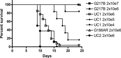
A G217B-derived strain, UC1, was then used for in vivo studies in mice. Studies with durations of 7 to 9 days were performed, with no unexpected deaths. During a long-term study examining compartmentalization and reactivation of H. capsulatum, 55% (33/60) of animals infected with 2 × 10⁶ yeast cells unexpectedly died in the first 3 weeks of the study. Because of this unusual occurrence, additional studies were performed to investigate the animal deaths. In the next series of studies, groups of C57BL/6 mice were infected with doses of UC1 organisms that varied between 2 × 10⁴ and 2 × 10⁶ yeast cells and followed for survival. Animals infected with >2 × 10⁵ yeast cells died between days 9 and 20. The hypervirulence of H. capsulatum UC1 compared to the parent strain H. capsulatum G217 was confirmed in survival studies (Fig. 1; Table 1). H. capsulatum UC1 was 100- to 1,000-fold more virulent than its parental H. capsulatum G217 strain.
FIG. 1.
Expression of hph results in lethality among mice infected with Histoplasma capsulatum. Groups of mice were infected intranasally with 2 × 10⁴ to 2 × 10⁷ yeast cells of H. capsulatum strain G217B (n = 8), strain G186AR (n = 8), the hygromycin-resistant G217B derivative UC1 (n = 16), or the hygromycin-resistant G186AR derivative UC2 (n = 8). Survival curves demonstrate the loss of animals infected with >2 × 10⁵ hygromycin-resistant H. capsulatum cells between days 9 and 20.
TABLE 1.
Fungal strains used and cumulative mortality
| H. capsulatum strain | Genotype | Cumulative mortality (%)a |
|---|---|---|
| G217B | Wild type | 0/12 (0) |
| G186AR | Wild type | 0/12 (0) |
| WU13 | G217B ura-41,zzz::[PCBP1-gfp hph] | 17/20 (85)b |
| WU15 | G217B ura5-Δ41 | |
| UC1 | G217B aaa::T-DNA-[PCBP1-gfp], loxP PGPD.hph TTrpC loxP | 58/86 (67.5) |
| UC2 | G186AR T-DNA-[PCBP1-gfp], loxP PGPD.hph TTrpC loxP | 8/8 (100) |
| UC3 | WU15 T-DNA-[PCBP1-gfp], loxP PGPD.hph TTrpC loxP | 11/20 (55)b |
| UC5 | G217B bbb::T-DNA-[PCBP1-gfp], loxP PGPD.hph TTrpC loxP | 6/6 (100) |
| UC16 | G217B T-DNA-loxP PGPD.ble TTrpC loxP | 0/12 (0) |
| UC17 | G217B T-DNA-loxP PGPD.hph TTrpC loxP | 6/6 (100) |
| UC18 | G217B ccc::T-DNA-PGPD.hph-1 TTrpC, loxP PGPD.ble TTrpC loxP | 0/12 (0) |
| UC19 | G217B ddd::T-DNA-PGPD.hph-1 TTrpC, loxP PGPD.ble TTrpC loxP | 0/6 (0) |
| UC24 | G217B eee::T-DNA-PGPD.hph TTrpC, loxP PGPD.ble TTrpC loxP | 5/6 (83.3) |
| UC25 | G217B fff::T-DNA-PGPD.hph TTrpC, loxP PGPD.ble TTrpC loxP | 5/6 (83.3) |
Associated with infection resulting from intranasal inoculation with 2 × 10⁶ yeast cells/animal.
Uracil auxotrophy complemented with plasmid pCR186.
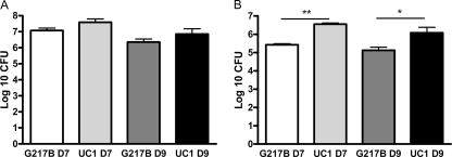
Organ cultures were performed to determine if UC1-infected mice died of overwhelming H. capsulatum infection. Organ cultures of animals infected with G217B or UC1 showed no significant difference in CFU (P > 0.05) in the lungs of mice sacrificed on day 7 or 9. The fungal burden in spleens of animals infected with UC1 exceeded that of wild-type G217B organisms at days 7 (P < 0.0001) and 9 (P < 0.05) (Fig. 2). The number of CFU present in both the lung and the spleen declined between days 7 and 9. Of note, neither of the numbers of organism isolated is usually associated with the demise of animals. Most often, death is accompanied by a level of ≥108 CFU per organ (1). Thus, it is unlikely that the apparent hypervirulence of UC1 could be attributed solely to an increased organism burden in animals infected with G217B-derived UC1 compared to the G217B strain of H. capsulatum.
FIG. 2.
Tissue fungal burden following Histoplasma capsulatum infection. Groups of mice were infected intranasally with 2 × 10⁶ yeast cells of H. capsulatum strain G217B or the hygromycin-resistant G217B derivative UC1. CFU in lungs (A) and spleens (B) were counted on day 7 (n = 13) or day 9 (n = 10) after infection. Data are means ± standard errors. *, P < 0 0.05; **, P < 0.0001.
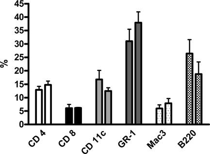
Exuberant inflammatory responses may cause immunopathology and result in the death of animals independent of organism burden. The phenotype of cells infiltrating into the lungs of animals infected with G217B and UC1 at day 7 was determined by fluorescence-activated cell sorter analysis. The lungs of animals infected with G217B and UC1 showed no difference in the number or proportion of CD4+ or CD8+ T cells, dendritic cells, neutrophils, macrophages, or B cells present on day 7 (Fig. 3).
FIG. 3.
Lung cell phenotype in Histoplasma capsulatum-infected animals. Mice were infected with H. capsulatum strain G217B or the hygromycin-resistant G217B derivative UC1, and on day 7 following infection lung homogenates were analyzed for CD4+ T cells, CD8+ T cells, dendritic cells, neutrophils, macrophages, and B cells. Data are means ± standard errors for four mice.
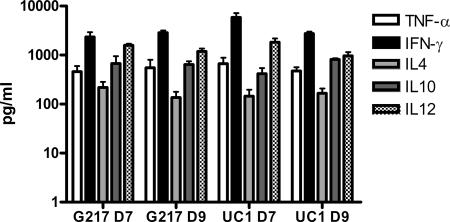
Cytokine analysis of lung and spleen homogenates provided additional evidence that the inflammatory response in animals infected with G217B was similar to that in animals infected with UC1. No significant differences in TNF-α, IFN-γ, IL-4, IL-10, or IL-12 levels were noted in the lungs or spleens at day 7 and day 9 despite infection with either G217B or UC1 (Fig. 4). Histological analysis of lungs from C57BL/6 mice sacrificed at day 7 after inoculation with sublethal inocula of either G217B or UC1 revealed severe pyogranulomatous pneumonia in all animals, with no significant difference discerned between animals infected with either strain (data not shown).
FIG. 4.
Cytokine profiles of splenic homegenates from Histoplasma capsulatum-infected animals. Mice were infected with H. capsulatum strain G217B or the hygromycin-resistant G217B derivative UC1, and lungs and spleens were harvested on day 7 and day 9 following infection. Levels of TNF-α, IFN-γ, IL-4, IL-10, and IL-12 were measured in the lung and spleen homegenates. Data are means ± standard errors for four mice.
We investigated if a correlation existed between the hypervirulence phenotype and the site of T-DNA integration into the H. capsulatum genome. A second independent isolate of GFP-expressing G217B, UC5, was studied. One hundred percent lethality between days 10 and 15 was similarly noted in C57BL/6 mice infected intranasally with a sublethal dose of 2 × 10⁶ yeast cells of UC5. The sites of T-DNA integration in UC1 and UC5 were then determined by sequencing TAIL-PCR-generated products. Despite the similar hypervirulent phenotypes of UC1 and UC5 in mice, sequence analysis revealed single unique sites of integration within the H. capsulatum genome for each strain (upstream of putative open reading frame HCAG 08014 and downstream of HCAG 07026, respectively). The increased virulence and unexpected deaths were also noted when UC3, which had been rendered prototrophic by complementation with the URA5-carrying plasmid pCR186, was used for infection.
Studies were designed to determine the role of GFP expression, hygromycin B expression, and AMT in the lethality. H. capsulatum G217B strain UC17, generated by the introduction of only the hph resistance cassette by AMT, was then used to infect C57BL/6 mice. Despite the lack of a GFP expression cassette, 100% of infected animals died between days 10 and 15, demonstrating that that the presence of the hph cassette was sufficient to cause the noted hypervirulence phenotype.
To determine if lethality was related to AMT, we generated a zeocin-resistant H. capsulatum G217B strain, UC16, by introduction of the S. hindustanus BLE gene under control of the A. nidulans GPD regulatory sequences by AMT. A single zeocin-resistant transformant was characterized and used to infect C57BL/6 mice at a dose of 2 × 10⁶ yeast cells via intranasal inoculation. All animals survived the challenge, and on sacrifice on day 21, organ culture of lungs and spleens demonstrated clearance of the infection. This suggested that the hypervirulence phenotype was related to expression of the hph gene and hygromycin resistance.
To further examine the role of AMT, hygromycin resistance, and GFP expression in the lethality noted in mice, C57BL/6 mice were infected with H. capsulatum strain WU13, a hygromycin-resistant GFP-expressing strain derived by non-A. tumifaciens-mediated random insertion. This uracil auxotroph was rendered prototrophic by introduction of the Podospora anserina ura5-carrying plasmid pCR186. As noted with other hygromycin-resistant strains, 85% of animals died between days 10 to 21 after infection (Table 1).
All studies described thus far had been performed on H. capsulatum with a G217B strain background. Similar studies were performed with sublethal doses of a hygromycin-resistant strain of G186AR H. capsulatum, UC2, generated by AMT. Eight of eight infected animals died by day 16 after inoculation, demonstrating that the hypervirulence was not restricted to the G217B strain of H. capsulatum (Fig. 1; Table 1).
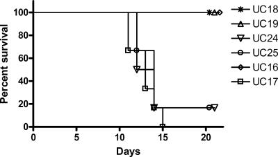
To explore the possibility that the virulence of H. capsulatum was associated with functional activity of the hygromycin phosphotransferase, site-directed mutagenesis was performed, resulting in the substitution of an alanine for the essential aspartic acid residue within the kinase activation domain of the hph gene product (2, 13). Introduction of the zeocin resistance cassette and a hygromycin resistance cassette containing the mutant hph gene into H. capsulatum G217B resulted in organisms (strains UC18 and UC19) that were resistant to zeocin but sensitive to hygromycin. A control strain was generated by the introduction of the zeocin resistance cassette and a hygromycin resistance cassette containing an active hph gene in H. capsulatum G217B (strains UC24 and UC25). UC24 and UC25 grew in the presence of hygromycin B concentrations of >300 μg/ml, while UC18 and UC19 were unable to grow in the presence of >150 μg/ml of hygromycin B. Cellular lysates of UC24 and UC25 demonstrated hygromycin B phosphotransferase activity by dot blot assay, while no activity was detected in lysates of UC18 and UC19. No hygromycin B phosphotransferase activity was detected in culture supernatants of UC18, UC19, UC24, or UC25. These strains were then characterized at a molecular level. All strains demonstrated hybridization patterns consistent with a single site of integration by Southern blot analysis (data no shown). The sites of T-DNA integration in UC18, UC19, UC24, and UC25 were determined by sequencing TAIL-PCR-generated products. The strains integrated within four distinct putative open reading frames: HCAG 06126, HCAG 04373, HCAG 00845, and HCAG 01994, respectively. PCR analysis using primers located upstream or downstream of the site of integration paired with left- and right-border primers revealed no evidence of genomic rearrangement associated with T-DNA integration. C57BL/6 mice infected with 2 × 10⁶ UC18 and UC19 organisms survived infection, demonstrating a course of disease seen following infection with wild-type G217B H. capsulatum until sacrifice on day 21, whereas five of six mice infected with either UC24 or UC25 died with median times to death of 12 and 14 days, respectively (Fig. 5).
FIG. 5.
Expression of hph results in lethality among mice infected with Histoplasma capsulatum. Groups of mice were intranasally infected with 2 × 10⁶ yeast cells of H. capsulatum strains UC16, expressing zeocin resistance; UC17, expressing hygromycin resistance; UC24 and UC25, expressing zeocin and hygromycin resistance; or UC18 and UC19, expressing zeocin resistance and a nonfunctional hygromycin phosphotransferase (n = 6 to 12 mice per strain). Survival curves demonstrate the loss of animals infected with hygromycin-resistant H. capsulatum between days 10 and 15.
DISCUSSION
Generation of GFP-expressing H. capsulatum in wild-type laboratory strains G217B and G186AR is an important tool for pathogenicity studies. GFP-positive ura5 gene deletions provide strains with better characterization than the UV-generated uracil auxtrophic strains, where the site of mutation(s) is unknown (20, 32). However, long-term studies in vivo with these strains unexpectedly led to lethality. The adverse outcomes were restricted to animal studies, and no differences were noted between wild-type and hygromycin-resistant strains in axenic or in vitro studies with macrophages.
Although small increases in organism burden were noted in animals infected with hygromycin-resistant H. capsulatum strains compared to wild-type strains, the number of organisms isolated is not sufficient to explain the deaths of mice (1). Typically, death from overwhelming infection is not observed with G217B in C57BL/6 mice until ∼108 CFU are present. Moreover, the burden of infection in mice infected with a strain of H. capsulatum that contains the hygromycin resistance gene was not that dissimilar from that with the wild type at days 7 and 9. Another possible explanation for these findings is an altered pathological response. This consideration is highly unlikely, since the pathology did not differ between the two groups and the characters of the inflammatory response as assessed by flow cytometry were similar.
The hypervirulence in animals was related to integration of a hygromycin resistance cassette comprising the A. nidulans GPD regulatory sequence, the E. coli hygromycin phosphotransferase gene, and the A. nidulans TrpC terminator region into the H. capsulatum genome. It was not related to the method of integration, as strains generated by both A. tumifaciens-mediated and non-A. tumifaciens-mediated random integration had a similar phenotype. Similarly, the site of integration did not appear to be critical. AMT exhibits little bias in site of integration, and thus while the site of integration was determined for only six strains, the probability that integration was at the same site in the other four strains is low (15). The hypervirulence was not related to the expression of GFP, as hygromycin-resistant strains without expression of GFP demonstrated similar lethality.
The observed hypervirulence was not limited to the G217B strain with which initial studies were performed but was similarly noted when H. capsulatum G186AR was rendered hygromycin resistant. Hygromycin resistance has been used as a selectable marker in fungi such as Aspergillus fumigatus, Cryptococcus neoformans, and Blastomyces dermatitidis, although many studies with these pathogens are of shorter duration (11, 12, 21, 30).
Prior studies have utilized hygromycin selection in the generation of H. capsulatum mutants and tagged strains. No virulence differences were noted in these studies despite the use of animal experiments (20, 25-27, 31). However, experiments with the animals studied in these reports were generally limited to durations of 7 to 10 days and thus may have been concluded before animals succumbed. In addition, most studies used hygromycin as a selectable marker in the generation of deletion strains with reduced virulence. In a background of reduced virulence, the lethality noted in this study associated with hph expression may not be manifest. We postulate, therefore, that the effect of the hygromycin resistance is likely to be manifest when the organism maintains its native virulence determinants. Thus, the effect of hygromycin resistance may also be observed when genes that do not perturb virulence traits are inserted. One prior study using hph-marked strains reported animal survival to 27 days postinfection (22). The DRKI-silenced strain generated in that study demonstrated markedly reduced virulence, which may have suppressed the hypervirulent phenotype. This study also utilized intratracheal inoculation of spores as a mode of infection, and the course of disease may be altered with this animal model. While the hypervirulence phenotype reported should not be extrapolated to other animal models without experimental evidence, the findings do raise a cautionary flag for other model systems.
The virulence is associated with the kinase activity of the hygromycin phosphotransferase (2, 19). Mutagenesis of the critical aspartic acid residue within the phosphorylation domain resulted in loss of hygromycin resistance, loss of phosphotransferase activity, and reversion to standard virulence. The increased virulence may be associated with phosphorylation of a bystander H. capsulatum protein or a mammalian host protein. Off-target phosphorylation by aminoglycoside antibiotic phosphotransferases has been previously reported in a number of mammalian systems but not in fungal organisms (3, 19). Studies to distinguish between these possibilities have not been performed. Hygromycin phosphotransferase is presumed to remain as an intracellular protein, suggesting the target to be an H. capsulatum protein. While the absence of hygromycin phosphotransferase activity in culture supernatants during in vitro growth may support this, no studies were performed to examine for cell surface activity or activity within the phagosome. Identification of phosphorylation substrates may be of importance in understanding the virulence of H. capsulatum.
While H. capsulatum proteins would seem the logical target of phosphorylation by the hph gene product, mammalian host proteins may be alternate targets. Compared to other mammalian fungal pathogens in which hygromycin resistance has been used as a selectable marker, H. capsulatum resides and proliferates within the macrophage phagosome (9, 23). It may be in this environment that hygromycin phosphotransferase may have access to host proteins. Although each pathogen is biologically unique, hygromycin B resistance has been successfully used in the intracellular bacterial pathogen Mycobacterium tuberculosis without reported effects (6).
These finding have significance for investigators studying H. capsulatum and perhaps other organisms. Few selectable markers have been characterized in H. capsulatum, and these results will promote systematic evaluation of alternative dominant selectable markers (18). Alternative strategies such as the use of Cre recombinase to delete the selectable marker from strains generated with loxP-flanked constructs have successfully been applied in H. capsulatum (data not shown). In H. capsulatum studies, hygromycin may still be used as a selectable marker if animal studies are not contemplated. In addition, while this phenomenon has not been recognized in other fungi, community awareness may lead to increased reporting of untoward effects.
Acknowledgments
We thank William Goldman, Chad Rappleye, and Russ Osguthorpe for H. capsulatum strains and plasmid constructs; Francisco Gomez for advice and support; and Reiko Tanaka and Holly Allen for technical assistance.
Footnotes
Published ahead of print on 14 September 2007.
REFERENCES
- 1.Allendoerfer, R., and G. S. Deepe, Jr. 1997. Intrapulmonary response to Histoplasma capsulatum in gamma interferon knockout mice. Infect. Immun. 65:2564-2569. [DOI] [PMC free article] [PubMed] [Google Scholar]
- 2.Boehr, D. D., P. R. Thompson, and G. D. Wright. 2001. Molecular mechanism of aminoglycoside antibiotic kinase APH(3′)-IIIa: roles of conserved active site residues. J. Biol. Chem. 276:23929-23936. [DOI] [PubMed] [Google Scholar]
- 3.Daigle, D. M., G. A. McKay, P. R. Thompson, and G. D. Wright. 1999. Aminoglycoside antibiotic phosphotransferases are also serine protein kinases. Chem. Biol. 6:11-18. [DOI] [PubMed] [Google Scholar]
- 4.Reference deleted.
- 5.Reference deleted.
- 6.De Voss, J. J., K. Rutter, B. G. Schroeder, H. Su, Y. Zhu, and C. E. Barry III. 2000. The salicylate-derived mycobactin siderophores of Mycobacterium tuberculosis are essential for growth in macrophages. Proc. Natl. Acad. Sci. USA 97:1252-1257. [DOI] [PMC free article] [PubMed] [Google Scholar]
- 7.Fickers, P., M. T. Le Dall, C. Gaillardin, P. Thonart, and J. M. Nicaud. 2003. New disruption cassettes for rapid gene disruption and marker rescue in the yeast Yarrowia lipolytica. J. Microbiol. Methods 55:727-737. [DOI] [PubMed] [Google Scholar]
- 8.Gaken, J., F. Farzaneh, C. Stocking, and W. Ostertag. 1992. Construction of a versatile set of retroviral vectors conferring hygromycin resistance. BioTechniques 13:32-34. [PubMed] [Google Scholar]
- 9.Gildea, L. A., G. M. Ciraolo, R. E. Morris, and S. L. Newman. 2005. Human dendritic cell activity against Histoplasma capsulatum is mediated via phagolysosomal fusion. Infect. Immun. 73:6803-6811. [DOI] [PMC free article] [PubMed] [Google Scholar]
- 10.Goldstein, A. L., and J. H. McCusker. 1999. Three new dominant drug resistance cassettes for gene disruption in Saccharomyces cerevisiae. Yeast 15:1541-1553. [DOI] [PubMed] [Google Scholar]
- 11.Hogan, L. H., and B. S. Klein. 1997. Transforming DNA integrates at multiple sites in the dimorphic fungal pathogen Blastomyces dermatitidis. Gene 186:219-226. [DOI] [PubMed] [Google Scholar]
- 12.Hua, J., J. D. Meyer, and J. K. Lodge. 2000. Development of positive selectable markers for the fungal pathogen Cryptococcus neoformans. Clin. Diagn. Lab. Immunol. 7:125-128. [DOI] [PMC free article] [PubMed] [Google Scholar]
- 13.Kocabivik, S., and M. H. Perlin. 1994. Amino acid substitutions within the analogous nucleotide binding loop (P-loop) of aminoglycoside 3′-phosphotransferase-II. Int. J. Biochem. 26:61-66. [DOI] [PubMed] [Google Scholar]
- 14.Kugler, S., B. Young, V. L. Miller, and W. E. Goldman. 2000. Monitoring phase-specific gene expression in Histoplasma capsulatum with telomeric GFP fusion plasmids. Cell Microbiol. 2:537-547. [DOI] [PubMed] [Google Scholar]
- 15.Lacroix, B., T. Tzfira, A. Vainstein, and V. Citovsky. 2006. A case of promiscuity: Agrobacterium's endless hunt for new partners. Trends Genet. 22:29-37. [DOI] [PubMed] [Google Scholar]
- 16.Lama, J., and L. Carrasco. 1992. Expression of poliovirus nonstructural proteins in Escherichia coli cells. Modification of membrane permeability induced by 2B and 3A. J. Biol. Chem. 267:15932-15937. [PubMed] [Google Scholar]
- 17.Liu, Y. G., and R. F. Whittier. 1995. Thermal asymmetric interlaced PCR: automatable amplification and sequencing of insert end fragments from P1 and YAC clones for chromosome walking. Genomics 25:674-681. [DOI] [PubMed] [Google Scholar]
- 18.Magrini, V., and W. E. Goldman. 2001. Molecular mycology: a genetic toolbox for Histoplasma capsulatum. Trends Microbiol. 9:541-546. [DOI] [PubMed] [Google Scholar]
- 19.Maio, J. J., and F. L. Brown. 1991. Gene activation mediated by protein kinase C in human macrophage and teratocarcinoma cells expressing aminoglycoside phosphotransferase activity. J. Cell Physiol. 149:548-559. [DOI] [PubMed] [Google Scholar]
- 20.Marion, C. L., C. A. Rappleye, J. T. Engle, and W. E. Goldman. 2006. An alpha-(1,4)-amylase is essential for alpha-(1,3)-glucan production and virulence in Histoplasma capsulatum. Mol. Microbiol. 62:970-983. [DOI] [PubMed] [Google Scholar]
- 21.McDade, H. C., and G. M. Cox. 2001. A new dominant selectable marker for use in Cryptococcus neoformans. Med. Mycol. 39:151-154. [DOI] [PubMed] [Google Scholar]
- 22.Nemecek, J. C., M. Wuthrich, and B. S. Klein. 2006. Global control of dimorphism and virulence in fungi. Science 312:583-588. [DOI] [PubMed] [Google Scholar]
- 23.Newman, S. L. 1999. Macrophages in host defense against Histoplasma capsulatum. Trends Microbiol. 7:67-71. [DOI] [PubMed] [Google Scholar]
- 24.Platt, S. G., and N. S. Yang. 1987. Dot assay for neomycin phosphotransferase activity in crude cell extracts. Anal. Biochem. 162:529-535. [DOI] [PubMed] [Google Scholar]
- 25.Rappleye, C. A., J. T. Engle, and W. E. Goldman. 2004. RNA interference in Histoplasma capsulatum demonstrates a role for alpha-(1,3)-glucan in virulence. Mol. Microbiol. 53:153-165. [DOI] [PubMed] [Google Scholar]
- 26.Rappleye, C. A., L. G. Eissenberg, and W. E. Goldman. 2007. Histoplasma capsulatum alpha-(1,3)-glucan blocks innate immune recognition by the beta-glucan receptor. Proc. Natl. Acad. Sci. USA 104:1366-1370. [DOI] [PMC free article] [PubMed] [Google Scholar]
- 27.Sebghati, T. S., J. T. Engle, and W. E. Goldman. 2000. Intracellular parasitism by Histoplasma capsulatum: fungal virulence and calcium dependence. Science 290:1368-1372. [DOI] [PubMed] [Google Scholar]
- 28.Soares, R. B., T. A. Velho, L. M. De Moraes, M. O. Azevedo, C. M. Soares, and M. S. Felipe. 2005. Hygromycin B-resistance phenotype acquired in Paracoccidioides brasiliensis via plasmid DNA integration. Med. Mycol. 43:719-723. [DOI] [PubMed] [Google Scholar]
- 29.Sorensen, M. S., M. Duch, K. Paludan, P. Jorgensen, and F. S. Pedersen. 1992. Measurement of hygromycin B phosphotransferase activity in crude mammalian cell extracts by a simple dot-blot assay. Gene 112:257-260. [DOI] [PubMed] [Google Scholar]
- 30.Sugui, J. A., Y. C. Chang, and K. J. Kwon-Chung. 2005. Agrobacterium tumefaciens-mediated transformation of Aspergillus fumigatus: an efficient tool for insertional mutagenesis and targeted gene disruption. Appl. Environ. Microbiol. 71:1798-1802. [DOI] [PMC free article] [PubMed] [Google Scholar]
- 31.Woods, J. P., E. L. Heinecke, and W. E. Goldman. 1998. Electrotransformation and expression of bacterial genes encoding hygromycin phosphotransferase and beta-galactosidase in the pathogenic fungus Histoplasma capsulatum. Infect. Immun. 66:1697-1707. [DOI] [PMC free article] [PubMed] [Google Scholar]
- 32.Worsham, P. L., and W. E. Goldman. 1988. Selection and characterization of ura5 mutants of Histoplasma capsulatum. Mol. Gen. Genet. 214:348-352. [DOI] [PubMed] [Google Scholar]
- 33.Xiang, C., P. Han, I. Lutziger, K. Wang, and D. J. Oliver. 1999. A mini binary vector series for plant transformation. Plant Mol. Biol. 40:711-717. [DOI] [PubMed] [Google Scholar]


