Abstract
Crimean-Congo hemorrhagic fever virus (CCHFV) causes severe human disease. The CCHFV medium RNA encodes a polyprotein which is proteolytically processed to yield the glycoprotein precursors PreGn and PreGc, followed by structural glycoproteins Gn and Gc. Subtilisin kexin isozyme-1/site-1 protease (SKI-1/S1P) plays a central role in Gn processing. Here we show that CCHFV-infected cells deficient in SKI-1/S1P produce no infectious virus, although PreGn and PreGc accumulated normally in the Golgi apparatus, the site of virus assembly. Only nucleoprotein-containing particles which lacked virus glycoproteins (Gn/Gc or PreGn/PreGc) were secreted. Complementation of SKI-1/S1P-deficient cells with a SKI-1/S1P expression vector restored release of infectious virus (>106 PFU/ml), confirming that SKI-1/S1P processing is required for incorporation of viral glycoproteins. SKI-1/S1P may represent a promising antiviral target.
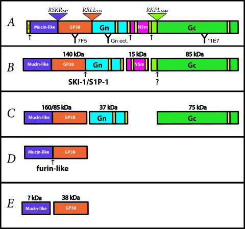
Crimean-Congo hemorrhagic fever virus (CCHFV) is a tick-borne virus endemic in many regions in Africa, Asia, and Europe. The CCHFV genome is composed of three negative-sense RNA segments (S, M, and L), which encode the nucleoprotein (N), surface glycoproteins (Gn and Gc), and RNA polymerase, respectively (6). The M segments of all viruses in the family Bunyaviridae are translated into polyproteins synthesized in the endoplasmic reticulum. Generally, these polyproteins are cotranslationally cleaved by the signal peptidase into the structural glycoproteins Gn and Gc (2, 7, 14) and, with the exception of the hantaviruses, a nonstructural protein (NSm) (1, 11, 15). The CCHFV M polyprotein has unique features. First, the amino-terminal region contains two extra domains of unknown function: the mucin-like and GP38 domains (Fig. 1) (21, 22). Second, Gn and Gc are not produced solely by signal peptidase cleavages since mature Gn and GP38 generation also requires the activity of cellular serine endoproteases (21, 27) related to the bacterial subtilisin commonly referred to as SKI-1 (23), or S1P (20), and furin (26).
FIG. 1.
Schematic representation of CCHFV M-encoded polyprotein domains and proteolytic processing. (A) Signal peptide (SP), mucin-like, GP38, Gn, NSm, and Gc M polyprotein domains are highlighted. Potential transmembrane domains (yellow) and signal peptidase cleavage sites are indicated by black arrows. Antibodies used in this study are indicated (with their binding regions in parentheses): 7F5 (PreGn/GP38), Gn ectodomain (Gn ect.) (Gn), and 11E7 (PreGc/Gc). Defined furin-like (RSKR247), SKI-1/S1P (RRLL519), and SKI-1/S1P-like (RKPL1040) cleavage sites are illustrated by inverted triangles. (B) The first proteolytic products are expected to occur cotranslationally or rapidly after the synthesis of the polyprotein. PreGn (140 kDa), NSm (15 kDa), and PreGc (85 kDa) are the results of these initial processing events. SKI-1/S1P and PreGc convertase will then cleave (indicated by arrows) PreGn and PreGc in the early secretory pathway. The PreGc cleavage also occurs early in the secretory pathway but the cognate protease (?) remains unidentified. (C) The activity of SKI-1/S1P and the PreGc convertase generates a nonstructural mucin-like GP38 protein of either 160 or 85 kDa, and the structural glycoproteins Gn (37 kDa) and Gc (75 kDa). (D) The mucin-like GP38 is further cleaved (arrow) by a furin-like protease in the late secretory pathway. (E) Furin-like enzyme cleavage results in production of a GP38 glycoprotein (38 kDa) and a mucin-like protein of unknown mass (? kDa).
The CCHFV (IbAr10200 strain) M polyprotein is rapidly cleaved into two glycoprotein precursors, PreGn (140 kDa) and PreGc (85 kDa) (Fig. 1) (22). The PreGn cleavage at the RRLL519↓ motif by SKI-1/S1P, which yields Gn (37 kDa), occurs early in the secretory pathway. GP38, a nonstructural protein, is the product of PreGn cleavage by both SKI-1/S1P and a furin-like protease (RSKR247↓) in the late secretory pathway (21). PreGc is cleaved into Gc at motif RKPL1040↓, which closely resembles that recognized by SKI-1/S1P, but the cognate protease remains uncharacterized. Clearly, CCHFV glycoprotein biogenesis is considerably more complex than that of viruses in other genera of the family Bunyaviridae. Interestingly, SKI-1/S1P was also shown to cleave the glycoprotein precursor (GPC) of two arenaviruses, Lassa virus (13) and lymphocytic choriomeningitis virus (3).
Proteolytic processing of viral glycoproteins is typically required for their fusogenic activity. However, lack of endoproteolysis of viral glycoproteins does not necessarily correlate with reduced infectivity. For instance, Ebola viruses displaying on their surface a mutant glycoprotein defective for furin cleavage replicate in cell culture (16) and in a nonhuman primate model (17) identically to virions displaying the wild-type cleaved glycoprotein. Here the biological significance of CCHFV PreGn conversion to Gn by SKI-1/S1P and its importance for virus infectivity were examined.
CCHFV growth in CHO-derived cells is independent of S2P activity.
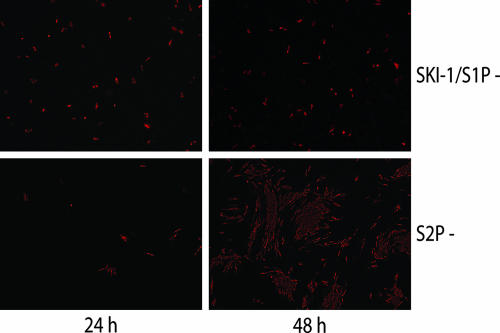
Cells deficient in SKI-1/S1P or S2P protease activity have been generated previously (8, 9, 19). Both SKI-1/S1P and S2P function in the normal cellular environment in a coordinated manner such that knockout of either protease produces similar defects in the proteolytic release of host cell transcription factors from the Golgi apparatus membranes (10, 18, 20, 24). These include the sterol-regulated element binding proteins (SREBPs) and activating transcription factor 6 (ATF6), which regulate cholesterol synthesis and unfolding protein response, respectively (20, 28). For this reason, S2P-deficient cells were included to serve as a control for the general effect of disruption of proteolytic release of host cell transcription factors from the Golgi apparatus membranes. Following infection with CCHFV, cells were stained for CCHFV antigens by indirect immunofluorescence assay (IFA). Both cell lines displayed CCHFV antigen-positive cells at 24 h postinfection (hpi), indicating that they are susceptible to CCHFV infection (Fig. 2). Examination of cell monolayers at 48 hpi and up to 5 days later (data not shown) showed no marked increase in the number of infected cells in cells lacking SKI-1/S1P activity, in contrast to results with S2P-deficient cells, where a substantial increase in virus antigen-positive cells was observed. These data indicate that while both cell lines are equally susceptible to initial CCHFV infection, release of infectious virus and further virus spread were inhibited in the SKI-1/S1P-deficient cells. The absence of virus growth defect in S2P-deficient cells indicates that the pleiotropic effect of SKI-1/S1P activity on host cell gene transcription was unlikely responsible for the virus lack of growth in the absence of SKI-1/S1P. Previously we had demonstrated that SKI-1/S1P-deficient cells do not produce any mature Gn (37 kDa) (27). Taken together, these data show that the processing of PreGn to Gn is likely required to generate infectious CCHFV.
FIG. 2.
CCHFV growth in the absence (−) of SKI-1/S1P or S2P. SRD12B cells (top, SKI-1/S1P−) or M19 cells (bottom, S2P−) were infected with CCHFV strain IbAr10200 at a multiplicity of infection of 0.5. Cells were fixed with 4% paraformaldehyde at the indicated times postinfection and irradiated with 2 × 10⁶ rads. Fixed cells were permeabilized with 0.5% Triton X-100 and processed by IFA with CCHFV hyperimmune mouse ascitic fluid developed against CCHFV IbAr10200, as described earlier (22), and anti-mouse Alexa 594 antibody (Invitrogen). Images were acquired with a 20× objective.
SKI-1/S1P is critical for the release of infectious virus.
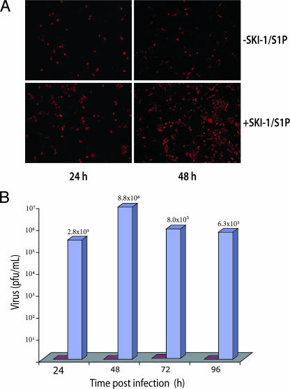
If SKI-1/S1P endoprotease activity was essential for CCHFV growth, then complementation of SKI-1/S1P-deficient cells with SKI-1/S1P should restore infectious virus release. Therefore, SKI-1/S1P-deficient cells were transiently transfected with a SKI-1 expression vector (or a control vector) by electroporation. Twenty-four hours later, transfected cells were infected with CCHFV, and viral antigen synthesis was monitored by IFA. At 24 hpi, no marked difference in the numbers of infected cells was observed in the presence or absence of SKI-1/S1P (Fig. 3A). However, cells transfected with the SKI-1/S1P expression plasmid showed a considerable increase in CCHFV-infected cells by 48 hpi, whereas no increase was observed in cells transfected with an empty vector. These data indicated that both S1P/SKI-1-expressing and nonexpressing cells were susceptible and supported CCHFV initial replication and the likelihood that S1P/SKI-1 was required for release of infectious virus and further virus spread. To examine whether infectious virus was released from these cells, the clarified cell supernatants were collected and virus titers determined by plaque assay. No infectious virus was recovered from cells lacking SKI-1/S1P (Fig. 3B). In contrast, SKI-1/S1P-expressing cells produced high virus titers, with a peak value of 8.8 × 10⁶ PFU/ml, 48 h after the initial infection. These results clearly indicated that SKI-1/S1P was strictly required for infectious CCHFV production.
FIG. 3.
Production of infectious CCHFV from SKI-1/S1P-deficient cells can be rescued by plasmid-expressed SKI-1/S1P. (A) SKI-1/S1P-deficient cells (SRD12B cells) were transiently transfected with control empty vector (top, −SKI-1/S1P) or with SKI-1/S1P-containing vector (bottom, +SKI-1/S1P) by use of a Nucleofector II electroporator, the H-014 program, and cell line Nucleofector solution T (Amaxa, Gaithersburg, MD). These transfected cells were infected 24 hpi and processed at the indicated times postinfection as described in the legend for Fig. 2. No obvious cytopathic effect was observed in the presence or absence of SKI-1/S1P (data not shown). (B) Infections were set up as described above. Infected cell supernatants were collected, clarified, and frozen in liquid nitrogen at the indicated times for subsequent titration by plaque assay on SW13 cell monolayers (22). Red bars indicate viral titers from −SKI-1/S1P-transfected and blue bars from +SKI-1/S1P-transfected cell supernatants.
Secretion of low levels of glycoprotein-deficient viral particles in the absence of SKI-1/S1P.
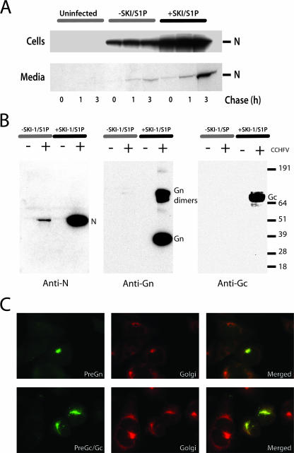
Analysis of other viral systems has demonstrated that disruption of viral glycoprotein cleavage can lead to the formation of virus particles decorated with uncleaved glycoprotein precursors on their surface (4, 5, 16, 25, 29). This situation applies to viral glycoproteins incompletely cleaved by furin-like convertases or to furin-resistant mutants. In contrast, only correctly SKI-1/S1P-processed mature glycoproteins (GP1 and GP2) are incorporated into arenavirus viral particles (12, 13). In order to determine if any viral proteins were secreted from CCHFV-infected cells, we analyzed the cell lysates and supernatants of infected SKI-1/S1P-deficient cells by pulse-chase and immunoprecipitation analyses in the presence or absence of SKI-1/S1P (Fig. 4A). Three to four times less labeled N protein was detected in lysates of SKI-1/S1P-deficient cells than in SKI-1/S1P-expressing cells at all time points examined (Fig. 4A). These findings are consistent with earlier results which demonstrated a lack of CCHFV production and spread in cells lacking SKI-1/S1P (Fig. 3). Nucleoprotein secretion into the supernatant increased over time in the presence or absence of SKI-1/S1P (Fig. 4A). However, five or eight times more N protein was secreted after 1 or 3 h chase, respectively, when SKI-1/S1P-expressing cells were compared with nonexpressing cells. These data indicated that a time-dependent increase in N secretion was observed in the absence of SKI-1/S1P activity but that the amount of N secreted was significantly smaller. As expected, and confirmed previously (27), uncleaved PreGn was detectable in the SKI-1/S1P-deficient cell lysates but not in supernatants (data not shown).
FIG. 4.
Glycoprotein-deficient viral particles are released in the infected cell supernatant of SKI-1/S1P-deficient cells. SRD12B cells stably expressing SKI-1/S1P were produced by transfection of the pIR-SKI-1/S1P vector with Lipofectamine 2000. Two days after transfection, cells were placed in Dulbecco's modified Eagle's medium-F-12 medium supplemented with 5% lipoprotein-deficient fetal bovine serum and penicillin-streptomycin for 14 days to select cells stably expressing SKI-1/S1P. The cell pools growing in the absence of exogenous cholesterol were used in all experiments. (A) SKI-1/S1P-deficient cells (−SKI/S1P) or SKI-1/S1P-deficient cells stably expressing SKI-1/S1P (+SKI-1/S1P) were infected with a multiplicity of infection (MOI) of 5. Twenty-four hpi, cells were pulsed with 100 μCi/ml of [35S]Cys for 30 min and chased for the indicated number of hours, as described previously (22). Triton X-100 was added to a concentration of 1% to cell supernatants prior to the immunoprecipitation with anti-N (5G2) MAb and protein A Sepharose (Amersham). Cell monolayers were solubilized with radioimmunoprecipitation assay buffer and immunoprecipitations carried out as described previously (22). All immunoprecipitated proteins were resolved with a NuPAGE gel system (3 to 8% bis-Tris gels) under reducing conditions. Protein bands were detected using phosphor screens and image digitized and quantitated using a Storm imager (GE Healthcare Biosciences) and ImageQuant software (GE Healthcare Biosciences). At the time of the pulse, we estimated by IFA that ∼15% of the SKI-1/S1P-deficient cells were infected, compared to ∼40% for cells expressing SKI-1/S1P (data not shown). (B) Confluent 175-cm2 flasks of SRD12B cells or SRD12B cells stably expressing SKI-1 were infected with CCHFV with an MOI of 5. Uninfected (−) and infected (+) cell supernatants were changed 24 h after the infection and collected at 48 hpi and viral particles purified as essentially as described previously (1). Purified viral pellets were solubilized in 100 μl of 2× NuPAGE sample buffer and gamma irradiated with 2 × 10⁶ rads prior to electrophoresis. A portion (20 μl) of the reduced samples was analyzed by Western blotting with chemiluminescence to detect CCHFV structural proteins N (9D5 MAb), Gn (Gn ectodomain polyclonal antibody), and Gc (11E7 MAb). The values at right are markers in kilodaltons. (C) SKI-1/S1P-deficient cells were infected and processed by IFA with antibodies 7F5 (PreGn/GP38) or 11E7 (PreGc/Gc) and Giantin (Golgi) from Covance (Berkeley, CA) 7 hpi. The green signal (anti-mouse Alexa 488) is specific for PreGn (top) or PreGc/Gc (bottom) and red (anti-rabbit Alexa 594) for Giantin (Golgi). Red and green digital images were overlaid (merged) to emphasize the colocalization of PreGn and PreGc/Gc with the cis and median Golgi cisternae marker. Images were acquired with a 100× objective.
The preceding data suggested that noninfectious N-containing viral particles are produced even in the absence of PreGn processing. To examine this, CCHFV-infected cell supernatants were pelleted through a sucrose cushion to partially purify any virus particles present, and viral pellets were examined for the presence of N, Gn, and Gc by Western blotting. N protein was detected in viral pellets even in the absence of SKI-1/S1P, albeit to a much lower level than in SKI-1/S1P-expressing cells (Fig. 4B). Despite good detection sensitivity, Gn (37 kDa), Gn dimers (70 kDa) (1), and Gc were observed only in SKI-1/S1P-expressing cells. In addition, no virus glycoprotein precursors (PreGn or PreGc) were detected in any of the virus pellets. These results strongly suggest that in the absence of SKI-1/S1P activity, low levels of CCHFV particles are produced, and that these are essentially naked nucleocapsid cores containing no or undetectable levels of mature Gn, Gc, or glycoprotein precursor forms.
PreGn and PreGc/Gc are localized in the Golgi apparatus.
A hallmark of members of the family Bunyaviridae is the targeting of their surface glycoproteins to the Golgi apparatus, where most viral assembly takes place. To determine if PreGn processing was required for the proper Golgi apparatus localization of the glycoproteins, we analyzed CCHFV-infected cells deficient in S1P/SKI-1 or transfected HeLa cells expressing a S1P/SKI-1 cleavage-resistant M polyprotein mutant (IRLL519) (27). IFA of these cells using PreGn/GP38- or PreGc/Gc-specific monoclonal antibodies (MAbs) showed that these proteins colocalized with a cis and median Golgi cisternae marker (Giantin) in both CCHFV-infected (Fig. 4C) and transfected (data not shown) cells. Since both PreGn and PreGc/Gc were localized in the Golgi apparatus in the absence of mature 37-kDa Gn, we concluded that glycoprotein trafficking was normal even in the absence of S1P/SKI-1 activity.
The fact that glycoprotein precursors were localized in the Golgi apparatus, the site of CCHFV assembly, but failed to be incorporated into particles suggests a selective mechanism for the incorporation of mature Gn and Gc into virions. An analogous situation has been reported for the GPCs of Lassa virus and lymphocytic choriomeningitis virus. The supernatants of SKI-1/S1P-deficient cells infected with these arenaviruses also contained a low abundance of N-containing noninfectious particles deficient in envelope GPC or mature cleavage products (GP1 and GP2) (12, 13). In contrast, it has been demonstrated for numerous other viruses that blockage of furin processing of glycoprotein precursors does not inhibit formation of virus particles (4, 5, 16, 29), suggesting that blockage of virion formation may be limited to glycoproteins cleaved by SKI-1/S1P. While unlikely, we cannot rule out that SKI-1/S1P could impact CCHFV budding in a manner unrelated to its protease activity.
SKI-1/S1P motifs can be identified in the deduced amino acid sequences of the glycoprotein precursors of not only all CCHFV strains sequenced to date but also Dugbe and Hazara viruses, the only other nairoviruses for which sequences are available. Therefore, SKI-1/S1P processing of PreGn is likely a shared feature of members of the genus Nairovirus and infectivity of these viruses may also be dependent on SKI-1/S1P. In contrast, the absence of such predicted sites in other genera of the family Bunyaviridae suggests that their growth is independent of SKI-1/S1P activity. In agreement, we observed that SKI-1/S1P-deficient cells are permissive to Rift Valley fever virus, a member of the genus Phlebovirus (data not shown).
CCHF is a rapidly progressing acute disease associated with high mortality. No vaccine is licensed currently, and limited antiviral therapeutic options exist. The strong dependence of CCHFV (and the hemorrhagic fever-associated arenaviruses) on SKI-1/S1P cleavage for production of infectious virus particles suggests that the development of SKI-1/S1P small-molecule inhibitors may represent a promising avenue for antiviral development.
Acknowledgments
We acknowledge César Albariño and Tom Ksiazek for critical reading of the manuscript and advice. We also thank Louis A. Altamura and Robert W. Doms (University of Pennsylvania, Philadelphia, PA) for technical advice and provision of the Gn ectodomain antipeptide antibody and the CCHFV IbAr10200 codon-optimized M-segment vector (pCAGGS-M). Mouse MAbs specific for N (9D5 and 5G2), PreGc/Gc (11E7), and PreGn/GP38 (7F5) were kindly provided by Jonathan Smith (formerly of the U.S. Army Medical Research Institute for Infectious Diseases [USAMRIID], Fort Detrick, MD). SRD12B and M19 cells were generously provided by J. L. Goldstein (University of Texas Southwestern Medical Center, Dallas, TX) and T. Y. Chang (Dartmouth Medical School, Hanover, NH), respectively. The pIRES2-EGFP plasmid containing human SKI-1/S1P (pIR-S1P/SKI-1) was generously provided by Nabil G. Seidah (Institut de Recherches Cliniques de Montréal, Montréal, Québec, Canada). We also express our gratitude to Cynthia Goldsmith and Angela Sanchez for technical help and biosafety level 4 training.
E.B. was supported by a CCID/ASM postdoctoral fellowship.
The findings and conclusions in this report are those of the authors and do not necessarily represent the views of the funding agency.
Footnotes
Published ahead of print on 26 September 2007.
REFERENCES
- 1.Altamura, L. A., A. Bertolotti-Ciarlet, J. Teigler, J. Paragas, C. S. Schmaljohn, and R. W. Doms. 2007. Identification of a novel C-terminal cleavage of Crimean-Congo hemorrhagic fever virus PreGN that leads to generation of an NSM protein. J. Virol. 81:6632-6642. [DOI] [PMC free article] [PubMed] [Google Scholar]
- 2.Andersson, A. M., L. Melin, R. Persson, E. Raschperger, L. Wikstrom, and R. F. Pettersson. 1997. Processing and membrane topology of the spike proteins G1 and G2 of Uukuniemi virus. J. Virol. 71:218-225. [DOI] [PMC free article] [PubMed] [Google Scholar]
- 3.Beyer, W. R., D. Popplau, W. Garten, D. von Laer, and O. Lenz. 2003. Endoproteolytic processing of the lymphocytic choriomeningitis virus glycoprotein by the subtilase SKI-1/S1P. J. Virol. 77:2866-2872. [DOI] [PMC free article] [PubMed] [Google Scholar]
- 4.de Haan, C. A., K. Stadler, G. J. Godeke, B. J. Bosch, and P. J. Rottier. 2004. Cleavage inhibition of the murine coronavirus spike protein by a furin-like enzyme affects cell-cell but not virus-cell fusion. J. Virol. 78:6048-6054. [DOI] [PMC free article] [PubMed] [Google Scholar]
- 5.Elshuber, S., S. L. Allison, F. X. Heinz, and C. W. Mandl. 2003. Cleavage of protein prM is necessary for infection of BHK-21 cells by tick-borne encephalitis virus. J. Gen. Virol. 84:183-191. [DOI] [PubMed] [Google Scholar]
- 6.Ergonul, O. 2006. Crimean-Congo haemorrhagic fever. Lancet Infect. Dis. 6:203-214. [DOI] [PMC free article] [PubMed] [Google Scholar]
- 7.Gerrard, S. R., and S. T. Nichol. 2007. Synthesis, proteolytic processing and complex formation of N-terminally nested precursor proteins of the Rift Valley fever virus glycoproteins. Virology 357:124-133. [DOI] [PMC free article] [PubMed] [Google Scholar]
- 8.Goldstein, J. L., R. B. Rawson, and M. S. Brown. 2002. Mutant mammalian cells as tools to delineate the sterol regulatory element-binding protein pathway for feedback regulation of lipid synthesis. Arch. Biochem. Biophys. 397:139-148. [DOI] [PubMed] [Google Scholar]
- 9.Hasan, M., C. Chang, and T. Chang. 1994. Somatic cell genetic and biochemical characterization of cell lines resulting from human genomic DNA transfections of Chinese hamster ovary cell mutants defective in sterol-dependent activation of sterol synthesis and LDL receptor expression. Somat. Cell Mol. Genet. 20:183-194. [DOI] [PubMed] [Google Scholar]
- 10.Kondo, S., T. Murakami, K. Tatsumi, M. Ogata, S. Kanemoto, K. Otori, K. Iseki, A. Wanaka, and K. Imaizumi. 2005. OASIS, a CREB/ATF-family member, modulates UPR signalling in astrocytes. Nat. Cell Biol. 7:186-194. [DOI] [PubMed] [Google Scholar]
- 11.Kormelink, R., M. Storms, J. Van Lent, D. Peters, and R. Goldbach. 1994. Expression and subcellular location of the NSM protein of tomato spotted wilt virus (TSWV), a putative viral movement protein. Virology 200:56-65. [DOI] [PubMed] [Google Scholar]
- 12.Kunz, S., K. H. Edelmann, J. C. de la Torre, R. Gorney, and M. B. Oldstone. 2003. Mechanisms for lymphocytic choriomeningitis virus glycoprotein cleavage, transport, and incorporation into virions. Virology 314:168-178. [DOI] [PubMed] [Google Scholar]
- 13.Lenz, O., J. ter Meulen, H.-D. Klenk, N. G. Seidah, and W. Garten. 2001. The Lassa virus glycoprotein precursor GP-C is proteolytically processed by subtilase SKI-1/S1P. Proc. Natl. Acad. Sci. USA 98:12701-12705. [DOI] [PMC free article] [PubMed] [Google Scholar]
- 14.Lober, C., B. Anheier, S. Lindow, H. D. Klenk, and H. Feldmann. 2001. The Hantaan virus glycoprotein precursor is cleaved at the conserved pentapeptide WAASA. Virology 289:224-229. [DOI] [PubMed] [Google Scholar]
- 15.Nakitare, G. W., and R. M. Elliott. 1993. Expression of the Bunyamwera virus M genome segment and intracellular localization of NSm. Virology 195:511-520. [DOI] [PubMed] [Google Scholar]
- 16.Neumann, G., H. Feldmann, S. Watanabe, I. Lukashevich, and Y. Kawaoka. 2002. Reverse genetics demonstrates that proteolytic processing of the Ebola virus glycoprotein is not essential for replication in cell culture. J. Virol. 76:406-410. [DOI] [PMC free article] [PubMed] [Google Scholar]
- 17.Neumann, G., T. W. Geisbert, H. Ebihara, J. B. Geisbert, K. M. Daddario-DiCaprio, H. Feldmann, and Y. Kawaoka. 2007. Proteolytic processing of the Ebola virus glycoprotein is not critical for Ebola virus replication in nonhuman primates. J. Virol. 81:2995-2998. [DOI] [PMC free article] [PubMed] [Google Scholar]
- 18.Raggo, C., N. Rapin, J. Stirling, P. Gobeil, E. Smith-Windsor, P. O'Hare, and V. Misra. 2002. Luman, the cellular counterpart of herpes simplex virus VP16, is processed by regulated intramembrane proteolysis. Mol. Cell. Biol. 22:5639-5649. [DOI] [PMC free article] [PubMed] [Google Scholar]
- 19.Rawson, R. B., D. Cheng, M. S. Brown, and J. L. Goldstein. 1998. Isolation of cholesterol-requiring mutant Chinese hamster ovary cells with defects in cleavage of sterol regulatory element-binding proteins at site 1. J. Biol. Chem. 273:28261-28269. [DOI] [PubMed] [Google Scholar]
- 20.Sakai, J., R. B. Rawson, P. J. Espenshade, D. Cheng, A. C. Seegmiller, J. L. Goldstein, and M. S. Brown. 1998. Molecular identification of the sterol-regulated luminal protease that cleaves SREBPs and controls lipid composition of animal cells. Mol. Cell 2:505-514. [DOI] [PubMed] [Google Scholar]
- 21.Sanchez, A. J., M. J. Vincent, B. R. Erickson, and S. T. Nichol. 2006. Crimean-Congo hemorrhagic fever virus glycoprotein precursor is cleaved by furin-like and SKI-1 proteases to generate a novel 38-kilodalton glycoprotein. J. Virol. 80:514-525. [DOI] [PMC free article] [PubMed] [Google Scholar]
- 22.Sanchez, A. J., M. J. Vincent, and S. T. Nichol. 2002. Characterization of the glycoproteins of Crimean-Congo hemorrhagic fever virus. J. Virol. 76:7263-7275. [DOI] [PMC free article] [PubMed] [Google Scholar]
- 23.Seidah, N. G., S. J. Mowla, J. Hamelin, A. M. Mamarbachi, S. Benjannet, B. B. Toure, A. Basak, J. S. Munzer, J. Marcinkiewicz, M. Zhong, J.-C. Barale, C. Lazure, R. A. Murphy, M. Chretien, and M. Marcinkiewicz. 1999. Mammalian subtilisin/kexin isozyme SKI-1: a widely expressed proprotein convertase with a unique cleavage specificity and cellular localization. Proc. Natl. Acad. Sci. USA 96:1321-1326. [DOI] [PMC free article] [PubMed] [Google Scholar]
- 24.Stirling, J., and P. O'Hare. 2006. CREB4, a transmembrane bZip transcription factor and potential new substrate for regulation and cleavage by S1P. Mol. Biol. Cell 17:413-426. [DOI] [PMC free article] [PubMed] [Google Scholar]
- 25.Strive, T., E. Borst, M. Messerle, and K. Radsak. 2002. Proteolytic processing of human cytomegalovirus glycoprotein B is dispensable for viral growth in culture. J. Virol. 76:1252-1264. [DOI] [PMC free article] [PubMed] [Google Scholar]
- 26.Thomas, G. 2002. Furin at the cutting edge: from protein traffic to embryogenesis and disease. Nat. Rev. Mol. Cell Biol. 3:753-766. [DOI] [PMC free article] [PubMed] [Google Scholar]
- 27.Vincent, M. J., A. J. Sanchez, B. R. Erickson, A. Basak, M. Chretien, N. G. Seidah, and S. T. Nichol. 2003. Crimean-Congo hemorrhagic fever virus glycoprotein proteolytic processing by subtilase SKI-1. J. Virol. 77:8640-8649. [DOI] [PMC free article] [PubMed] [Google Scholar]
- 28.Ye, J., R. B. Rawson, R. Komuro, X. Chen, U. P. Dave, R. Prywes, M. S. Brown, and J. L. Goldstein. 2000. ER stress induces cleavage of membrane-bound ATF6 by the same proteases that process SREBPs. Mol. Cell 6:1355-1364. [DOI] [PubMed] [Google Scholar]
- 29.Zhang, X., M. Fugere, R. Day, and M. Kielian. 2003. Furin processing and proteolytic activation of Semliki Forest virus. J. Virol. 77:2981-2989. [DOI] [PMC free article] [PubMed] [Google Scholar]


