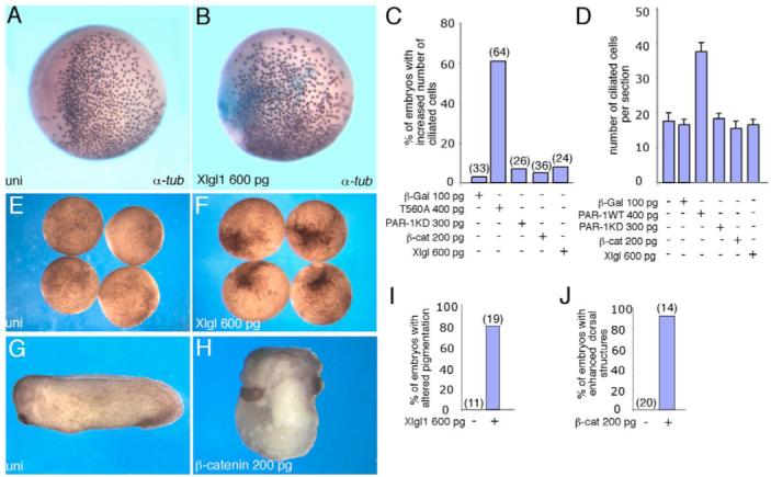
Fig. 4. Lack of effect of β-catenin and LGL on ciliated cell development.

In situ hybridization with α-tubulin probe is shown. For experimental details, see Fig. 1 legend. (A) Uninjected embryo. (B) LGL1 (Xlgl1) RNA has no effect on α-tubulin-expressing cells. (C,D) Quantification of the effects of T560A, PAR1, PAR1-KD, β-catenin and LGL1 RNAs on ciliated cell development, presented as frequencies of affected embryos (C) and numbers of ciliated cells per section (D). In C, numbers of embryos per group are shown above bars. Data are representative of four different experiments. (E,F,I) LGL1 RNA, used in B, altered ectoderm pigmentation in 79% of injected embryos (n=19; F) as compared with uninjected controls (E). (I) Quantification of the results in E and F. (G,H,J) Marginal zone-injected β-catenin RNA dorsalized 92% of injected embryos (n=14), characterized by enlarged head and cement gland and truncated or missing tail (H), as compared with uninjected siblings (G). (J) Quantification of the results in G,H.
