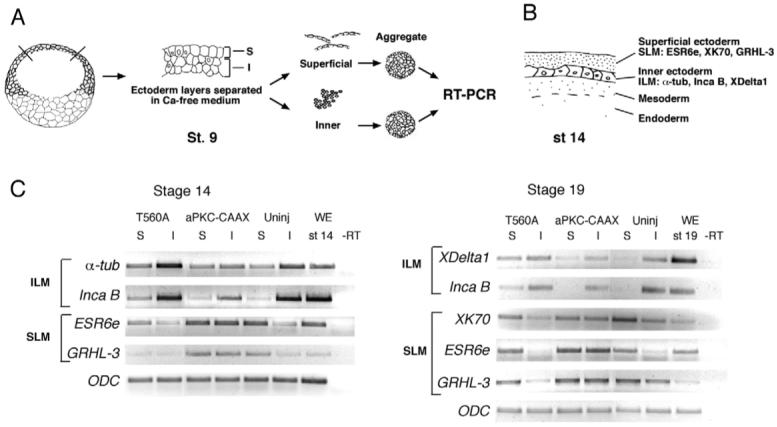
Fig. 7. Opposite effects of PAR1 and aPKC on gene expression in separated ectoderm cell layers.

(A) Experimental scheme for the layer separation assay. Two-cell embryos were injected with T560A or aPKC-CAAX mRNA. Animal pole explants were dissected from injected or uninjected embryos at stage 9, superficial (S) and inner (I) cell layers were separated based on their different abilities to dissociate in a calcium- and magnesium-free buffer and allowed to reaggregate. Cell aggregates were cultured until sibling embryos reached stage 14 or stage 19. RNA was prepared from aggregate lysates and analyzed by semi-quantitative RT-PCR. (B) A schematic section of the frog embryo at stage 14 shows superficial and inner layers of non-neural ectoderm with distinct sets of molecular markers. SLM, superficial layer markers; ILM, inner layer markers. (C) Stage 14 aggregates: T560A RNA upregulates the inner layer markers α-tubulin and inca B and downregulates the superficial layer markers ESR6e and grhl3 in both inner and outer layer explants. aPKC-CAAX has a complementary effect. Stage 19 aggregates: T560A RNA upregulates the inner layer markers XDelta-1 and inca B and downregulates XK70 in the superficial layer. aPKC-CAAX downregulates XDelta-1 and inca B in the superficial layer and upregulates XK70, ESR6e and grhl3 in inner cell aggregates. ODC is a control for loading. Uninj, no RNA injection; WE, whole embryo; −RT, no reverse transcriptase control. The analysis of two representative sets of cDNAs from several independent experiments is shown.
