As June 9 turned into June 10, crowds kept surging toward the Hachik-o area west of Shibuya station. Shibuya has long been a boisterous focal point for Tokyo youth, but this was something else. Japan had just won their Group H match against Russia, and Tokyo was celebrating as if the World Cup was theirs. The honking horns and chants drowned out the megaphones of the massively outnumbered police. Yet, by the standards of most countries, an extraordinary level of order prevailed. The huge crowd was content to take over the intersection, leaping and cavorting, only when the pedestrian lights were green. A change to red, a few police whistles, and the multitude moved politely to the side.
Figure .
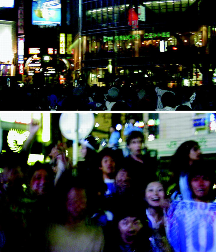
Celebration in Tokyo. But will youthful energy be enough to change the scientific career structure in Japan?
That cultural ambivalence—setting youthful exuberance against an obedience toward elders and authority—is being played out elsewhere in Japanese society, notably in the world of cell biology. Younger scientists are demanding, and in some cases receiving, more autonomy at an earlier stage of their careers. Yet, just as the crowds in Shibuya are unlikely to match past celebrations in Rome or Rio de Janeiro, it seems that Japanese science is under no threat, any time soon, of becoming another United States.
The virtue of stability
If the United States is the land of opportunity and immigration, with a science funding and career policy to match, then Japan is much closer to some European countries. As in some of these countries, with their longer histories and lower levels of immigration, hierarchy takes a more prominent role. Within the laboratory of a full professor, there are students (who, more often than not, do not receive financial support), joshu (a term that covers the United States equivalent of both postdoctoral fellow and assistant professor), and usually a single associate professor. In total, most Japanese researchers spend at least eight or ten years in a single lab after completing their Ph.D. Some of that time they may be called an associate professor, but they are still beholden to the full professor in charge of the laboratory.
Just how beholden they are is dependent on the individual relationship between the junior scientist and full professor. At stake is a long list of rights, each one of which is largely under the control of the full professor. That list includes the ability of the joshu or associate professor to acquire their own grants, use all or most of their own grant money, obtain space for additional personnel or specialized equipment, hire a technician, attend and present at meetings, and assume senior authorship on papers. In the United States, many of these issues are either assumed or negotiated during a job search, when the junior investigator (if he or she has several job offers) has some power.
But the Japanese system is not without its advantages for younger researchers. Setting up a new laboratory in the United States can be a traumatic experience for even the most successful of postdocs. So many new responsibilities—finding a job, moving, getting funding, learning how to be a boss, and directing research projects without any oversight—come on top of one another. In Japan, many of these skills are learned in a more gradual manner, or with the guiding hand of a full professor or the other joshu in the same laboratory. Furthermore, the joshu have an assured position that, in theory, lasts as long as the full professor has a laboratory.
In this more nurturing environment, research can be more daring. “The U.S. has flexibility and opportunity,” says Nobutaka Hirokawa (Tokyo University, Tokyo, Japan). “But we have stability.” He says that the projects tackled by joshu or associate professors can be riskier and have longer-term pay-offs because of the high job security. In his own laboratory, he says that only the stability of the personnel has allowed him to build up a laboratory that can tackle such a unique combination of biophysics, structure, and basic biology.
And the inevitability of change
But even if the current system is wise, change is coming. Earlier Japanese administrations threatened to convert universities into “Independent Administrative Institutions” (IAIs), and now the government of Prime Minister Junichiro Koizumi is planning even more extensive changes. University reform fits right in with Koizumi's plans to privatize everything from road construction to the management of post-office savings accounts.
Many of these reforms are in question as Koizumi's approval rating drops and his own party turns against him. But not all reform targets are equal. There is the savings account system—a handy slush fund favored by powerful politicians—and then there are universities. The latter have far fewer powerful people lobbying to prevent their reform, and there is a widespread sentiment that some form of change is needed.
One radical proposal, which was announced on March 26th of this year, involves stripping all university employees of their status as public servants. As an independent agency, each university would now receive a block of money that could be distributed with a greater degree of autonomy. Salaries and the rules attached to them could be determined locally. This would free professors from public service rules that have prevented them from founding, spending time at, or being paid by biotech companies. In addition, university pay scales may be made more flexible, perhaps increasing the mobility of a notoriously static workforce by allowing recruiting between universities and from overseas.
Figure .
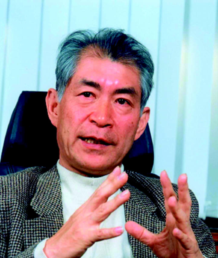
Tasuku Honjo, the master of class switch recombination, believes faculty will be hired at younger ages.
The age at which researchers become independent will also be determined locally. “I think probably all universities will encourage younger faculty to be independent,” says Tasuku Honjo (Kyoto University, Kyoto, Japan). “I think that's a good thing, and it's a good way to attract bright young people from other universities.”
Those changes may come just in time. According to one Japanese researcher based in the United States, until recently a Japanese researcher doing a postdoc in the United States would inevitably return to Japan to take up a research associate position in a full professor's laboratory. But now, the researcher says, “young Japanese people are getting good positions in the U.S. Japan is now starting to lose many good talents, and they have to start thinking about it seriously.”
The test case
Figure .
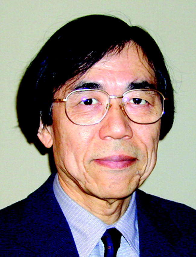
Having founded the field of cadherin biology, Masatoshi Takeichi is now directing a new style of institute, the CDB.
The hiring of younger independent faculty is already underway outside of the university system. A notable example is at the RIKEN Center for Developmental Biology (CDB) in Kobe, Japan, where Masatoshi Takeichi (formerly at Kyoto University, Kyoto, Japan) is now Director. “Japanese universities have a very hierarchical system,” he says. “But these labs [at the CDB] are completely independent.”
The CDB system pairs this early hiring with a reevaluation after five years, in a process similar to the tenure review in other countries. “That's quite a revolutionary situation in Japan,” says Yasushi Hiraoka (Kansai Advanced Research Center, Kobe, Japan). “If it's a success, other institutes may follow.” Other researchers are skeptical, however, as at other RIKEN institutes in the past the organization has failed to follow through on early promises of strict evaluation and firing.
Figure .
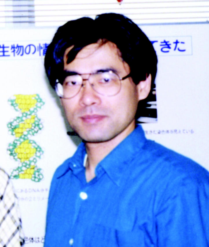
Yasushi Hiraoka feels that the CDB may be the start of a new trend.
The CDB is also an example of a second trend in Japanese biological science: the funding of large, concentrated research efforts. Although the individual laboratories at the CDB are standard in size, the institute itself, just months after its official opening, boasts 27 laboratories. The funds for all those shiny facilities were distributed by a few powerful government bureaucrats. As CDB Director, Takeichi is in charge of this very “top-down” funded Institute, even though he remains a fierce proponent of the peer-review system of “bottom-up” funding practiced in the universities.
Many young researchers in Japan welcome the arrival of the CDB. But top-down funding has also been on the increase in individual laboratories, and here it has provoked concern. “Some people who are productive get even more,” says one researcher. “There is a huge distance between the rich and poor scientists. Famous, important people—too senior—are doing all the judging.” The result, says the researcher, is that “it's harder for young people to get grants, and it's harder to get grants in smaller areas.”
Cultural considerations
The liberation (or desertion) of universities by the Japanese government means that the potential for local institutions to undertake large initiatives like the creation of the CDB should be greater. But change may not come so quickly.
“Japanese faculty tend to reach a consensus, instead of one person taking leadership and responsibility,” says Hiraoka. “We need a bureaucratic effort to persuade people.”
The hiring of younger faculty may run up against this and other cultural considerations. “We cannot import the overseas system directly, because of cultural differences,” says Toshio Yanagida (Osaka University, Osaka, Japan). For example, he says, “even if young scientists get huge money, they cannot get great jobs.”
The reason? “Human relationships between old people and young people are still important in Japan,” he says. “Even if the government financially supports the young scientists, they have to organize these human relationships.”
In the end, says Yanagida, “the university is a member of Japanese society.” It is not yet clear how far that society—with its culture based on both wildly imaginative innovation and a deep conservatism—is willing to follow Koizumi in his quest for openness, accountability, and efficiency. And then there is the risk that comes with any change. As Honjo points out, not all universities may survive the reforms. “Freedom is nice,” he says, “but this is also freedom to collapse.”
Probably the only sure thing is that the conflicts, even if unspoken, will continue. Of course, such conflicts are not restricted to science. When I returned to my hotel room in Shibuya, the joyous scenes from outside were being shown on the late-night news. At their conclusion, the elderly newscaster shifted uncomfortably and disapprovingly in his immaculate suit and said a tight-lipped, “Hai; So desu” (loosely translated as “Yes, well, there it is”), before moving onto the next news item. The revolution in Japan may be televised, but not with any great enthusiasm. ▪
The antibody of death
Kyoto University has a number of researchers whose careers started off with an extraordinary bang. Eisuke Nishida isolated MAP kinase, Shin-Ichi Nishikawa (now moving to the RIKEN Center for Developmental Biology in Kobe, Japan) isolated stem cells, and Shuh Narumiya found that his favorite toxin shut down a certain protein called Rho, thus halting cytokinesis and the formation of actin stress fibers and focal adhesions.
Figure .
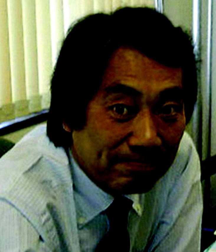
Shin Yonehara
Shin Yonehara, meanwhile, characterized an antibody to a peculiar protein called Fas. He was looking for an antibody that would inhibit the interferon pathway, but his antibody killed cells outright. Yonehara was intrigued. But “at that time,” he says, “almost all researchers did not believe this phenomenon,” and the antibody sat on a shelf for five years. Eventually, Yonehara dusted the project off, found that the killing was a specific, regulated process rather than nonspecific toxicity, and published a paper that has now been cited over 1,100 times. “My curious antibody,” he says, “turned out to be meaningful.”
The Fas apoptotic pathway has now been well picked over, but Yonehara thinks there are exciting branches still to be discovered. There are distinct differences between mice lacking Fas (which are defective in apoptosis, and get cancer and autoimmune diseases) and mice lacking downstream components such as caspase 8 or Fadd (which die early with developmental problems). This suggests that death and developmental pathways may be talking to one another in yet unimagined ways.
“Many researchers think the analysis of Fas signaling is finished, but I think a new phase is coming now,” says Yonehara. “I'm very excited, as much as the time when I found Fas.” ▪
References:
Yonehara, S., et al. 1989. J. Exp. Med. 169:1747–1756.
Imai, Y., et al. 1999. Nature. 398:777–785.
How to make meiosis
Yoshinori Watanabe has almost single-handedly explained why meiosis I—the business end of meiosis—is different from both meiosis II and its close relative mitosis. Only in meiosis I do homologous chromosomes undergo reductional segregation, and only in meiosis I do sister chromatids stay together. The fission yeast protein that defines this unusual segregation behavior, according to Watanabe, is Rec8.
Figure .
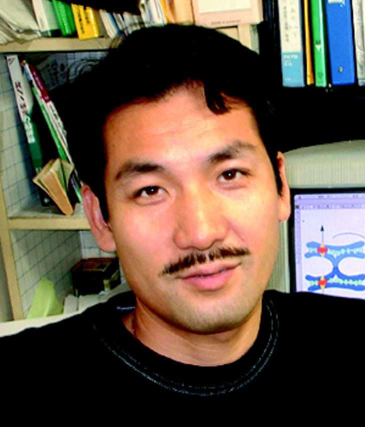
Yoshinori Watanabe
Watanabe has worked on Rec8 first as a postdoctoral fellow with Paul Nurse (ICRF, London, England) and more recently as an associate professor in the laboratory of Masayuki Yamamoto (Tokyo University, Tokyo, Japan). The Rec8 cohesin protein is a meiotic counterpart to the mitotic cohesin Rad21/Scc1. But, whereas Rad21/Scc1 only binds to the outer, repetitive sequence areas of the centromere, Rec8 also binds to the inner, unique sequence elements. This Rec8 is needed to make the two sister kinetochores attach to the same spindle pole and to keep them together throughout meiosis I.
If Rec8 is absent, sister chromatids undergo an equational (mitotic-like) segregation during meiosis I. Watanabe found that the same was true if meiosis was triggered in G2 cells, unless Rec8 expression was also delayed in these cells. Thus, the expression of Rec8 during a preceding S phase is what defines the identity of meiosis I. ▪
Reference:
Watanabe, Y., et al. 2001. Nature. 409:359–363.
Brain circuits by ablation
To most of us, the brain is too vast and impenetrable for sensible study, but Shigetada Nakanishi (Kyoto University, Kyoto, Japan) is not most of us. After cloning some of the major excitatory glutamate receptors used in the nervous system, Nakanishi is now ablating individual cell types to decipher the wiring logic underlying everything from vision to motion.
Figure .
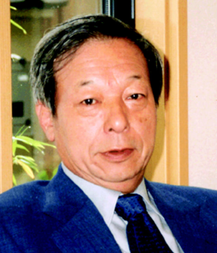
Shigetada Nakanishi
The ablation process involves transgenically expressing a receptor in a particular cell type, and then adding a complex of antireceptor antibody fused to a toxin. With this technique Nakanishi has, for example, demonstrated that starburst amacrine cells in the retinal network help to generate signals responsive to light moving from one direction but not another.
He has also proposed a logical circuit that might help the cells to achieve this directional selectivity. In this scheme, cells exposed to light from the nonfavored direction first send a signal to an inhibitory starburst interneuron. By the time the original neuron sends a direct positive signal to its final target, the interneuron has already shut things down. But if light is moving in the favored direction, the impulse in the sensing cell first fires off the direct, positive signal. Only when the impulse gets further down the axon is the indirect (and ultimately negative) signal sent, and by then the positive signal has already had its effect.
Nakanishi has used similar experiments to propose long-range circuits involved in motion and other higher brain functions. He is currently measuring direct readouts of the electrophysiology to confirm some of these circuit theories. ▪
Reference:
Yoshida, K., et al. 2001. Neuron. 30:771–780.
Outward growth
Tadashi Uemura (Kyoto University, Kyoto, Japan) is a busy man. He is interested in how cells decide to make actin-based outgrowths, but he is investigating not one but three major aspects of this process.
Figure .
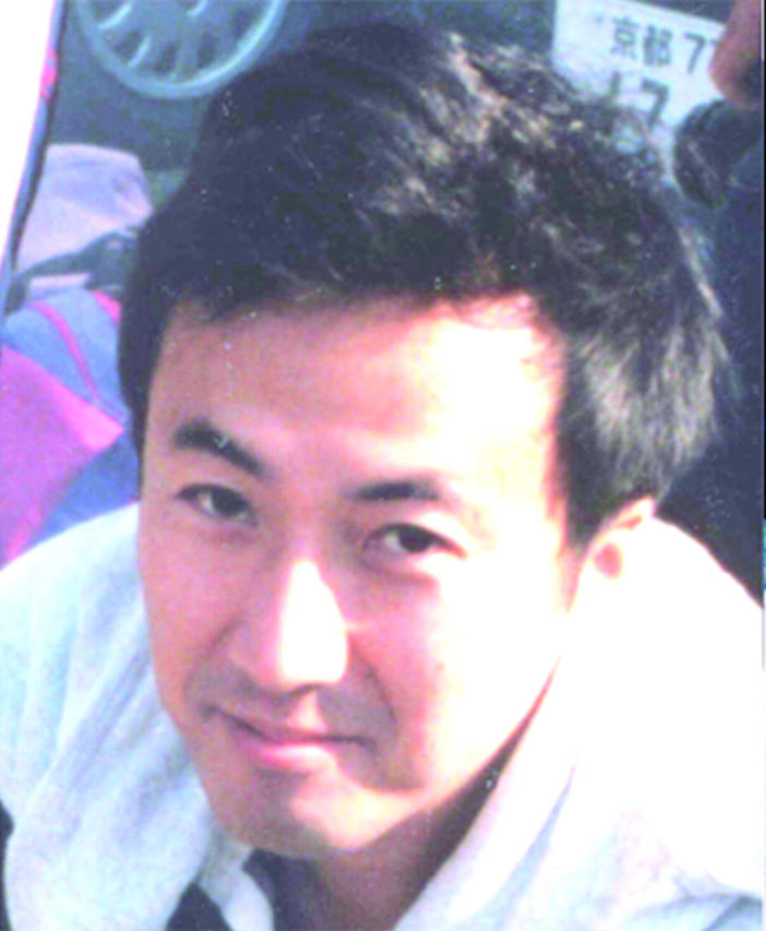
Tadashi Uemura
The initial step is the decision about where an outgrowth will be made. Uemura has found that Flamingo (Fmi), a seven-pass transmembrane cadherin, is necessary to restrict fly hairs to the distal edge of wing cells. Uemura is now working out the complicated interplay between this adhesion molecule and both the Frizzled extracellular signal and Dishevelled intracellular signal in deciding this problem of planar cell polarity. (Another set of polarity determinants is being studied by Fumio Matsuzaki [now at the RIKEN Center for Developmental Biology, Kobe, Japan], who has found that Prospero, Miranda, Lethal giant larvae [Lgl] and Lethal discs large [Dlg] are all required on the basal side of neural stem cells to direct asymmetric divisions.)
Figure .
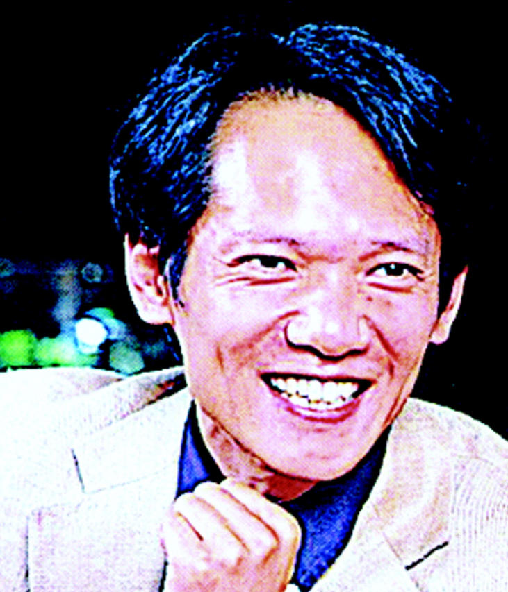
Fumio Matsuzaki
Correct outgrowth of the fly hair requires Slingshot (SSH), which Uemura has found is a phosphatase that acts on ADF (actin depolymerizing factor)/cofilin. If SSH is lost, the inhibitory LIM kinase phosphorylation of cofilin remains in place, and cofilin can no longer depolymerize actin, leading to an overabundance of disorganized actin filaments.
The factors controlling the extent and shape of outgrowths are even more obscure. Uemura is investigating this process in neurons. He hopes to understand how dendritic trees attain their characteristic shapes, and what prevents certain dendritic trees from crossing each other. ▪
References:
Niwa, R., et al. 2002. Cell. 108:233–246.
Oshiro, T., et al. 2000. Nature. 408:593–596.
Microscopic motor mechanics
Shinji Kamimura's work has, literally, a solid foundation. His microscope is seated on a 2 m × 2 m × 2 m concrete block that is located in, but vibrationally isolated from, the basement of a Tokyo University research building. He uses the microscope to push the limits of sensitivity in detecting motion of flagellar dynein motors.
Figure .
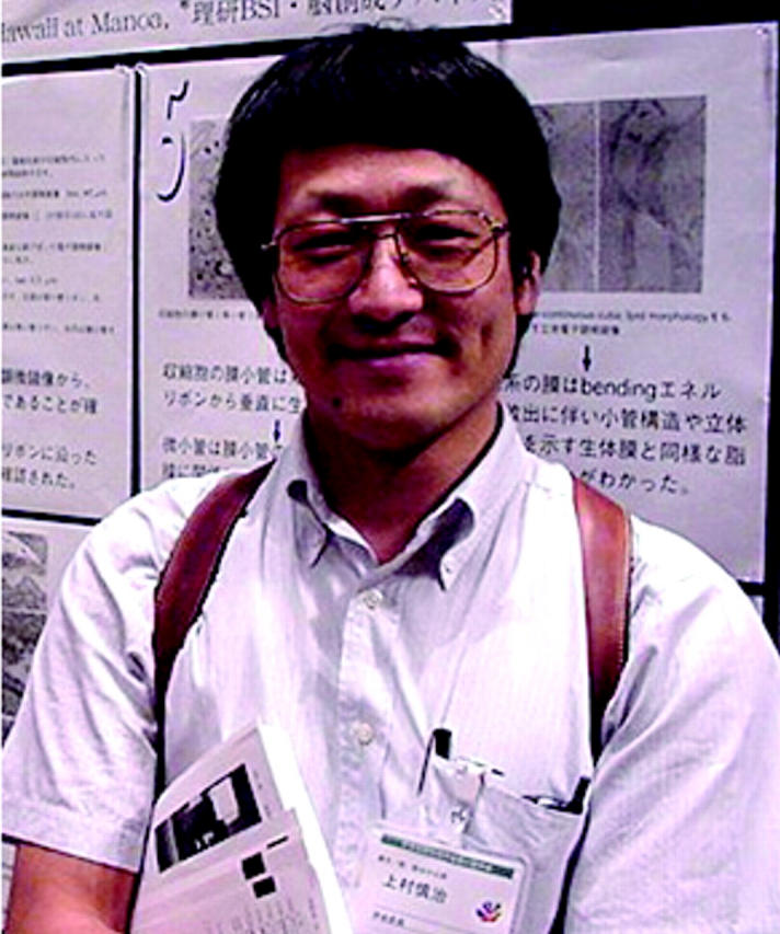
Shinji Kamimura
The theoretical resolving power of a light microscope is ∼200 nm; with image-processing techniques, this can be pushed to ∼50 nm. But Kamimura has used quadrant photodiode detection of attached microbeads to push the limit below the nanometer mark. Motors displace the attached microbeads, and bead movement is detected as a change in light reaching each of the four quadrants of a photodiode. (Bead movement can also be detected by attaching a probe from an atomic force microscope. The probe produces an electrical current when it is deformed by bead movement.)
Kamimura has published a study at 1-nm and 1-kHz resolution, and is now aiming for 0.1 nm and 1 kHz. Dynein motion occurs at ∼100 Hz, so 1-kHz sampling should give him a good picture of the inner workings of a flagellum.
In a nearby laboratory at Tokyo University, Yoshikazu Suzuki and Kazuo Sutoh have been concentrating on the motions of myosin. They attached GFP and BFP to the amino- and carboxy-terminal ends of a myosin head, respectively, and looked for changes in the level of fluorescence resonance energy transfer (FRET) between the two protein domains. The BFP is attached to the motor protein's lever arm, so as the protein went through its ATP hydrolysis and release cycle the BFP swung relative to the GFP. This increased or decreased the level of FRET.
Figure .
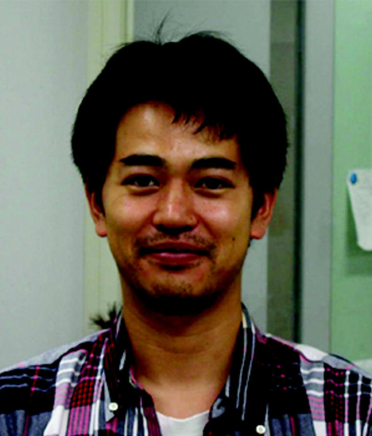
Yoshikazu Suzuki
Suzuki (now at the Biomolecular Engineering Research Institute, Suita, Osaka) and Kamimura have now set up a microscope that can detect an extensive spectrum from a single fluorescent protein. Thus, each motor protein is represented not by a discrete dot of one wavelength, but by a streak running from high to low wavelengths. As the motor moves, the researchers hope to detect a fluctuation in the ratio of high-to-low wavelength emissions. This would be a readout of the increase and decrease in FRET during swings of the lever arm, and thus constitute a visualization of the inner workings of a myosin motor in action. ▪
References:
Suzuki, Y., et al. 1998. Nature. 396:380–383.
Suzuki, Y., et al. 2002. FEBS Lett. 512:235–239.
Kinesins for every occasion
Kinesins first appeared to Nobutaka Hirokawa as short leg-like structures on vesicles in electron microscope (EM) images. At Tokyo University (Tokyo, Japan), he went on to isolate and characterize multiple kinesin genes and proteins, including those that transport synaptic vesicles, mitochondria, protein complexes needed for the cilia that determine left/right asymmetry, and scaffolding proteins for neurotransmitter receptors and the mannose-6-phosphate receptor.
Figure .
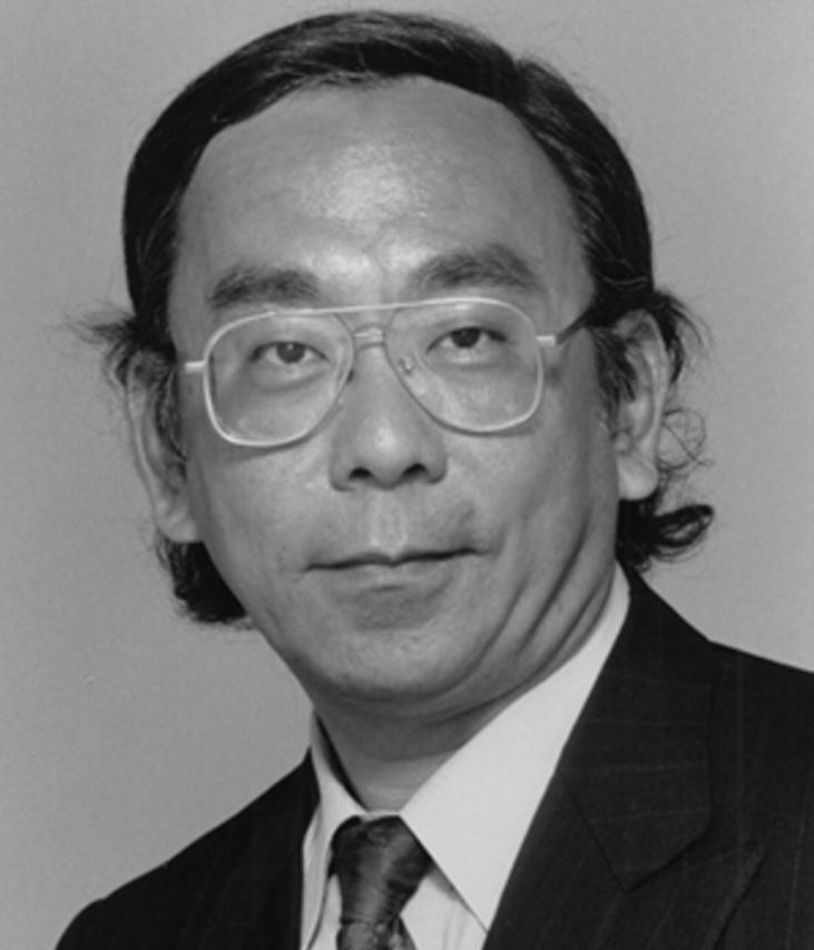
Nobutaka Hirokawa
He has also ventured back into structural analysis. Using cryo-EM and x-ray crystallography, he has developed a model for the biased Brownian motion of both the monomeric kinesin motor KIF1A and, by extension, that of other kinesin motors. He believes that a lysine-rich loop of KIF1A grabs onto the flexible carboxy-terminal tail of tubulin. A rotation of the protein then biases the protein so that the lysine-rich loop will tend to contact the next tubulin monomers in front of it. A similar nucleotide-induced rotation may underlie the motion of other kinesin motors. ▪
Reference:
Kikkawa, M., et al. 2001. Nature. 411:439–445.
Random proteins; random brains
When Toshio Yanagida was young, he judged the challenges of computer science as insufficient. “The principles of the operation of the transistor were known at that time,” he says. “I wanted to research new machine principles. I wanted to study the molecular machine.”
Figure .
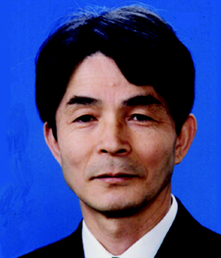
Toshio Yanagida
The molecular machine of choice was the muscle motor myosin. Yanagida was one of the first in Japanese bioscience to get hefty funds for a single project, and he used that money to develop microscopes and force-detection equipment with unprecedented sensitivities.
When all that technical power was focused on myosin, the results were unexpected. “The underlying mechanism of biological machines is essentially different from [that of] man-made machines,” says Yanagida. The key realization was that biological machines operate at energies similar to thermal noise. “Whereas man-made machines operate very accurately, and at much higher energies,” says Yanagida, “we have found that biological machines do not overcome noise but instead use it.”
Yanagida believes that myosin moves because it is buffeted by Brownian motion, and the only function of the ATP hydrolysis cycle is to bias that movement in one particular direction. This idea was originally heretical, but has recently been finding more support in the motor community.
The motor studies continue at Osaka University (Osaka, Japan), but Yanagida now has a new interest based at the Kansai Advanced Research Center (KARC, Kobe, Japan). He is moving from individual motor proteins to entire human brains, but believes that similar principles will be sustained. “There is a big gap between a motor protein and a human brain,” he says. “But biological systems are composed of molecular machines. In such a dynamic system, fluctuation of the elements will play an essential role.”
Figure .
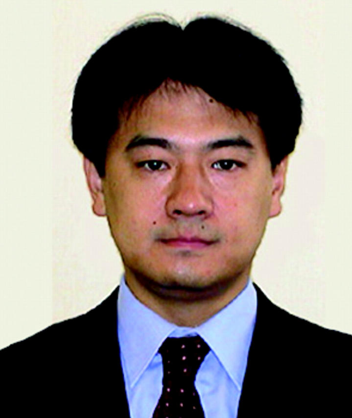
Tsutomu Murata
Initially he is studying a set of visual stimuli that can be seen in one of two ways. The Necker cube, for example, can be interpreted as projecting out of the paper and to the right, or into the paper and to the left. These mutually exclusive interpretations of an unchanging single object can be reported by a human subject as they alternate in the person's consciousness.
He feels that the pattern of this alternation will mirror the situation with myosin. “There is a stochastic process in consciousness,” he says. “I am sure that in the brain system that fluctuation plays an essential role.”
Tsutomu Murata, who is working on the brain project, thinks that some form of noise may be necessary for the brain to do its job properly. “To have a coherent percept, you need a very flexible ability to bind,” he says. “Using fluctuation, you can try various potential combinations of the information. Without significant fluctuations your percepts are fixed.”
Yanagida's proposals in brain science seem certain to be as controversial as his earlier work in myosin movement. But that won't stop him from aiming for the ultimate goal with which he entered science. “I am serious,” he says. “My dream is to find the emerging principle of the brain-style computer.” ▪
Reference:
Tanaka, H., et al. 2002. Nature. 415:192–195.
Rings of cleavage
It's old news that an actomyosin ring squeezes one cell into two during cytokinesis, but the details of that process still remain obscure. Issei Mabuchi (Tokyo University, Tokyo, Japan) has been taking a closer look with fission yeast cells and frog eggs.
Figure .
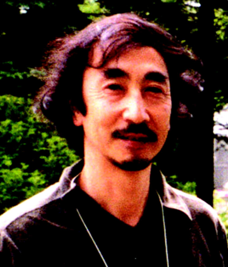
Issei Mabuchi
His group has found that myosin-II dots appear first at the center of a fission yeast cell, and that the dots then convert to a ring. Accumulation does not require F-actin, and similar myosin dot accumulation occurs during cleavage furrow formation of frog eggs.
Actin accumulates next. An actin aster forms in the central cortex of the fission yeast cell, and one actin cable extends from the aster to encircle the cell, in a process that requires formin proteins possibly downstream of Rho. Other actin cables then coalesce with the primary cable.
Now that Mabuchi has a sequence of events, he is looking for the mechanisms that control those events. He believes that accumulation of myosin dots requires myosin dephosphorylation, which liberates myosin from an intramolecular inhibition. He is also looking after dot formation to determine which actin-modulating proteins function in actin cable formation. ▪
Reference:
Arai, R., and I. Mabuchi. 2002. J. Cell Sci. 115:887–898.
Loosening the tight junctions
Shoichiro Tsukita's discovery of first occludin and then claudins at tight junctions (TJs) has allowed a molecular analysis of these structures, which are responsible for sealing one tissue compartment from another. Now, mouse knockouts are helping him to determine how TJs are used to mediate the barrier function needed in a particular tissue.
Figure .
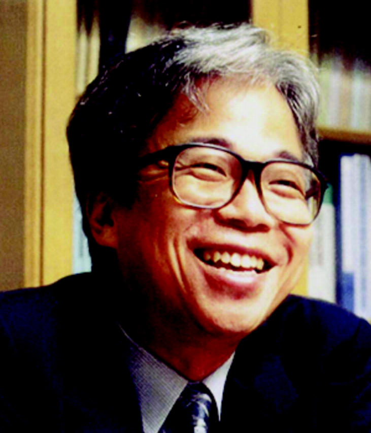
Shoichiro Tsukita
Recently, for example, Tsukita and Mikio Furuse have found that TJs are required in the epidermis to prevent dehydration. TJs were thought to be essential only in simple epithelia such as the gut, rather than in the stratified epithelium of the skin. Tsukita is also interested in claudin function in worms, and in determining whether any of the claudins are necessary for establishing the blood–brain barrier, which is responsible for keeping most drugs from accessing the central nervous system.
Reference:
Furuse, M., et al. 2002. J. Cell Biol. 156:1099–1111.
Everybody can clone
Teruhiko Wakayama (RIKEN Center for Developmental Biology, Kobe, Japan) has a simple recipe for mouse cloning. First, you extract a nucleus. “Not so difficult,” he says. Then you inject that nucleus into an enucleated oocyte. “A little bit difficult,” he says.
Figure .
Teruhiko Wakayama
But compared with some other things, cloning is Wakayama's preferred pursuit. “Molecular biology, I cannot see; so I don't like molecular biology,” he says. “I like micromanipulation because I can see it.”
He pioneered the micromanipulation method of cloning at the University of Hawaii (Honolulu, HI) with Ryuzo Yanagimachi. “I like many science fiction [stories],” says Wakayama. “So I like cloning.” But Yanagimachi had no interest in cloning experiments, and initially Wakayama set aside his ideas. “Six years ago I believed cloning was impossible,” he says.
But then Dolly came along. Cloning was back on the table. Still, Yanagimachi was reticent. “He said you cannot stay here if you do cloning experiments,” says Wakayama. “So I did [them] in secret.” When Cumulina, the first cloned mouse, arrived, everything came out into the open. “After that [Yanagimachi] got wildly excited and very much supported and helped me,” says Wakayama.
The initial success has been tempered somewhat by the hard, and largely unsuccessful, slog ever since, in which Wakayama has been attempting to increase the efficiency of cloning. He has tried using nuclei from different cell types, adding various chemicals (DMSO helps somewhat), and activating oocytes at different times or only after serial transfer. But the results have barely shifted. “In 1997 our success rate was 2%,” he says. “Now, in 2002, the success rate is still the same. Sometimes I think it's impossible.”
The primary difficulty with cloning efficiency, which may also explain the widespread placental abnormalities and birth defects seen in cloning experiments, has been ascribed to one of two problem areas. Either the DNA is not being subjected to the correct reprogramming—a resetting of the DNA that normally occurs during sperm and egg development—or there are problems with imprinting.
If the latter is true, the problems may be harder to fix. Imprinting involves differential marks (leading to differential expression) on maternal versus paternal DNA, and the likelihood of being able to fix this complex pattern is remote when dealing with a complete, diploid set of chromosomes. Reprogramming, however, might be tractable. As yet, neither Wakayama nor other cloning researchers can be sure which problem is the primary one.
For now, Wakayama is still pushing the technical issues, and may look at possible reprogramming factors. Either approach will take a lot of mice. Luckily, the very new RIKEN institute has a vast, high-tech mouse facility, with robots that collect mouse cages for automatic cleaning and replenishing. As one of the robots suddenly jerks into motion and drags its cargo around a corner, Wakayama turns to watch it. “That's great,” he says. “I've never seen that before.” ▪
References:
Wakayama, T., et al. 1998. Nature. 394:369–374.
Wakayama, T., et al. 2001. Science. 292:740–743.


