Abstract
The Sec1 family of proteins is proposed to function in vesicle trafficking by forming complexes with target membrane SNAREs (soluble N-ethylmaleimide-sensitive factor [NSF] attachment protein [SNAP] receptors) of the syntaxin family. Here, we demonstrate, by using in vitro binding assays, nondenaturing gel electrophoresis, and specific neurotoxin treatment, that the interaction of syntaxin1A with the core SNARE components, SNAP-25 (synaptosome-associated protein of 25 kD) and VAMP2 (vesicle-associated membrane protein 2), precludes the interaction with nSec1 (also called Munc18 and rbSec1). Inversely, association of nSec1 and syntaxin1A prevents assembly of the ternary SNARE complex. Furthermore, using chemical cross-linking of rat brain membranes, we identified nSec1 complexes containing syntaxin1A, but not SNAP-25 or VAMP2. These results support the hypothesis that Sec1 proteins function as syntaxin chaperons during vesicle docking, priming, and membrane fusion.
Keywords: membrane fusion, synaptic vesicles, exocytosis, SNARE, synapse
Introduction
Two of the most intensively studied membrane transport processes are the fusion of Golgi apparatus-derived vesicles with the plasma membrane in yeast and the secretion of neurotransmitter from the mammalian presynaptic nerve terminal ( Bennett and Scheller 1994; Jahn and Südhof 1994; Rothman 1994). These membrane fusion events are mediated by a set of structurally and likely functionally related proteins called SNAREs (soluble N-ethylmaleimide-sensitive factor [NSF] attachment protein [SNAP] receptors; Bennett 1995). These proteins include vesicle-associated membrane proteins (VAMP) 1 and 2 (synaptobrevin), a synaptosome-associated protein of 25 kD (SNAP-25), and syntaxin1A and B in mammals; and their yeast orthologues Snc1 and 2, Sec9, and Sso1 and 2, respectively ( Bennett and Scheller 1994; Brennwald et al. 1994). The pairing of SNAREs across membranes to form four-stranded helical bundles is a late event in the fusion process, perhaps driving the actual mixing of the lipid bilayers ( Poirier et al. 1998; Sutton et al. 1998; Weber et al. 1998; Chen et al. 1999).
In addition to SNAREs, a critical component of the fusion process is the Sec1p protein, known in mammals as munc18, nSec1, or rbSec1 ( Hata et al. 1993; Garcia et al. 1994; Pevsner et al. 1994b). This soluble protein is critical for vesicle trafficking in yeast and is proposed to play an important role in the exocytosis process. Loss of Sec1 function results in an accumulation of vesicles at specific steps in secretion ( Novick and Scheckman 1979; Ossig et al. 1991; Cowles et al. 1994), and overexpression of Sec1 in Drosophila melanogaster results in a reduction of synaptic transmission ( Schulze et al. 1994; Wu et al. 1998). Biochemical studies conducted with the mammalian proteins showed that nSec1 from brain extracts, bacterially expressed recombinant, or in vitro translated nSec1, binds directly to syntaxin ( Hata et al. 1993; Garcia et al. 1994; Hodel et al. 1994; Pevsner et al. 1994b). Further investigations demonstrated that when syntaxin is bound to nSec1, the binding to either VAMP or SNAP-25 is inhibited ( Pevsner et al. 1994a).
A model for Sec1/syntaxin function was suggested from an early study demonstrating that binding of the NH2-terminal region of syntaxin to its own COOH-terminal H3 domain resulted in inhibition of the interaction with VAMP and SNAP-25 ( Calakos et al. 1994). It was proposed that syntaxin could adopt a closed conformation to which Sec1 bound and stabilized, inhibiting syntaxin from interacting with the other SNAREs ( Pevsner et al. 1994a). Further, it was proposed that interactions with Rab proteins or their effectors were responsible for dissociating nSec1 from syntaxin, allowing syntaxin to “open up” and interact with the SNAREs. Over the last several years, a variety of studies have confirmed and extended these initial results ( Dulubova et al. 1999; Peterson et al. 1999). For example, nuclear magnetic resonance studies of the NH2-terminal domain of syntaxin revealed a three-helical structure, containing a surface groove to which the COOH-terminal H3 domain of syntaxin might bind in forming the closed conformation ( Fernandez et al. 1998; Dulubova et al. 1999).
In contrast to the binding and structural studies performed with the brain proteins, a recent investigation of the interaction of yeast Sec1p with SNAREs came to different conclusions ( Carr et al. 1999). In their study, immunoprecipitations and binding studies lead the authors to conclude that Sec1p binds to the heterotrimeric core fusion complex comprised of Snc1p, Sec9p, and Sso1p, not to monomeric Sso1p as would be expected from the mammalian studies. Importantly, however, the complexes under investigation were not purified to homogeneity. Instead, the experiments were performed with extracts of yeast and were analyzed by immunoblotting. Thus, it is possible that the biochemical associations observed were not direct protein–protein interactions, but were mediated through intervening molecules. The alternative explanation is that there is a fundamental difference between the yeast and mammalian sec1 proteins, in spite of the fact that the other components of the membrane fusion pathway appear to have similar functions.
At the time of the initial studies on the nSec1/syntaxin interaction, relatively little was known about the core fusion complex. After further investigation, we conclude that the binding of nSec1 to syntaxin does indeed result in a stable complex that is unable to associate with VAMP and SNAP-25 to form the core fusion complex. We also conclude that nSec1 does not bind to the heterotrimeric fusion complex. These data further support the hypothesis that, as the vesicle interacts with the target membrane, a signal is transmitted, possibly through a Rab or Rab effector, to the Sec1/syntaxin complex, resulting in conformational changes that would allow or possibly facilitate formation of the core fusion complex.
Materials and Methods
Plasmids and Plasmid Construction
Plasmids encoding the syntaxin1A (amino acids 4–266), SNAP-25 (amino acids 1–206, with the four cysteines between amino acids 85–92 mutated to alanines), SNAP-25 NH2-terminal domain (amino acids 1–82) and COOH-terminal domain (amino acids 141–206), VAMP2 cytoplasmic domain (amino acids 25–94), and nSec1 (amino acids 1–594) were prepared as described previously ( Pevsner et al. 1994b; Yang et al. 1999). All proteins were expressed as glutathione S-transferase (GST)-fusion proteins.
Protein Expression and Purification
GST-fusion proteins were expressed in Escherichia coli AB1899 cells and purified using glutathione agarose as described previously ( Yang et al. 1999).
Preparation of the Binary and Ternary SNARE Complexes and nSec1/syntaxin1A Complex
Binary SNARE complex and ternary SNARE complex were formed by mixing approximately equal molar amounts of syntaxin1A and SNAP-25 with or without VAMP2 at 4°C overnight. nSec1/syntaxin1A complex was formed by incubating equal molar amounts of nSec1 and syntaxin1A for 1 h at 4°C. Complexes were purified as described previously ( Yang et al. 1999) in 150 mM NaCl, 20 mM Tris-HCl, pH 8, and 5 mM β-ME.
In Vitro Binding Assay
GST-fusion proteins incorporating nSec1 were prepared as described above. nSec1 was eluted from glutathione-agarose beads using 10 mM reduced glutathione in 150 mM NaCl, 20 mM Tris-HCl, pH 8. The protein was then purified by gel filtration as described with 0.02% Tween-20, and reconjugated onto glutathione-agarose beads. For nSec1/syntaxin1A beads, GST-nSec1 was incubated with syntaxin1A for 1 h at 4°C. The complex was then purified by gel filtration and conjugated onto glutathione-agarose beads. The nSec1 or nSec1/syntaxin1A bound beads (10 μl) were incubated with approximately equal molar amount of the indicated recombinant proteins for 120 min at 4°C in the buffer above with 0.05% Tween-20 and 5 mM β-ME. After washing, proteins on the beads were solubilized in 10 μl of sample buffer and subjected to electrophoresis. Protein bands were visualized by Coomassie blue staining.
Electrophoretic Procedures
SDS resistance was tested as previously described ( Yang et al. 1999). Native gel electrophoresis was performed using 8–25% Phast gels with native buffer strips (Pharmacia Biotech, Inc.). Gels were stained with Coomassie blue or transferred onto nitrocellulose and probed with indicated antibodies.
Neurotoxin Treatment
Equal molar amounts (0.5 μmol) of each protein (combination of nSec1/syntaxin1A, SNAP-25, and VAMP2, or syntaxin1A, SNAP-25, and VAMP2) were incubated for 1 h at 4°C and the mixtures (10 μl/reaction) were treated with 300 nM of either botulinum toxin D or E at 25°C for 30 min in 150 mM NaCl and 20 mM Tris-HCl, pH 8. The untreated samples were incubated in the same buffer without adding toxins. As a control, the protein monomers, syntaxin1A, SNAP-25, VAMP2, and the nSec1/syntaxin1A complex were treated with toxins in the same way (data not shown). The reactions were terminated by adding protein sample buffer and then subjected to SDS-PAGE analysis.
Brain Membrane Cross-linking Assay
Rat brain membranes were extracted with 1% Triton X-100 and the remaining insoluble material was sedimented at 100,000 g for 1 h. The Triton X-100 extract was incubated with 1 mM noncleavable cross-linked disuccinimidyl suberate (DSS; Pierce Chemical Co.) for 2 h on ice and quenched with 100 mM glycine for 30 min. A second portion of membranes was incubated first with DSS and then quenched before being mixed with SDS-containing sample buffer. The cross-linked membranes were separated on 16% SDS-PAGE and transferred onto nitrocellulose paper for Western blotting.
Results
Exclusive Interactions of nSec1/Syntaxin1 and SNAREs
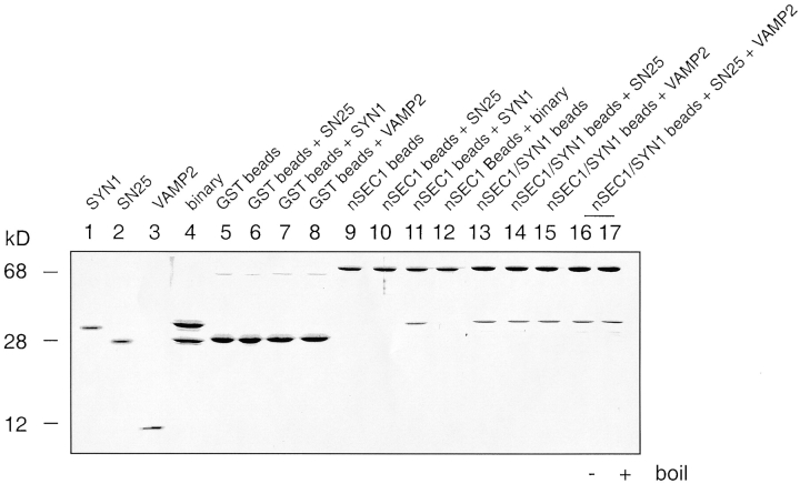
Previously, it was shown that nSec1 interacts with syntaxin1A with high affinity and that this complex prevents syntaxin from binding the target SNARE, SNAP-25, in vitro ( Pevsner et al. 1994a). However, it was never determined whether the binary association of syntaxin and SNAP-25 could prevent the nSec1/syntaxin interaction. To test this possibility, we incubated purified syntaxin1A, SNAP-25, or binary SNARE complex (syntaxin1A/SNAP-25) with GST-nSec1–bound glutathione-agarose beads. nSec1-bound agarose beads retained syntaxin, but not SNAP-25 ( Fig. 1, lanes 10 and 11), consistent with previous results. The nSec1/syntaxin1A interaction, however, was prevented by syntaxin1A/SNAP-25 binary association ( Fig. 1, lane 12). As a control, GST beads did not retain any SNAREs ( Fig. 1, lanes 5–8). Furthermore, we tested the ability of nSec1 to prevent assembly of the complete ternary SNARE complex. The glutathione-agarose beads conjugated with the nSec1/syntaxin1A complex were incubated with either SNAP-25, VAMP2, or both. The results showed that the high-affinity nSec1/syntaxin1A interaction completely prevented binding among the SNAREs ( Fig. 1, lanes 14 and 15) and assembly of the ternary SNARE complex ( Fig. 1, lanes 16 and 17). These results indicate that association of syntaxin with SNAREs in either binary or ternary complexes and the association of syntaxin with nSec1 are mutually exclusive.
Figure 1.
Among proteins of the SNARE complex, only free syntaxin interacts with nSec1. nSec1 (nSEC1) and all SNARE complex components, syntaxin1A (SYN1), SNAP-25 (SN25), and VAMP2, were purified as monomeric species by affinity chromatography. SYN1/SNAP-25 (binary) and nSec1/SYN1 complexes were assembled and further purified by size-exclusion chromatography. The bead-binding assays were performed by prebinding purified GST-tagged nSec1 or nSec1/SYN1 onto glutathione-agarose beads. The beads were then incubated with the indicated components overnight at 4°C. After washing, sample buffer was added to the beads and the proteins were separated on a 16% SDS-polyacrylamide gel. Molecular mass markers are indicated on the left in kD. Apparent molecular weights: monomeric nSec1, 70 kD; SYN1, 31 kD; SN25, 25 kD; and VAMP2, 10 kD.
Syntaxin Bound to nSec1 Does Not Form SNARE Complexes, and nSec1 Does Not Associate with the Preassembled SNARE Complex
To test the possibility that SNARE complexes act as receptors for nSec1 ( Carr et al. 1999), either purified syntaxin or nSec1/syntaxin1A complex was incubated with SNAP-25 and VAMP2 in solution. Free syntaxin, along with the other SNAREs, formed a SDS-resistant complex, as previously reported ( Fig. 2 A). Similarly, the nSec1-bound syntaxin was capable of forming an SDS-resistant SNARE complex ( Fig. 2 A). This nSec1/syntaxin1A-derived SNARE complex required all SNARE protein components for SDS resistance (data not shown) and dissociated into individual monomers after boiling ( Fig. 2 A), indicating that the complex is identical to the ternary SNARE complex. To test whether nSec1 remains associated with the SDS-resistant SNARE complex, we separated the protein complexes on a nondenaturing gel. SNAP-25, nSec1/syntaxin1A, and syntaxin1A ran as distinct bands on the native gel, whereas nSec1 and the positively charged VAMP2 did not enter the gel ( Fig. 2 B, lanes 1–5). Surprisingly, in contrast to the results obtained by SDS gel electrophoresis, the native gel showed that the mixture of nSec1/syntaxin1A, SNAP-25, and VAMP2, instead of running as a unique complex, ran as individual components ( Fig. 2 B, lane 6). In the absence of nSec1, the mixture of SNAREs ran as a unique band that represents the assembled complex ( Fig. 2 B, lane 7), indicating that this assay is capable of detecting assembled SNARE complex. Therefore, we conclude that the ability of the nSec1/syntaxin1A complex to form SNARE complexes in the presence of SDS is due to the SDS-induced dissociation of the nSec1/syntaxin1A complex, which allows the released syntaxin to bind the other SNAREs. Furthermore, we tested for the possible association between nSec1 and the preassembled SNARE complex using a bead binding assay. As shown in Fig. 3, nSec1 beads failed to retain any of the ternary SNARE complex.
Figure 2.

nSec1-bound syntaxin does not form SNARE complexes. A, Syntaxin1A/nSec1 complex (SYN1/nSEC1), syntaxin1A (SYN1), SNAP-25 NH2- and COOH-terminal domains (SN25), and VAMP2 were purified by affinity and size-exclusion chromatography. Either SYN1, SN25 and VAMP2 (left), or SYN1/nSec1, SN25, and VAMP2 (right) were combined and incubated for 1 h at 4°C. For each combination, sample buffer (final concentration of 2% SDS) was added, and half of the mixture was boiled, whereas the other half was kept at room temperature. The samples were then separated on a 12% SDS-polyacrylamide gel and visualized by Coomassie blue staining. Molecular mass markers are indicated on the left in kD. Apparent molecular weights: nSec1, 70 kD; and SYN1, 31 kD. SN25 NH2-terminal domain, SN25 COOH-terminal domain, and VAMP2 ran as 12 kD or below. SDS-resistant bands are marked by asterisks. Using full-length SNAP-25 gave similar results, with the SDS-resistant complex running larger. B, Native gel migration of full-length SN25, VAMP2, SYN1/nSEC1, nSec1, SYN1, and mixtures of SYN1/nSEC1, SN25, and VAMP2, or SYN1, SN25, and VAMP2. Note that the mixture of SYN1/nSEC1, SN25, and VAMP2, instead of running as a unique complex, ran as individual monomers. Neither VAMP2 nor nSec1 entered the native gel.
Figure 3.
nSec1 does not bind the ternary SNARE complex. Both GST and GST-nSec1 (nSEC1) were purified by affinity chromatography. Ternary SNARE complex (marked by an asterisk) was formed by mixing syntaxin1A, full-length SNAP-25, and VAMP2, and purified by size-exclusion chromatography. The purified GST or GST-nSec1 was incubated with glutathione-agarose beads for 1 h and washed. The beads were then incubated with purified ternary SNARE complex overnight at 4°C. The beads were washed and sample buffer (final concentration of 2% SDS) was added, and half of the mixture was boiled whereas the other half was kept at room temperature. Protein were separated on a 16% SDS-polyacrylamide gel. Molecular mass markers are indicated on the left in kD.
nSec1 Prevents Syntaxin from Forming a Neurotoxin-Resistant SNARE Complex
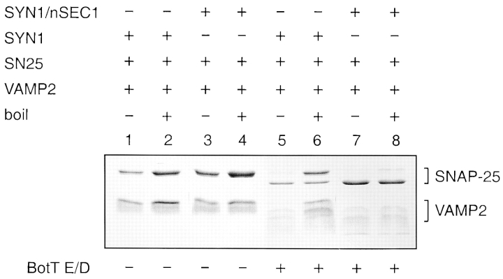
Assembly of binary and ternary SNARE complexes results in protection from neurotoxin digestion, whereas free SNAP-25 and VAMP2 are specifically cleaved by botulinum toxin E or D, respectively ( Hayashi et al. 1994). To further characterize whether both free syntaxin1A and prebound nSec1/syntaxin1A complex were able to form stable, neurotoxin-protected SNARE complexes in solution, we incubated either free syntaxin ( Fig. 4, lanes 1, 2, 5, and 6) or prebound nSec1/syntaxin ( Fig. 4, lanes 3, 4, 7, and 8) with SNAP-25 and VAMP2, followed by treatment with either botulinum toxin E or D. Without toxin treatment ( Fig. 4, lanes 1–4), both free syntaxin and nSec1/syntaxin1A were capable of forming thermally sensitive SNARE complexes as assayed by SDS-PAGE. As shown in Fig. 2, the nSec1/syntaxin complex is dissociated by SDS, followed by SNARE complex formation. Upon boiling in the presence of SDS, the complexes dissociated into SNARE monomers, as demonstrated by the increase in the density of both the SNAP-25 and VAMP2 protein bands. With either botulinum toxin E or D treatment, the complexes formed by syntaxin1A, SNAP-25, and VAMP2 were greatly protected from digestion, as intact SNAP-25 and VAMP2 bands were observed after boiling ( Fig. 4, lanes 5 and 6). In contrast, treatment of nSec1/syntaxin1A, SNAP-25, and VAMP2 with botulinum toxin E or D resulted in digestion of SNAP-25 and VAMP2 into products with higher electrophoretic mobility ( Fig. 4, lanes 7 and 8), indicating that neither SNAP-25 nor VAMP2 were protected as part of an assembled SNARE complex. These results further indicate that prebound nSec1/syntaxin1A, SNAP-25, and VAMP2 are unable to assemble into a complex in solution without addition of SDS.
Figure 4.
Association between nSec1 and syntaxin prevents toxin-resistant SNARE complex formation. Either syntaxin1A (SYN1; lanes 1, 2, 5, and 6) or nSec1/syntaxin1A complex (SYN1/nSEC1; lanes 3, 4, 7, and 8) was combined with full-length SNAP-25 (SN25) and VAMP2. The mixtures were incubated for 1 h at 4°C. Then, either botulinum toxin E to digest SNAP-25 (BotT E; upper panel of lanes 5–8) or botulinum toxin D to digest VAMP2 (BotT D; lower panel of lanes 5–8) was added and incubated 30 min at 25°C. After incubation, sample buffer (final concentration of 2% SDS) was added, and half of the mixture was boiled, whereas the other half was kept at room temperature. Proteins were then separated on a 16% SDS-polyacrylamide gel and visualized by Coomassie blue staining.
In Vivo nSec1 Is Present in a Complex Containing Syntaxin1A, but Not SNAP-25 or VAMP2
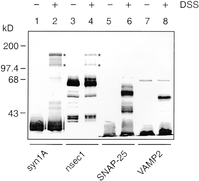
Next, to investigate the in vivo interactions between nSec1 and the SNARE proteins, we cross-linked detergent-solubilized brain extracts with the noncleavable cross-linked DSS. The extracts were analyzed by Western blotting with antibodies against either nSec1 or the different SNAREs, as indicated in Fig. 5. Cross-linking generated two higher molecular mass complexes (apparent molecular weights: 105 and 140 kD), both of which were immunoreactive with antisyntaxin1A and anti-nSec1. Significantly, however, these two bands were not recognized by either anti-SNAP-25 or anti-VAMP2. Furthermore, chemically cross-linking purified rat brain membrane before solubilization with detergent gave the same results (data not shown). Therefore, in the mammalian case, we see no evidence for the existence of assembled SNARE complexes bound to nSec1.
Figure 5.
Chemical cross-linking of rat brain membrane reveals complexes containing syntaxin1A and nSec1, but not VAMP2 or SNAP-25. Rat brain membrane detergent extracts were incubated with or without DSS (1 mM) for 2 h on ice. Lysates (20 μg/lane) were analyzed by Western blot with the antisyntaxin1A, anti-nSec1, anti–SNAP-25, or anti-VAMP2 antibodies as indicated. Asterisk-labeled complexes are described in Results. There are also two major unidentified cross-linked bands with molecular weights ranging from 45–60 kD recognized by anti–SNAP-25 antibody. There is one major 56-kD cross-linked band recognized by anti-VAMP2 antibody corresponding to synaptophysin/VAMP2 complex as previously demonstrated ( Calakos and Scheller 1994). Molecular mass markers are indicated on the left in kD.
Discussion
Using recombinant proteins purified to homogeneity, we have investigated the interactions between nSec1 and the SNAREs. Several observations, obtained from in vitro binding assays, nondenaturing gel electrophoresis, and specific neurotoxin treatment, suggest that the interaction of syntaxin1 with the core SNARE components, SNAP-25 and VAMP2, precludes the interaction of syntaxin1 with nSec1. Inversely, association of nSec1 and syntaxin1 prevents assembly of the binary and ternary SNARE complexes. Furthermore, chemical cross-linking of rat brain membranes revealed nSec1 complexes containing syntaxin1, but not SNAP-25 or VAMP2. These data are consistent with the hypothesis that syntaxin adopts an inaccessible closed conformation when bound to nSec1 that must be actively dissociated or conformationally altered to become eligible to form fusion complexes. In contrast to this hypothesis, a recent study using yeast concluded that Sec1p binds only to preformed SNARE complexes ( Carr et al. 1999). However, associations of membrane proteins in detergents can result in complexes that bind via hydrophobic membrane anchors, making assignment of stoichiometries difficult. NSF dissociation of SNARE complexes would likely reduce associations via membrane anchors as well. Carr et al. 1999 further speculated that Sec1p functions to promote exocytosis after SNARE complexes have been assembled. If both the conclusions from the yeast study and this work are true, there is likely to be a fundamental difference between the yeast and mammalian Sec1 proteins, despite the fact that other components of the membrane fusion pathway appear to be similar. Alternative explanations include the possibility that the binding affinity of the mammalian fusion complex and nSec1 is too weak to be detected in our assays. However, in yeast, Carr et al. 1999 failed to detect any binding between Sec1p and Ssop, whereas the mammalian counterparts interact with very high affinity (Kd = 10 nM). Perhaps in the yeast study, the Sec1p is already bound to Ssop or another protein and is, therefore, inhibited from binding to recombinant Sso1p. Most interestingly, perhaps Sec1p can adopt two conformations, one with a high affinity for syntaxin and another with a high affinity for the SNARE complex. The mammalian and yeast Sec1 proteins may, for unknown reasons, favor these alternative conformations. Characterization of the possible fundamental differences between different Sec1 proteins in yeast and mammals should be performed with either recombinant or purified yeast proteins to confirm direct protein–protein interactions. In addition, the yeast complexes need to be purified to be certain of the precise protein composition and stoichiometry.
When the vesicle arrives at the acceptor membrane, a series of events, beginning with target recognition and culminating with membrane fusion, ensues. Our data support a model in which the Sec1/syntaxin complex is at least one component of the target membrane receptor for the vesicle. Consistent with earlier hypothesis, we favor the idea that the Sec1/syntaxin complex is recognized by the vesicle, perhaps through a Rab/Rab effector interaction, and that conformational changes then occur that allow syntaxin to unfold to an open state. The molecular events leading to these proposed recognition events and conformational changes are not known and represent one of the most important issues in the membrane trafficking field. The open state of syntaxin is suggested to then interact with other SNAREs, leading to the formation of core complexes and membrane fusion. The question of whether Sec1 is totally dissociated from the SNARE complex or may remain bound deserves further investigation.
Acknowledgments
We thank Y.A. Chen for preparation of botulinum toxin D and E and for helpful discussions. We also thank M. Poirier and M. Bennett for plasmid constructs.
Footnotes
Abbreviations used in this paper: DSS, disuccinimidyl suberate; GST, glutathione S-transferase; NSF, N-ethylmaleimide-sensitive factor; SNAP, soluble NSF attachment protein; SNAP-25, synaptosome-associated protein of 25 kD; SNARE, SNAP receptor; VAMP, vesicle-associated membrane protein.
References
- Bennett M.K. SNAREs and the specificity of transport vesicle targeting. Curr. Opin. Cell Biol. 1995;7:581–586 . doi: 10.1016/0955-0674(95)80016-6. [DOI] [PubMed] [Google Scholar]
- Bennett M.K., Scheller R.H. A molecular description of synaptic vesicle membrane trafficking. Annu. Rev. Biochem. 1994;63:63–100 . doi: 10.1146/annurev.bi.63.070194.000431. [DOI] [PubMed] [Google Scholar]
- Brennwald P., Kearns B., Champion K., Keranen S., Bankaitis V., Novick P. Sec9 is a SNAP-25-like component of a yeast SNARE complex that may be the effector of Sec4 function in exocytosis. Cell. 1994;79:245–258 . doi: 10.1016/0092-8674(94)90194-5. [DOI] [PubMed] [Google Scholar]
- Calakos N., Scheller R.H. Vesicle-associated membrane protein and synaptophysin are associated on the synaptic vesicle. J. Biol. Chem. 1994;269:24534–24537 . [PubMed] [Google Scholar]
- Calakos N., Bennett M.K., Peterson K.E., Scheller R.H. Protein–protein interactions contributing to the specificity of intracellular vesicular trafficking. Science. 1994;263:1146–1149 . doi: 10.1126/science.8108733. [DOI] [PubMed] [Google Scholar]
- Carr C.M., Grote E., Munson M., Hughson F.M., Novick P.J. Sec1p binds to SNARE complexes and concentrates at sites of secretion. J. Cell Biol. 1999;146:333–344 . doi: 10.1083/jcb.146.2.333. [DOI] [PMC free article] [PubMed] [Google Scholar]
- Chen Y.A., Scales S.J., Patel S.M., Doung Y.C., Scheller R.H. SNARE complex formation is triggered by Ca2+ and drives membrane fusion. Cell. 1999;97:165–174 . doi: 10.1016/s0092-8674(00)80727-8. [DOI] [PubMed] [Google Scholar]
- Cowles C.R., Emr S.D., Horazdovsky B.F. Mutations in the VPS45 gene, a SEC1 homologue, result in vacuolar protein sorting defects and accumulation of membrane vesicles. J. Cell Sci. 1994;107:3449–3459 . doi: 10.1242/jcs.107.12.3449. [DOI] [PubMed] [Google Scholar]
- Dulubova I., Sugita S., Hill S., Hosaka M., Fernandez I., Sudhof T.C., Rizo J. A conformational switch in syntaxin during exocytosisrole of munc18. EMBO (Eur. Mol. Biol. Organ.) J. 1999;18:4372–4382 . doi: 10.1093/emboj/18.16.4372. [DOI] [PMC free article] [PubMed] [Google Scholar]
- Fernandez I., Ubach J., Dulubova I., Zhang X., Sudhof T.C., Rizo J. Three-dimensional structure of an evolutionarily conserved N-terminal domain of syntaxin 1A. Cell. 1998;94:841–849 . doi: 10.1016/s0092-8674(00)81742-0. [DOI] [PubMed] [Google Scholar]
- Garcia E.P., Gatti E., Butler M., Burton J., De Camilli P. A rat brain Sec1 homologue related to Rop and UNC18 interacts with syntaxin. Proc. Natl. Acad. Sci. USA. 1994;91:2003–2007 . doi: 10.1073/pnas.91.6.2003. [DOI] [PMC free article] [PubMed] [Google Scholar]
- Hata Y., Slaughter C.A., Sudhof T.C. Synaptic vesicle fusion complex contains unc-18 homologue bound to syntaxin. Nature. 1993;366:347–351 . doi: 10.1038/366347a0. [DOI] [PubMed] [Google Scholar]
- Hayashi T., McMahon H., Yamasaki S., Binz T., Hata Y., Sudhof T.C., Niemann H. Synaptic vesicle membrane fusion complexaction of clostridial neurotoxins on assembly. EMBO (Eur. Mol. Biol. Organ.) J. 1994;13:5051–5061 . doi: 10.1002/j.1460-2075.1994.tb06834.x. [DOI] [PMC free article] [PubMed] [Google Scholar]
- Hodel A., Schafer T., Gerosa D., Burger M.M. In chromaffin cells, the mammalian Sec1p homologue is a syntaxin 1A-binding protein associated with chromaffin granules. J. Biol. Chem. 1994;269:8623–8626 . [PubMed] [Google Scholar]
- Jahn R., Südhof T.C. Synaptic vesicles and exocytosis. Annu. Rev. Neurosci. 1994;17:219–246 . doi: 10.1146/annurev.ne.17.030194.001251. [DOI] [PubMed] [Google Scholar]
- Novick P., Scheckman R. Secretion and cell surface growth are blocked in a temperature-sensitive mutant of Saccharomyces cerevisiae . Proc. Natl. Acad. Sci. USA. 1979;76:1858–1862 . doi: 10.1073/pnas.76.4.1858. [DOI] [PMC free article] [PubMed] [Google Scholar]
- Ossig R., Dascher C., Trepte H.H., Schmitt H.D., Gallwitz D. The yeast SLY gene products, suppressors of defects in the essential GTP-binding Ypt1 protein, may act in endoplasmic reticulum-to-Golgi transport. Mol. Cell. Biol. 1991;11:2980–2993 . doi: 10.1128/mcb.11.6.2980. [DOI] [PMC free article] [PubMed] [Google Scholar]
- Peterson M.R., Burd C.G., Emr S.D. Vac1p coordinates Rab and phosphatidylinositol 3-kinase signaling in Vps45p-dependent vesicle docking/fusion at the endosome. Curr. Biol. 1999;9:159–162 . doi: 10.1016/s0960-9822(99)80071-2. [DOI] [PubMed] [Google Scholar]
- Pevsner J., Hsu S.C., Braun J.E., Calakos N., Ting A.E., Bennett M.K., Scheller R.H. Specificity and regulation of a synaptic vesicle docking complex Neuron. 13 1994. 353 361a [DOI] [PubMed] [Google Scholar]
- Pevsner J., Hsu S.C., Scheller R.H. n-Sec1a neural-specific syntaxin-binding protein Proc. Natl. Acad. Sci. USA. 91 1994. 1445 1449b [DOI] [PMC free article] [PubMed] [Google Scholar]
- Poirier M.A., Xiao W., Macosko J.C., Chan C., Shin Y.K., Bennett M.K. The synaptic SNARE complex is a parallel four-stranded helical bundle. Nat. Struct. Biol. 1998;5:765–769 . doi: 10.1038/1799. [DOI] [PubMed] [Google Scholar]
- Rothman J.E. Mechanisms of intracellular protein transport. Nature. 1994;372:55–63 . doi: 10.1038/372055a0. [DOI] [PubMed] [Google Scholar]
- Schulze K.L., Littleton J.T., Salzberg A., Halachmi N., Stern M., Lev Z., Bellen H.J. rop, a Drosophila homolog of yeast Sec1 and vertebrate n-Sec1/Munc-18 proteins, is a negative regulator of neurotransmitter release in vivo. Neuron. 1994;13:1099–1108 . doi: 10.1016/0896-6273(94)90048-5. [DOI] [PubMed] [Google Scholar]
- Sutton R.B., Fasshauer D., Jahn R., Brunger A.T. Crystal structure of a SNARE complex involved in synaptic exocytosis at 2.4 A resolution. Nature. 1998;395:347–353 . doi: 10.1038/26412. [DOI] [PubMed] [Google Scholar]
- Weber T., Zemelman B.V., McNew J.A., Westermann B., Gmachl M., Parlati F., Sollner T.H., Rothman J.E. SNAREpinsminimal machinery for membrane fusion. Cell. 1998;92:759–772 . doi: 10.1016/s0092-8674(00)81404-x. [DOI] [PubMed] [Google Scholar]
- Wu M.N., Littleton J.T., Bhat M.A., Prokop A., Bellen H.J. ROP, the Drosophila Sec1 homolog, interacts with syntaxin and regulates neurotransmitter release in a dosage-dependent manner. EMBO (Eur. Mol. Biol. Organ.) J. 1998;17:127–139 . doi: 10.1093/emboj/17.1.127. [DOI] [PMC free article] [PubMed] [Google Scholar]
- Yang B., Gonzalez L., Jr., Prekeris R., Steegmaier M., Advani R.J., Scheller R.H. SNARE interactions are not selective. Implications for membrane fusion specificity. J. Biol. Chem. 1999;274:5649–5653. doi: 10.1074/jbc.274.9.5649. [DOI] [PubMed] [Google Scholar]


