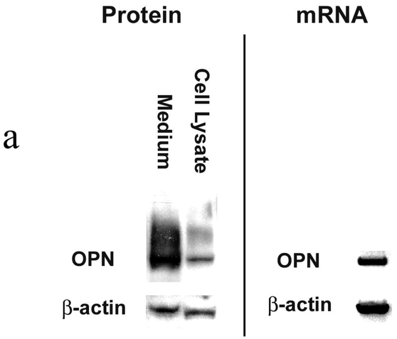
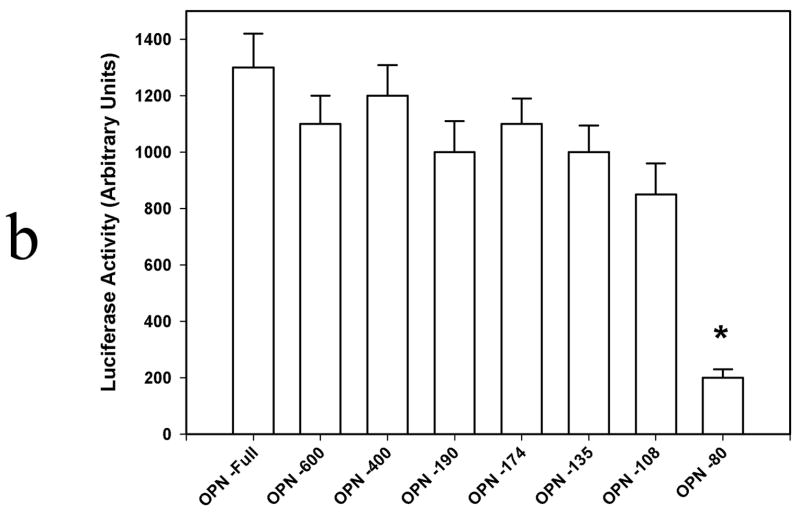
Figure 1. OPN expression in SW480 colon cancer cells.

Figure 1a. OPN protein and mRNA expression.
Western blot analysis was performed using cell lysate and concentrated culture media from SW480 cells. Confirmation of OPN mRNA expression was determined using Northern blot analysis. Blots are representative of three experiments.
Figure 1b. Transient transfection analysis of OPN promoter constructs.
5′-Deletion fragments of the human OPN promoter tested were: OPN −80 (nt −80 to nt +86), OPN −108 (nt −108 to nt +86), OPN −135 (nt −135 to nt +86), OPN −174 (nt −174 to nt +86), OPN −190 (nt −190 to nt +86), OPN −400 (nt −400 to nt +86), and OPN -Full (−2098 to +86). Data are expressed as mean ± SEM of three experiments.
(* p<0.01 vs OPN −108, OPN −135, OPN −174, OPN −190, OPN −400, and OPN –Full)

