

Abstract
Fragile X syndrome, which is caused by expansion of a (CGG)n repeat in the FMR1 gene, occurs in approximately 1:3500 males and causes mental retardation/behavioral problems. Smaller (CGG)n repeat expansions in FMR1, premutations, are associated with premature ovarian failure and fragile X-associated tremor/ataxia syndrome. An FMR1-sizing assay is technically challenging because of high GC content of the (CGG)n repeat, the size limitations of conventional PCR, and a lack of reference materials available for test development/validation and routine quality control. The Centers for Disease Control and Prevention and the Association for Molecular Pathology, together with the genetic testing community, have addressed the need for characterized fragile X mutation reference materials by developing characterized DNA samples from 16 cell lines with repeat lengths representing important phenotypic classes and diagnostic cutoffs. The alleles in these materials were characterized by consensus analysis in nine clinical laboratories. The information generated from this study is available on the Centers for Disease Control and Prevention and Coriell Cell Repositories websites. DNA purified from these cell lines is available to the genetics community through the Coriell Cell Repositories. The public availability of these reference materials should help support accurate clinical fragile X syndrome testing.
Fragile X syndrome (FXS) is the most common inherited cause of mental retardation, with an incidence in males of approximately 1 in 3500 (for a recent review, see http://genetests.org). Clinical features in males include mental retardation, specific physical characteristics (enlarged testes, large ears, and long face), and behavioral abnormalities, sometimes including autism spectrum disorder. Affected females, with an incidence of approximately 1 in 8000, have mild mental retardation. Our knowledge of the spectrum of phenotypes associated with expansion of the FMR1 gene now also includes premature ovarian failure1 and fragile X (FX)-associated tremor/ataxia syndrome.2,3,4,5 Thus, genetic testing for FX mutations is important at all life stages, prenatally to adulthood. FMR1-associated phenotypes are usually caused by expansion of an unstable (CGG)n repeat sequence in the 5′ untranslated region of the gene. There are essentially four allelic forms of the repeat length: normal (5 to 44 repeats), gray zone (also referred to as either intermediate or borderline, 45 to 54 repeats), premutation (55 to ∼200 repeats), and full mutation (>∼200 to 230 repeats). The boundaries between genotypic classes are currently defined by the American College of Medical Genetics 2005 Technical Standards and Guidelines for FX Molecular Testing (http://www.acmg.net/Pages/ACMG_Activities/stds-2002/fx.htm).
GeneTests (http://www.genetests.org) lists approximately 100 laboratories that offer clinical FMR1 testing, 70% of which are in North America. In the future, adult carrier and/or newborn diagnostic screening may become the standard of care. The American Society for Reproductive Medicine and the National Institute for Child Health and Human Development together with the scientific community, advocacy groups, professional societies, and industry are developing standardized clinical definitions, terminology, and testing recommendations for FXS. Among other recommendations, these groups suggested that FMR1 testing should be offered to women diagnosed with premature ovarian failure, reproductive or fertility problems associated with elevated basal FSH levels, and those with a low response to gonadotropin stimulation (http://www.fmr1pof.com/pdf/FMR1_Meeting_Guidelines.pdf). Similarly for fragile X-associated tremor/ataxia syndrome, guidelines for testing individuals with tremor, ataxia, and parkinsonian features of unknown etiology for FXS are being established. These recommendations, together with other potential recommendations for carrier and newborn screening by professional organizations, could increase testing volumes and the number of testing laboratories.
A DNA fragment-sizing assay of the triplet repeat region of the FMR1 gene is technically challenging. The high GC content of the (CGG)n repeat complicates conventional PCR amplification by reducing efficiency. The large size of some premutations and most full mutations also presents significant obstacles to effective PCR amplification. Simultaneous or reflex Southern analysis for some patient samples has limited resolution. Although Strom et al6 have described a capillary method that may largely overcome the need for conventional Southern analysis, the possible broad application of this method is unproven. Until recently, there were no commercially available reagents, such as probes and size calibration reference material, for FMR1 analysis. Clinical laboratories have developed in-house assays, or laboratory-developed assays (LDAs). Validation of LDAs has relied on publicly available cell lines, usually from the Coriell Cell Repositories, residual patient specimens, and voluntary interlaboratory comparisons. Importantly, the submitter defines the expected FMR1 allele sizes of these cell lines, and the Repository does not routinely verify the sizes. Thus, if the expected values are incorrect, then patient samples, interlaboratory comparisons, and test validations could be evaluated using reference materials with inaccurate allele sizes. Similarly, the use of incorrectly sized residual patient samples as reference materials could cause chronic errors in clinical laboratories. The performance of clinical laboratories in molecular genetic proficiency tests co-administered by the College of American Pathologists (CAP) and the American College of Medical Genetics demonstrates disparity among laboratories, due in part to the lack of FMR1 reference materials available for routine quality control, proficiency testing, and test development.
The National Institute of Standards and Technologies (NIST) has produced a standard reference material (SRM) for FXS testing, SRM 2399 (https://srmors.nist.gov/view_detail.cfm?srm=2399). SRMs are highly characterized, high-order reference materials that are produced in relatively limited quantities. The FX SRM consists of a set of nine amplicons with a variety of CGG repeat lengths. However, as for all SRMs, the FX SRM is intended for occasional use for assay validation or as a calibration material but not as a daily use reference or QC material.7
Although the NIST SRM is a useful starting point for assay validation, genomic DNAs with known allele lengths are the most critical reference materials for LDA validation because they most closely resemble patient specimens. It is imperative that clinical laboratories have access to characterized materials to provide the most accurate assessments of allele lengths in patient samples. In addition, publicly available DNA samples are often the source of CAP proficiency samples. Due to the clinical implications of the different size categories of premutations and all full mutations, the clinical laboratory must provide an accurate and precise assessment of FMR1 (CGG)n allele length for patient samples. To date, there has not been any organized evaluation of the publicly available reference materials that clinical laboratories can use to develop and validate their assays or to perform lot testing and daily QC.
The Centers for Disease Control and Prevention-based Genetic Testing Reference Materials Coordination Program (GeT-RM), the Association for Molecular Pathology, and the clinical genetics community initiated a project to obtain and characterize reference materials for FXS testing to address the lack of available characterized reference materials. Herein, we present a study performed by nine clinical laboratories in which the (CGG)n repeat size was measured in DNA samples derived from 16 cell lines containing clinically relevant FMR1 alleles in the normal and premutation range. The laboratories used both in-house and a common, research-use-only (RUO) platform to determine the allele size. These characterized FX reference materials are publicly available for use by clinical laboratories to ensure accuracy of FXS genetic testing.
Materials and Methods
Recruitment of Laboratories
Volunteers were solicited at a meeting convened by the Centers for Disease Control and Prevention entitled “Developing QC Materials for Genetic Testing” held in conjunction with the Association for Molecular Pathology annual meeting in Los Angeles in November 2004 (http://www.phppo.cdc.gov/dls/genetics/qcmaterials/default.aspx). At this meeting, leaders from academic/commercial laboratories, government, and industry recommended that FX reference materials be developed and characterized. Several individuals volunteered to form a consortium effort, later nicknamed the “Fragile Xperts”, sponsored jointly by the Association for Molecular Pathology Clinical Practice Committee and the Centers for Disease Control and Prevention-based GeT-RM Program. Of these Fragile Xperts, the Scientific Directors of nine clinical molecular genetics laboratories agreed to characterize reference materials (Table 1).
Table 1.
Participating Clinical Laboratories
| Laboratory | Scientific director |
|---|---|
| ARUP | Elaine Lyon |
| Baylor College of Medicine | Benjamin Roa |
| Emory University School of Medicine | Kasinathan Muralidharan |
| Genzyme Genetics | Elizabeth Rohlfs |
| Mayo Clinic | W. Edward Highsmith, Jr. |
| Ohio State University | Thomas Prior |
| Oregon Health & Science University | C. Sue Richards |
| University of Virginia | Lawrence Silverman |
| Virginia Commonwealth University | Andrea Ferreira-Gonzalez |
Reference Material Characterization Plan
The Fragile Xperts designed a consensus characterization study of DNA isolated from cell lines with normal, intermediate, and premutation FMR1 alleles representing important phenotypic classes and diagnostic cutoffs. We suspected that sequencing, the gold standard for validated reference materials, would not be feasible for longer FMR1 alleles, because of their high GC content and repetitive nature. In addition, the size limitations of conventional PCR would probably prove inadequate to amplify fragments with 100 or more CGG repeats. Thus, we executed a consensus characterization using a variety of PCR-based LDAs. Because we did not know, in advance, whether the variability in the LDAs would preclude consensus, we performed two characterization studies in parallel. In addition to their PCR LDA, each of the nine laboratories also assayed the DNA samples using a set of common RUO reagents, developed by Celera Diagnostics (Alameda, CA) and referred to throughout this study as the “common platform”. These RUO reagents were prepared from research reagent lots that were identical in composition to the analyte specific reagent (ASR) for FMR1 and ASR Primers for Gender that are now commercially available from Abbott Molecular (Des Plaines, IL). The common aspect among laboratories using this platform is only based on the RUO PCR reagents, since each laboratory used their own unique combination of general reagents, equipment, and instrumentation.
To validate the common platform, each of the nine laboratories tested the NIST SRM 2399 using their PCR LDA and the common platform. These samples were assayed only once. Each laboratory tested five previously sequenced Coriell control DNAs and nine previously tested in-house DNA samples, once on their PCR LDA and three times on the common platform. The samples were analyzed in batches of 14, on three separate runs and three separate days. These platform validation data are not shown.
To test the specimens of interest, each laboratory determined the sizes of the FMR1 alleles from DNA derived from 16 experimental samples and five previously sequenced DNA samples (Tables 2 and 3) using both their own PCR LDA (Table 4) and the common platform. Each laboratory tested each of the 21 samples three separate times on each of the two assays, for a total of 126 possible data points per laboratory. The DNA samples were distributed directly to the laboratories from the Coriell Repositories and were blinded with respect to sex and submitter's estimate of the allele size(s).
Table 2.
Consensus Data for Length of FMR1 Repeat in 5 Male Control Coriell Cell Lines Used in the Consensus and Values Obtained
| Cell line | Allele length (previously determined) | Platform | Pooled meana | SEM | Pooled varianceb | Consensus lengthc |
|---|---|---|---|---|---|---|
| NA07174 | 30 | In-house | 29.7 | 0.49 | 1.6 | 30 |
| Common | 30.1 | 0 | 0.2 | |||
| CD00014 | 56 | In-house | 55.8 | 0.54 | 0.5 | 56 |
| Common | 55.4 | 0.17 | 0.3 | |||
| NA06892 | 93 | In-house | 85.8 | 1.1 | 0.3 | 86 |
| Common | 86.5 | 0.105 | 0.2 | |||
| NA06906 | 96 | In-house | 96.7 | 2.04* | 1.2 | No Consensus |
| Common | 98.8 | 0.86 | 4.9** | No Consensus | ||
| NA06891 | 118 | In-house | 117.9 | 1.55* | 0.3 | No Consensus |
| Common | 116.8 | 0.131 | 25.4** | No Consensus |
Pooled mean: weighted average of triplicate group means.
Pooled variance: weighted average of group variances.
Consensus length: consensus value for (CGG)n allele lengths, based on the data from nine laboratories. Consensus is reached within either assay type (LDA or common) when the SEM and pooled variance are not significant.
Significant variation at the 5% level of probability;
significant variation at the 1% level of probability.
Table 3.
Consensus Data for Length of FMR1 Repeat in 16 Experimental Coriell Cell Lines Used in the Consensus and Values Obtained
| Cell line | Sex | Submitter's estimate of allele length | Platform | Allele | Pooled meana | SEM | Pooled varianceb | Consensus lengthc |
|---|---|---|---|---|---|---|---|---|
| NA07538 | F | 29/29 | In-house | 1 | 29.2 | 0.16 | 0.2 | 29 |
| 2 | 0.21 | 29 | ||||||
| Common | 1 | 29.1 | 0.06 | 0 | 29 | |||
| 2 | 0.526 | 29 | ||||||
| NA20238 | F | 29/30 | In-house | 1 | 28.9 | 0.202 | 1.2 | 29 |
| 2 | 29.9 | 0.133 | 0.1 | 30 | ||||
| Common | 1 | 29.0 | 0.033 | 0 | 29 | |||
| 2 | 30.1 | 0.048 | 0.1 | 30 | ||||
| NA20244 | M | 41 | In-house | 1 | 40.9 | 0.205 | 0.1 | 41 |
| Common | 1 | 40.9 | 0.069 | 0 | 41 | |||
| NA20243 | F | 29/41 | In-house | 1 | 28.8 | 0.104 | 0 | 29 |
| 2 | 40.7 | 0.187 | 0 | 41 | ||||
| Common | 1 | 29.1 | 0.057 | 0 | 29 | |||
| 2 | 40.9 | 0.057 | 0 | 41 | ||||
| NA20235 | F | 29/45 | In-house | 1 | 28.9 | 0.119 | 0 | 29 |
| 2 | 44.6 | 0.259 | 0.2 | 45 | ||||
| Common | 1 | 29 | 0 | 0 | 29 | |||
| 2 | 44.8 | 0.104 | 0.1 | 45 | ||||
| NA20232 | M | 46 | In-house | 1 | 45.6 | 0.273 | 0.2 | 46 |
| Common | 1 | 45.7 | 0.09 | 0 | 46 | |||
| NA20234 | F | 31/46 | In-house | 1 | 30.8 | 0.145 | 0.2 | 31 |
| 2 | 45.7 | 0.241 | 0.2 | 46 | ||||
| Common | 1 | 31 | 0 | 0 | 31 | |||
| 2 | 45.8 | 0.076 | 0.2 | 46 | ||||
| NA20230 | M | 54 | In-house | 1 | 53.3 | 0.336 | 0.2 | 53 |
| Common | 1 | 53.1 | 0.144 | 0.3 | 53 | |||
| NA20236 | F | 31/55 | In-house | 1 | 30.9 | 0.123 | 0.1 | 31 |
| 2 | 53.3 | 0.333 | 0 | 53 | ||||
| Common | 1 | 31 | 0.05 | 0.1 | 31 | |||
| 2 | 53.2 | 0.181 | 0 | 53 | ||||
| NA20231 | M | 79 | In-house | 1 | 75.7 | 0.524 | 0.5 | 76 |
| Common | 1 | 76.5 | 0.163 | 0.4 | 76 | |||
| NA20240 | F | 30/85 | In-house | 1 | 29 | 0.134 | 0.1 | 30 |
| 2 | 80.3 | 0.585 | 0.6 | 80 | ||||
| Common | 1 | 30.1 | 0.053 | 0.1 | 30 | |||
| 2 | 80.7 | 0.107 | 0.2 | 80 | ||||
| NA20233 | M | 100 | In-house | 1 | 113.8 | 2.11* | 2.9 | No Consensusd |
| Common | 1 | 117.2 | 0.087 | 0.2 | 117 | |||
| NA20242 | F | 30/100 | In-house | 1 | 29.9 | 0.14 | 0.1 | 30 |
| 2 | 72.3 | 0.567 | 0.8 | 73 | ||||
| Common | 1 | 30.1 | 0.051 | 0 | 30 | |||
| 2 | 72.7 | 0.133 | 0.2 | 73 | ||||
| NA20237 | M | 120 | In-house | 1 | 103.4 | 2.93* | 59.2 | No Consensus |
| Common | 1 | 100.2 | 1.66* | 61.9 | No Consensus | |||
| NA20241 | F | 30/120 | In-house | 1 | 28.9 | 0.132 | 0.1 | 29 |
| 2 | 109.4 | 2.79** | 1.1 | No Consensus | ||||
| Common | 1 | 29.1 | 0.051 | 0 | 29 | |||
| 2 | 93.6 | 3.11** | 79.7 | No Consensus | ||||
| NA20239 | F | 23/200 | In-house | 1 | 20 | 0.154 | 0.2 | 20 |
| 2 | 183 | 22.54** | NAe | No Consensus | ||||
| Common | 1 | 20.2 | 0.1 | 0.2 | 20 | |||
| 2 | 192.7 | 6.69** | 0.1 | No Consensus |
Pooled mean: weighted average of triplicate group means.
Pooled variance: weighted average of group variances.
Consensus length: consensus value for (CGG)n allele lengths, based on the data from nine laboratories. Consensus is reached within either assay type (LDA or common) when the SEM and pooled variance are not significant.
No Consensus: SEM, pooled variance, or both are significant.
NA, only a single laboratory reported sizes for this allele.
Significant variation at the 5% level of probability;
significant variation at the 1% level of probability.
Table 4.
Summary of Laboratory-Developed Methodologies for Sizing of Normal and Premutation FMR1 Alleles
| span Lab ID | Primers | Fragment sizing instrument or method | Matrix | Label | Allele sizing calculation | Reference |
|---|---|---|---|---|---|---|
| 1 | F 5′-6FAM-GGAACAGCGTTGATCACGTGACGTGGTTTC-3′ | ABI 310 | POP6 | 6-FAM | (bp-113)/3 | 10 |
| R 5′-GGGGCCTGCCCTAGAGCCAAGTACCTTGT-3′ | ||||||
| 2 | F 5′-GACGGAGGCGCCCGTGCCAGG-3′ | PAGE and phosphoimaging | 6% polyacrylamide (19:1), 7 mol/L urea | 32P-dCTP incorporated during PCR | (bp-113)/3 | 11 |
| R 5′-TCCTCCATCTTCTCTTCAGCCCT-3′ | ||||||
| 3 | F 5′-6FAM-TGACGGAGGCGCCGCTGCCAGGGGGCGTGC-3′ | ABI 377 | 7% polyacrylamide (19:1), 8.3 mol/L urea | 6-FAM | (bp-69)/3 | 12 |
| R 5′-GAGAGGTGGGCTGCGGGCGCTCGAGGCCCA-3′ | ||||||
| 4 | F 5′-AGGCGCTCAGCTCCGTTTCGGTTTCACTTC-3′ | ABI 3100 | POP6 | 6-FAM | (bp-147)/3 | 13 |
| R 5′-6FAM-GTGGGCTGCGGGCGCTCGAGG-3′ | ||||||
| 5 | F 5′-6TETGCTCAGCTCCGTTTCGGTTTCACTTCCGGT-3′ | ABI 377 | 6% polyacrylamide (19:1), 6 mol/L urea | TET | (bp-218)/3 | 14 |
| R 5′-AGCCCCGCACTTCCACCACCAGCTCCTCCA-3′ | ||||||
| 6 | F 5′-GCTCAGCTCCGTTTCGGTTTCACTTCCGGT-3′ | ABI 3100 | POP4 | 6-FAM | (bp-221)/3)+4 | 15 |
| R 5′-6FAM-AGCCCCGCACTTCCACCACCAGCTCCTCCA-3′ | ||||||
| 7 | F 5′-GCTCAGCTCCGTTTCGGTTTCACTTCCGGT-3′ | PAGE and autoradiography | 6% polyacrylamide (19:1), 7 mol/L urea | 32P-dCTP incorporated during PCR | (bp-218)/3 | 15 |
| R 5′-AGCCCCGCACTTCCACCACCAGCTCCTCCA-3′ | ||||||
| 8 | F 5′-GACGGAGGCGCCGCTGCCAGG-3′ | PAGE and autoradiography | 6% polyacrylamide (19:1), 7 mol/L urea | 32P-(CGG)5 probe | (bp-62)/3 | 15 |
| R 5′-GTGGGCTGCGGGCGCTCGAGG-3′ | ||||||
| 9 | F 5′-6FAM-GTGACGGAGGCGCCGCTGCCA-3′ | ABI 3100 | POP4 | 6-FAM | [bp-18)/3]+3 | 15 |
| R 5′-AGCTCCTCCATCTTCTCTTCAGCCCTGCTA-3′ |
R, reverse; F, forward; PAGE, polyacrylamide gel electrophoresis.
DNA Samples
NIST Standard Reference Material (SRM 2399) and Controls
The NIST Fragile X Human DNA Triplet Repeat Standard (SRM 2399, described at https://srmors.nist.gov/view_detail.cfm?srm=2399) was used in platform validation. The nine components of this SRM consists of nine amplicons, A through I, that contain 20, 30, 41, 51, 60, 73, 93, 96, and 118 FMR1 (CGG)n repeats, respectively. Previously NIST-sequenced DNA samples derived from five Coriell FX cell lines8,9 (Kristy Richie, personal communication) were used as controls in this study (Table 2).
Coriell DNAs
The 16 experimental cell lines were chosen based on the submitter's estimate of the FMR1 allele lengths and their clinical relevance (Table 3). Fifteen of the cell lines listed in Table 3 were submitted (by S.L.S.) to the National Institute of General Medical Sciences Human Genetic Cell Repository, one of the Coriell Cell Repositories, for this study. Sample NA07538 was a preexisting Coriell cell line. Samples were received as frozen vials of Epstein-Barr virus-transformed B-lymphocyte cell lines. We were not able to calculate the number of population doublings since the inception of culture for these samples because no history was available.
All samples were placed into culture and expanded to yield approximately 2 × 10⁸ total viable cells. The culture medium was antibiotic-free to increase the likelihood that contamination would be readily detected. The cell suspension was dispersed in 40 1-ml vials at 5 × 10⁶ viable cells per vial. Cultures were cryopreserved in heat-sealed borosilicate glass ampules and stored in liquid nitrogen (liquid phase). Successful cultures were free from bacterial, fungal, and mycoplasmal contamination and were viable after cryopreservation, as evidenced by a doubling of the cell number within four days of recovery.
PCR LDAs
The PCR LDAs used by the nine laboratories are summarized in Table 4.10,11,12,13,14,15 Gender determination was not included in this validation plan. Of note, each laboratory used a unique combination of amplification primers, equipment, and detection methods. In general, PCR amplification was followed by either capillary or denaturing polyacrylamide gel electrophoresis (Figure 1 and Table 4). Amplification products were detected using fluorescence-labeled primers (six of nine laboratories), a radioisotope-labeled primer (one of nine laboratories), and incorporation of a radioisotope-labeled deoxynucleoside-5′-triphosphate (one of nine laboratories) or a radioactive probe (one of nine laboratories). To size-fractionate the amplicons, six laboratories used genetic analysis instruments (ABI 377, 2; ABI 310, 1; ABI 3100, 3) and associated software, whereas three laboratories used labeled sequencing gels and analyzed amplicon size visually against a size marker. The allele-sizing calculations for each LDA, which are based on primer location, are also summarized in Table 4.
Figure 1.
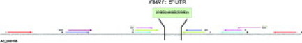
Location of PCR primers flanking the CGG repeat region in the 5′ untranslated region (5′ UTR) of the FMR1 gene. The CGG trinucleotide repeats with the interrupted AGG repeats are indicated by the green box. Calculation of the number of CGG repeats is specific to the primers used. The forward and reverse primers used by each laboratory, numbers 1 through 9, are indicated. The direction of the arrows indicates the direction of DNA synthesis from the primers.
Common Platform Method
Each laboratory used a set of common RUO reagents, obtained from Celera Diagnostics, as described below. These reagents were provided to the consortium as RUO reagents to legally permit the manufacturer to furnish a starting protocol and to provide training to the sites. This allowed the laboratories to use an assay that was consistent between laboratories and avoided the need to develop nine different additional LDAs based on ASRs. Thus, since “RUO” is a regulatory classification and not associated with product quality or performance, the use of RUO reagents herein does not negatively impact the results of this study.
Three μl of genomic DNA (10 ng/μl, 30 ng total) were added to 17 μl of PCR Master Mix (13 μl of High GC PCR Buffer), 0.6 μl (10 μmol/L) of Gender Primers (sequence not shown), 0.8 μl (10 μmol/L) of FX Primers (forward primer 5′-GACGGAGGCGCCGCTGCCAGG-3′ and reverse primer 5′GTGGGCTGCGGGCGCTCGAGG-3′), 1.2 μl of TR PCR Enzyme Mix, and 1.4 μl of DNase/RNase-free water and amplified as described in Table 5. Three μL of CleanUp Enzyme mix (Abbott/Celera, Abbott Park, IL) was then added to 2 μl of PCR product and incubated in a thermal cycler at 75°C for 10 minutes. To prepare for electrophoresis, 10 μl of electrophoresis mix [7 μl of Hi-Di formamide; Applied Biosystems (ABI), Foster City, CA] and 3 μl of ROX 1000 Size Standard (Abbott/Celera) were mixed with 5 μl of the purified PCR products and loaded onto the laboratory instrument for electrophoretic size analysis. Although the laboratories used several different instruments for allele sizing (Table 4), an example of the parameters for the ABI PRISM 3100 and the POP6 polymer is shown in Table 6; two sizing runs were automatically performed sequentially for each sample, one for short sequences (5 to 70 repeats) and one for alleles greater than 71 repeats. The instrument was configured to automatically perform both sizing runs serially (short run first) for all samples within the test batch. Fragment data were collected using the ABI Data Collection and analyzed using ABI GeneMapper Software. For each software package, the version used was dependent on the model of the capillary electrophoresis system, as outlined in Table 4. GeneMapper evaluates data by creating a sizing curve for each sample and assigning a fragment size to each of the CGG repeat PCR products. Although the Gender Primers were included in the PCR amplification, the samples were not analyzed with respect to sex.
Table 5.
Amplification Conditions for the Common Platform
| Temperature | Time (min:sec) | Cycles |
|---|---|---|
| 98.5°C | 0:10 | 15 |
| 58.0°C | 1:00 | |
| 75.0°C | 6:00 | |
| 98.5°C Auto X; 0.1°C/cycle* | 0:10 | 15 |
| 56.0°C | 1:00 | |
| 75.0°C | 6:00 | |
| 4.0°C | Hold |
+0.1 each cycle.
Table 6.
ABI 3100 Genetic Analyzer Electrophoresis Conditions for the Common Platform Using the POP6 Polymer and a 36 cm Array and Data Collection Software v2.0
| Parameter | Data collection |
|---|---|
| Shorter [5–70 (CGG)n] repeats | |
| Running temperature | 60°C |
| Cap fill volume | NA |
| Current tolerance | NA |
| Run current | NA |
| Voltage tolerance | NA |
| Pre-run voltage | 15 kV |
| Pre-run time | 180 seconds |
| Injection voltage | 10 kV |
| Injection time | 1 second |
| Run voltage | 15 kV |
| Number of steps | 10 nK |
| Voltage step interval | 60 seconds |
| Data delay time | 300 seconds |
| Run time | 3000 seconds |
| Longer [71–230 (CGG)n] repeats | |
| Running temperature | 60°C |
| Cap fill volume | NA |
| Current tolerance | NA |
| Run current | NA |
| Voltage tolerance | NA |
| Pre-run voltage | 15 kV |
| Pre-run time | 180 seconds |
| Injection voltage | 15 kV |
| Injection time | 22 seconds |
| Run voltage | 15 kV |
| Number of steps | 10 nK |
| Voltage step interval | 60 seconds |
| Data delay time | 300 seconds |
| Run time | 5600 seconds |
Southern Analysis
Two laboratories performed Southern analysis of DNA from early and late passages of the 16 Coriell cell lines to detect instabilities (changes in allele size, methylation, and any evidence of mosaicism) that may have arisen in culture. For early-passage DNA, one vial of approximately 5 × 10⁶ viable cells was thawed and cultured to yield approximately 2 × 10⁷ viable cells (approximately two population doublings) and 100 μg of DNA. For late-passage DNA, a second vial of 5 × 10⁶ viable cells was thawed and cultured to yield approximately 4 × 10⁹ viable cells (approximately 10 population doublings) and 20 mg of DNA. Both laboratories used modifications of the Southern analysis method.16 One laboratory digested the DNA samples with EcoRI and the methylation-sensitive NruI and probed with StB 12.3.16 The other laboratory digested the samples with EcoRI and the methylation-sensitive enzyme XhoI and probed with an in-house clone derived from a PCR product.
DNA Sequencing of the FMR1 (CGG)n Repeat
DNA sequence analysis was performed at NIST. The following primers (Operon Technologies, Inc., Alameda, CA) amplified a PCR product containing the repeat and 222 bp of flanking sequences: forward primer, 5′-GCTCAGCTCCGTTTCGGTTTCACTTCCGGT-3′; reverse primer, 5′-AGCCCCGCACTTCCACCACCAGCTCCTCCA-3′. DNA samples were amplified using the GC-Rich PCR System (Roche Diagnostics, Indianapolis, IN). One μL of genomic DNA (∼300 to 360 ng) was amplified in a 50-μl reaction volume containing 1.5 M GC-Rich Resolution Solution, 1 μl of deoxynucleoside-5′-triphosphate mix (10 mmol/L each), 1 μl (10 μmol/L) of each primer, and 2.0 units of GC-Rich enzyme in a reaction buffer containing 1.5 mmol/L MgCl2. Thermal cycling conditions, using an ABI 9700 thermal cycler, were as follows: 98.5°C, 5 seconds; 56°C; 4 minutes; 69°C, 6 minutes for 14 cycles, followed by 98.5°C, 5 seconds; 56°C with autoextension of 0.1°C per cycle, 4 minutes; 69°C, 6 minutes for 15 cycles. PCR products were gel purified using a filter column QIAquick gel extraction kit (Qiagen, Valencia, CA). The amplification step was performed once per sample. Purified DNA samples were cycle sequenced using ABI BigDye Terminator sequencing kit, version 1.1. Samples were prepared using 1 μl of the forward primer (10 μmol/L), 1 to 2 μl of PCR product, 2 μl of dimethyl sulfoxide, 8 μl of Enzyme Mix, and 9 to 10 μl of water for a final volume of 22 to 23 μl. Cycle sequencing reactions were performed on an ABI GeneAmp 9700 PCR System using the following conditions: 95°C, for 3 minutes, followed by 45 cycles of 53°C, 30 seconds; 68°C, 2 minutes; 98°C, 30 seconds. The DNA products were purified by passage through Performa Gel Filtration Cartridges, (Edge BioSystems, Gaithersburg, MD). The samples were then vacuum dried and reconstituted in 20 μl of ABI Template Suppression Reagent. DNA sequencing was performed in triplicate using an ABI PRISM Model 310 Genetic Analyzer with the POP6 polymer system and 47 cm × 50 μm capillaries.
DNA sequence data were analyzed with Sequencing Analysis Software, version 3.3 (DNASTAR Inc., Madison, WI), and the Lasergene 6.1. SeqMan II software (DNASTAR Inc.) was used to determine the quality scores. Briefly, the quality score of a peak was calculated based on the shape and height of each peak and was adjusted relative to the maximum height of any peak in the entire sequence. Taller, sharper peaks receive the highest scores. The heights of any underlying peaks are subtracted from the highest peak's score. Scores are not assigned to gaps. An average quality score, calculated from a 100-nucleotide region of each sample, was determined for each sample and accepted when greater than 40.17
Statistical Analysis
Each laboratory submitted up to three replicates per DNA sample for each of the two platforms. The data from all of the laboratories were collected, checked for error (eg, sample swap or incorrect transcription) and organized in a relational database. The database was created using MySQL (MySQL Inc. Seattle, WA), which supported automated validation of the data. Specific data that were not uniformly reported were then manually curated. Multivariate analysis was used to analyze the data. SEM is an estimate of variation between laboratories for each cell line and platform. SEM was calculated for each DNA sample and each platform and was used as an estimate of variance between the pooled mean of the data from all laboratories. The pooled variance, which is the estimate of variance between replicates for each laboratory, was calculated as the weighted average of the group variances. Analysis of variance was used to determine the hypothesis of equality for each allele on each platform. We also used the pooled mean and SEM to determine the consensus length for each allele. Consensus of allele length is reached within either assay type (LDA or common) when the SEM and pooled variance are not significant.
Results
Our goal was to provide clinically relevant reference materials for amplification-based assays of the FMR1 gene. We chose cell lines from an academic research laboratory (Stephanie Sherman, Emory University) based on estimated allele size, attempting to include alleles at or near the phenotypic boundaries described in the American College of Medical Genetics guidelines. For all phenotypic categories, matched male and female pairs with the larger allele in common were chosen (Tables 2 and 3). We also chose two female cell lines with the most prevalent allele (29 repeats), one homozygous (29/29) and one with a second allele that was larger by only a single repeat (29/30). As a point of reference, both among laboratories and between platforms, we also genotyped five male “controls” that were characterized in earlier studies.
Consensus Study
We performed a consensus characterization study for a range of FMR1 normal and premutation allele sizes. Knowing that length determination by sequencing is problematic for GC-rich regions, particularly for larger FMR1 alleles,8 we chose a consensus genotyping approach. We also believed it was important to assess the genotypes in these materials in a diagnostic setting, using current clinical assays. Each of the nine laboratories characterized the repeat length of DNA from 16 cell lines using their clinical LDA, as well as a common platform. The consensus data for these materials are shown in Tables 2 and 3. The pooled means were tested for statistically significant differences on the basis of the SEM and the pooled variance; we considered a pooled mean to be a consensus value when neither of these statistics was significant at the 5% level of probability.
We achieved consensus for only three of the five controls, with allele lengths from 30 to 86 (Table 2). We did not achieve consensus for two of the controls that were previously shown by DNA sequence analysis to have allele lengths of 96 and 118.
We achieved consensus using both the LDAs and the common platform for 12 of 16 experimental cell lines (Table 3). The consensus values range from 29 to 80 repeats in both male and female samples. In addition, the laboratories distinguished a homozygous female with 29 repeats from a heterozygous female with allele lengths of 29 and 30. We reached consensus on the common platform, but not on the LDAs, for one male DNA sample (117 repeats, NA20233). We did not achieve consensus using either the LDAs or the common platform for three DNA samples with the largest estimated allele sizes: a male with an estimated 120 repeat allele (NA20337) and two heterozygous female samples with estimated premutations of 120 and 200 repeats (NA20241 and NA20239, respectively). Consensus was achieved on both platforms for the smaller allele (29 and 20 repeats, respectively) in these two samples.
For DNA derived from cell lines of male patients, the consensus value was at or within one repeat of the submitter's estimate in three of six samples for which we achieved consensus (41 to 54 repeats in cell lines NA20244, NA20232, and NA20230). The consensus values differed by three or more repeats in the remaining male samples. Similar results were achieved for female DNA samples, for which most alleles of 53 or less were within one repeat of the submitter's estimate, and greater differences were observed for larger alleles, with the exception of the heterozygous line NA20239, for which the consensus value of the smaller allele was three repeats less20 than the submitter's estimate.23
Stability Studies
We assessed the stability of each of the experimental cell lines in mass culture after two (early) and ten (late) population doublings by Southern analysis. Both testing laboratories obtained the same results, and the data from one are presented in Figure 2. There were no obvious increases or decreases in allele size between the early and late passages, and all of the cell lines had the expected methylation patterns. Importantly, in one cell line, NA20237, we detected the development of a smaller premutation between the early and late passage, indicative of mosaicism.
Figure 2.

FMR1 Southern analysis of early (E) and late (L) passage in culture from 15 Coriell cell lines, performed by laboratory 4; NA07538 is an established Coriell cell line and is included in this analysis only for completeness. For early-passage DNA, one vial of approximately 5 × 10⁶ viable cells was thawed and cultured to yield approximately 2 × 10⁷ viable cells (approximately two population doublings) and 100 μg of DNA. For late-passage DNA, a second vial of 5 × 10⁶ viable cells was thawed and cultured to yield approximately 4 × 10⁹ viable cells (approximately 10 population doublings) and 20 mg of DNA. Five mg of DNA from each sample was digested with EcoRI and NruI, size fractionated on an 0.8% agarose gel, transferred to a positively charged nylon membrane in the presence of alkali, and probed with a clone from a normal individual that contains sequence located in the fragment containing the (CGG)n repeat. Probe was generated by amplification of a 1031-bp fragment generated by amplification with forward (6730-FXF 5′-CTTCTCAGTTGGATACCAGCA-3′) and reverse (XhoI-REV 5′-CCACCGGAAGTGAAAACCG-3′) primers and then subcloned using the TA-clone kit (Invitrogen, Carlsbad CA).
Sequencing of Male Cell Lines
The three control samples for which we achieved consensus were confirmed by sequence analysis at NIST (Table 7). For two of these with smaller alleles (30 repeats, NA07174; and 56 repeats, CD00014), the values based on sequencing exactly matched the consensus values. The sequencing-based value for the premutation control, NA06892 (88 or 89 repeats), was within two to three repeats of the consensus value (86 repeats). We note that repeated sequencing analysis of NA06892 consistently yielded values of 88 or 89 repeats, but the exact number could not be determined. In addition, we were not able to determine the full length of the control with the largest allele, NA06891 (118 repeats), by sequencing (data not shown).
Table 7.
Comparison of Male FMR1 Allele Lengths Based on Consensus and Sequencing
| Cell line | Allele length based on consensus | Allele length based on multiple sequencing assays | Quality score |
|---|---|---|---|
| Control | |||
| NA07174 | 30 | 30, 30, 30 | 51.7 |
| CD00014 | 56 | 56, 56, 56 | 45.6 |
| NA06892 | 86 | 88/89* 88/89, 88/89 | 41.7 |
| Experimental | |||
| NA20244 | 41 | 41, 41, 41 | 56.4 |
| NA20232 | 46 | 46, 46, 46 | 56.6 |
| NA20230 | 53 | 54, 54, 54 | 52.9 |
| NA20231 | 76 | 78, 78, 78 | 45.9 |
| NA20233 | 117 | >100,† >100 | 50.1 |
Could not resolve exact size.
Only two sequencing reactions were read.
DNA from the five male experimental cell lines for which we achieved consensus (Table 7) was also sequenced. The allele lengths based on sequencing values exactly matched the consensus values for two cell lines (NA20244, 41 repeats; and NA20232, 46 repeats). The length, based on sequencing for NA20230, was within one repeat of the consensus value (54 vs. 53, respectively). For NA20231, the repeat length based on sequencing, 78, was two repeats larger than the consensus value.76 We were not able to determine the exact length of the largest consensus allele by sequencing (NA20233, 117 repeats) but did confirm that this allele is greater than 100 repeats.
The average QSs for each sequencing reaction met the acceptance criteria (> 40). This was true even for the samples with longer alleles, for which we could not determine the exact size (NA20233 and NA06892). However, the QSs were determined by examination of peak height and shape and normalized over 100-bp increments of each sequence with more than 100 bp. Thus, within each of these increments for all cell lines, the sequence was acceptable.
Discussion
Genetic testing for FXS is technically challenging. Affected individuals with full mutations have allele lengths greater than 200 CGG repeats, which is beyond the amplification power of conventional PCR methods. Molecular analysis excludes a diagnosis of FXS in the majority of symptomatic males, when normal-length alleles are detected and mosaicism is not present. The detection of premutations is of obvious clinical importance for identification of carriers, as well as risk assessment of these individuals with regard to development of premature ovarian failure and fragile X-associated tremor/ataxia syndrome. The clinical laboratory must be able to 1) accurately size alleles using PCR for clinically relevant phenotypic boundaries, 2) discriminate normal and intermediate alleles, and 3) accurately determine the size of premutations up to 100 repeats to predict the risk of expansion to a full mutation through female meiosis and thus accurately assess reproductive risk. Clinicians rely on the accurate sizing by laboratories.
The CAP FX proficiency experience of the last few years demonstrates that clinical laboratories may report more consistent data in the lower repeat range but less consistent data for expanded alleles. For example, a problematic sample in a 2005 CAP survey, MGLA, was a premutation male for whom laboratories reported a range of 67 to 128 repeats (modal value of 90 repeats); only half the laboratories reported 88 to 92 repeats (V.M.P., unpublished review of participant summaries, 1999 to present; CAP Proficiency Testing Surveys, MGLA-B 1999–2006A). Given that the risk of full expansion in a carrier female for a premutation of less than 70 repeats is 5%, but rises to 100% for a 128-repeat allele, accurate sizing of premutations has profound implications for genetic counseling of carrier females.18
Clinical laboratories use multiple LDAs (PCR and Southern analyses), either simultaneously or reflexively, because no single method can detect all allele lengths. Although the size limitations of PCR vary among LDAs, many use PCR to determine the length of “smaller” alleles that extend from the normal into the premutation range.
We report herein a consortium effort to characterize PCR-amplifiable reference materials in the normal and premutation size range for FMR1 molecular diagnosis. Each of the nine participating laboratories characterized the repeat length of DNA from 16 cell lines using their clinical PCR-based LDA. We limited our efforts to alleles in the normal to premutation size range since resolution of full mutations by Southern analysis is poor. Since the probability of failure due to interlaboratory variability was unknown to us, the laboratories also characterized the test samples using a set of common RUO reagents. Even though we refer to the use of these reagents as a “common platform”, each laboratory applied their own unique combination of general reagents (eg, POP4, POP6, POP7), instruments (eg, ABI 377, ABI 3100, ABI 310), software-driven (ABI instruments) or visual-based (PAGE assays) amplicon sizing, and equipment (thermal cyclers, centrifuges, etc). Before analysis, the data were inspected to exclude bias based on the different in-house methods or commonalities of methods (eg, instrument or software) (analysis not shown). This lack of bias is also underscored by the similarity of the pooled means between the LDAs and the common platform for amplicons up to 93 repeats. This is not entirely unexpected since the total amplicon length varied only from ∼300 to ∼500 bps among the different LDAs and the common platform.
Consensus was based on statistical analysis of repeat length data from the laboratories. We used the estimate of pooled variance to arrive at the nearest whole number value from each of the platforms used by the laboratories. Although DNA sequencing is often considered to be the gold standard, we chose not to rely on sequencing alone because of the known limitations of GC-rich and repetitive regions. Moreover, to simulate conditions in which these samples would be used, we wanted to use actual clinical assays to characterize these reference materials. A reference material representing the cutoff between premutation and full mutation, 200 to 230 repeats, would also be useful, but the presence of methylation would contribute more to a diagnosis of an affected patient than allele size.
We tested five banked cell lines that had been previously characterized by DNA sequence analysis and achieved consensus for only three of these, with allele lengths between 30 and 86 repeats. Consensus was not reached for two of these controls with larger allele lengths, previously reported as 96 and 118. It is possible, but not proven, that mass culture of the cell lines with larger repeats enhanced inherent instability, but we did not perform any additional characterization. It has been demonstrated with other models that triplet repeats can be unstable in culture.19,20 This observation reinforces the need to use highly characterized reference materials that have been analyzed using actual clinical methods. Based on our data, we recommend that laboratories continue to use NA07174 (30 repeats), CD00014 (56 repeats), and NA06892 (86 repeats) as reference materials, as they performed well in our studies.
Consensus was achieved on both platform types for 11 of 16 experimental samples and one additional sample using the common platform. These 12 samples thus represent the FMR1 allele sizes at the clinical boundaries that can be amplified, using current technology. Of these, we characterized normal-length alleles in DNA from one male (NA20244, 41 repeats), a homozygous female (NA07538, 29/29 repeats), and two heterozygous females, one of whom had a single repeat difference between alleles (NA20238, 29/30; and NA20243, 29/41). We have consensus data for three heterozygous females with one allele in the intermediate range (NA20235, 29/45; NA20234, 31/46; NA20236 31/55) and two males with intermediate alleles (NA20232, 46 repeats; and NA20230, 54 repeats). Importantly, we verified premutations in two males (NA20231, 76 repeats; and NA20233, 117 repeats, common platform only) and in two heterozygous females (NA20242, 30/73 repeats; and NA20240, 30/80 repeats).
Consensus was not reached for some of the samples with larger premutation alleles, eg, NA20237, a male sample with an estimated allele size of 120 repeats. In contrast, we did reach consensus on the common platform for a male sample with a similar allele size, 117 repeats. It is likely that our success with one and not the other is due to the development of mosaicism between early and late mass culture, which we detected for NA20237 in the Southern analysis. Since size alone cannot account for the development of instability, we recommend that stability be assessed using Southern analysis for new lots of DNA produced after mass culture. More generally, every new lot of any DNA-based reference material must be re-characterized, although probably not to the same extent as in the current study.
In our view, the 12 consensus DNA samples are sufficient for verification of a PCR assay designed to detect amplifiable FMR1 alleles. However, it may also be desirable to locate and characterize a reference material with 100 repeats, since the risk of a maternal expansion to a full mutation is virtually 100% for a premutation of this size.12 Our collection includes alleles that are 80 and 117 repeats, and it is not necessary to use PCR to accurately size premutations larger than 117 repeats based on the clinical implications discussed above. However, we look forward to new technology that will allow us to distinguish between very large premutations and small full mutations.
DNA sequencing of GC-rich regions is problematic because of polymerase pause and introduction of stutter. We were able to reliably sequence alleles as large as 90 repeats (Table 7). However, our sequencing results were variable for two samples that have similar and larger consensus allele lengths (NA20233 and NA06891, respectively). Based on our data, the upper size limit for (CGG)n repeat sequencing is likely to be between approximately 90 and 120 repeats.
Currently, more than 1000 genetic tests are offered world wide (http://www.genetsts.org). The vast majority of these are small volume assays, for which reference materials do not exist. In the absence of publicly available reference materials, laboratories use a variety of alternate materials such as residual patient specimens for QC, test development, test validation, and proficiency tests. The genetics community has long recognized that many more QC and reference materials should be made available to support the accuracy of genetic testing21 (see also http://www.phppo.cdc.gov/dls/pdf/genetics/dyncor.pdf). To make more materials available, the GeT-RM and the genetics community are collaborating to develop characterized genomic DNA materials for use as reference materials for genetic testing. These reference materials are intended for routine use to verify the accuracy and precision of the amplification portion of a FX assay. Reference materials differ from other materials such as SRMs or certified reference materials, which are more highly characterized, are expensive, and are intended for more occasional use such as assay calibration and/or validation.
The genomic reference materials characterized in this project will be useful for quality control, proficiency testing, test development, and research and should help to support the accuracy of FXS genetic testing. This is especially important for alleles that are close to the diagnostic cutoffs (normal, intermediate, and premutation) because inaccurate sizing could have an impact on the clinical interpretation. In addition, these studies validate our voluntary community-based approach to reference material development and may serve as a model for similar projects in the future.
DNA samples purified from these cell lines, as well as other materials developed by GeT-RM, are publicly available from the Coriell Cell Repositories (http://ccr.coriell.org/nigms/). More information about the program and available QC and reference materials can be found on the GeT-RM website (http://www.phppo.cdc.gov/dls/genetics/qcmaterials/default.aspx).
Acknowledgements
We thank Celera Diagnostics, in particular, Michael Zoccoli, for reagents, training on the common platform, and generous support of this project and the Association for Molecular Pathology. We also thank Mary Steele-Williams for coordinating the administrative activities of this project.
Footnotes
M.A.J. and C.S.R. are supported, in part, by Celera, Alameda, CA. The collection of biological samples and establishment of cell lines was supported, in part, by the National Institutes of Health [grants R01 HD29909 and National Center for Research Resources grant M01 RR00039 (S.L.S.)]. The laboratories at ARUP Laboratories, Baylor College of Medicine, Emory University School of Medicine, Genzyme Genetics, Mayo Clinic, The Ohio State University, Oregon Health & Science University, Quest Diagnostics, University of Virginia Health Sciences Center, and Virginia Commonwealth University offer clinical fragile X testing on a fee for service basis.
Standard of practice is not being defined by this article, and there may be alternatives.
The 2005–2007 Association for Molecular Pathology (AMP) Clinical Practice Committee consisted of Jean Amos Wilson, Aaron Bossler, Deborah Dillon, Michelle Dolan, Julie Gastier-Foster, Dan Jones, Elaine Lyon (Chair 2005–2006), Victoria M. Pratt (Chair 2007), Antonia Sepulveda, Kathleen Stellrecht, and Daynna A. Wolff.
Certain commercial equipment, instruments, materials, or companies are identified in this paper to specify the experimental procedure. Such identification does not imply recommendation or endorsement by the Centers for Disease Control and Prevention, the US Department of Health and Human Services, or the US National Institute of Standards and Technology (NIST), nor does it imply that the materials or equipment identified are the best available for this purpose. As this paper is a contribution of NIST, it is not subject to copyright.
Current address of K.M.: Quest Diagnostics, Nichols Institute, Chantilly, VA. Current address of E.M.C.: Baylor College of Medicine, Houston. TX. Current address of J.P.J.: Cipher Systems, Crofton, MD. Current address of B.B.R.: Myriad Genetic Laboratories Inc., Salt Lake City, UT. Current address of T.S.: Amicus Therapeutics, Cranbury, NJ.
Address reprint requests to the Association for Molecular Pathology (AMP), 9650 Rockville Pike, Bethesda, MD 20814. E-mail: amp@asip.org
References
- 1.Sherman SL. Premature ovarian failure in the fragile X syndrome. Am J Med Genet. 2000;97:189–194. doi: 10.1002/1096-8628(200023)97:3<189::AID-AJMG1036>3.0.CO;2-J. [DOI] [PubMed] [Google Scholar]
- 2.Hagerman RJ, Leehey M, Heinrichs W, Tassone F, Wilson R, Hills J, Grigsby J, Gage B, Hagerman PJ. Intention tremor, parkinsonism, and generalized brain atrophy in male carriers of fragile X. Neurology. 2001;57:127–130. doi: 10.1212/wnl.57.1.127. [DOI] [PubMed] [Google Scholar]
- 3.Hagerman RJ, Hagerman PJ. The fragile X permutation: into the phenotypic fold. Curr Opin Genet Dev. 2002;12:278–283. doi: 10.1016/s0959-437x(02)00299-x. [DOI] [PubMed] [Google Scholar]
- 4.Jacquemont S, Hagerman RJ, Leehey M, Grigsby J, Zhang L, Brunberg JA, Greco C, Des Portes V, Jardini T, Levine R, Berry-Kravis E, Brown WT, Schaeffer S, Kissel J, Tassone F, Hagerman PJ. Fragile X premutation tremor/ataxia syndrome: molecular, clinical, and neuroimaging correlates. Am J Hum Genet. 2003;72:869–878. doi: 10.1086/374321. [DOI] [PMC free article] [PubMed] [Google Scholar]
- 5.Jacquemont S, Hagerman RJ, Leehey MA, Hall DA, Levine RA, Brunberg JA, Zhang L, Jardini T, Gane LW, Harris SW, Herman K, Grigsby J, Greco CM, Berry-Kravis E, Tassone F, Hagerman PJ. Penetrance of the fragile X-associated tremor/ataxia syndrome in a premutation carrier population. JAMA. 2004;291:460–469. doi: 10.1001/jama.291.4.460. [DOI] [PubMed] [Google Scholar]
- 6.Strom CM, Huang D, Li Y, Hantash FM, Rooke J, Potts SJ, Sun W. Development of a novel, accurate, automated, rapid, high-throughput technique suitable for population-based carrier screening for Fragile X syndrome. Genet Med. 2007;9:199–207. doi: 10.1097/gim.0b013e31803d3ac9. [DOI] [PubMed] [Google Scholar]
- 7.Emons H, Fajgelj A, van der Veen AMH, Watters R. New definitions on reference materials. Accred Qual Assur. 2006;10:576–578. [Google Scholar]
- 8.O'Connell C, Atha DH, Jakupciak JP, Amos JA, Richie KL. Standardization of PCR amplification for fragile X trinucleotide repeat measurements. Clin Genet. 2002;61:13–20. doi: 10.1034/j.1399-0004.2002.610103.x. [DOI] [PubMed] [Google Scholar]
- 9.Bernacki SH, Beck JC, Stankovic AK, Williams LO, Amos J, Snow Bailey K, Farkas DH, Friez MJ, Hantash FM, Matteson KJ, Monaghan KG, Muralidharan K, Pratt VM, Prior TW, Richie KL, Rohlfs EM, Schaefer FV, Shrimpton AE, Spector EB, Stolle CA, Strom CM, Thibodeau SN, Cole EC, Goodman BK, Stenzel TT. Genetically characterized positive control cell lines derived from residual clinical blood samples. Clin Chem. 2005;51:2013–2024. doi: 10.1373/clinchem.2005.048694. [DOI] [PubMed] [Google Scholar]
- 10.Chong SS, Eichler EE, Nelson DL, Highes MR. Robust amplification and ethidium-visible detection of the fragile X syndrome CGG repeat using Pfu polymerase. Am J Med Genet. 1994;51:522–526. doi: 10.1002/ajmg.1320510447. [DOI] [PubMed] [Google Scholar]
- 11.Pergolizzi RG, Erster SH, Goonewardena P, Brown WT. Detection of full fragile X mutation. Lancet. 1992;339:271–272. doi: 10.1016/0140-6736(92)91334-5. [DOI] [PubMed] [Google Scholar]
- 12.Wang Q, Green D, Bobrow M, Matthew CG. A rapid non-radioactive screening test for fragile X mutations at the FRAXA and FRAXE loci. J Med Genet. 1995;32:170–173. doi: 10.1136/jmg.32.3.170. [DOI] [PMC free article] [PubMed] [Google Scholar]
- 13.Tarlton J. Detection of FMR1 trinucleotide repeat expansion mutations using Southern blot and PCR methodologies. In: Potter N, editor. Neurogenetics: Methods and Protocols (Methods in Molecular Biology, vol. 217) Humana Press Inc; Totowa NJ: 2003. pp. 29–39. [DOI] [PubMed] [Google Scholar]
- 14.Verkerk AJ, Pieretti M, Sutcliffe JS, Fu YH, Kuhl DP, Pizzuti A, Reiner O, Richards S, Victoria MF, Zhang FP, Eussen BE, van Ommen G-JB, Blonden LAJ, Riggens GJ, Chastain JL, Kunst CB, Gaijarrd H, Caskey CT, Nelson DL, Oostra BA, Warren ST. Identification of a gene (FMR-1) containing a CGG repeat coincident with a breakpoint cluster region exhibiting length variation in fragile X syndrome. Cell. 1991;65:905–914. doi: 10.1016/0092-8674(91)90397-h. [DOI] [PubMed] [Google Scholar]
- 15.Fu YH, Kuhl DP, Pizzuti A, Pieretti M, Sutcliffe JS, Richards S, Verkerk AJ, Holden JJ, Fenwick RG, Jr, Warren ST, Oostra BA, Nelson DL, Caskey CT. Variation of the CGG repeat at the fragile X site results in genetic instability: resolution of the Sherman paradox. Cell. 1991;67:1047–1058. doi: 10.1016/0092-8674(91)90283-5. [DOI] [PubMed] [Google Scholar]
- 16.Oberle I, Rousseau F, Heitz D, Kretz C, Devys D, Hanauer A, Boue J, Bertheas MF, Mandel JL. Instability of as 550-base pair DNA segment and abnormal methylation in fragile X syndrome. Science. 1991;252:1097–1102. doi: 10.1126/science.252.5009.1097. [DOI] [PubMed] [Google Scholar]
- 17.Allex CF, Shavlik JW, Blattner FR. Neural network input representations that produce accurate consensus sequences from DNA fragment assemblies. Bioinformatics. 1999;15:723–728. doi: 10.1093/bioinformatics/15.9.723. [DOI] [PubMed] [Google Scholar]
- 18.Nolin SL, Brown WT, Glicksman A, Houck GE, Jr, Gargano AD, Sullivan A, Biancalana V, Brondum-Nielsen K, Hjalgrim H, Holinski-Feder E, Kooy F, Longshore J, Macpherson J, Mandel JL, Matthijs G, Rousseau F, Steinbach P, Vaisanen ML, von Koskull H, Sherman SL. Expansion of the fragile X CGG repeat in females with premutation or intermediate alleles. Am J Hum Genet. 2003;72:454–464. doi: 10.1086/367713. [DOI] [PMC free article] [PubMed] [Google Scholar]
- 19.Manley K, Pugh J, Messer A. Instability of the CAG repeat in immortalized fibroblast cell cultures from Huntington's disease transgenic mice. Brain Res. 1999;835:74–79. doi: 10.1016/s0006-8993(99)01451-1. [DOI] [PubMed] [Google Scholar]
- 20.Gomes-Pereira M, Fortune MT, Monckton DG. Mouse tissue culture models of unstable triplet repeats: in vitro selection for larger alleles, mutational expansion bias and tissue specificity, but no association with cell division rates. Hum Mol Genet. 2001;10:845–854. doi: 10.1093/hmg/10.8.845. [DOI] [PubMed] [Google Scholar]
- 21.Williams LO, Cole EC, Lubin IM, Iglesias NI, Jordan RL, Elliot LE. Quality assurance in human molecular genetics testing- status and recommendations. Arch Pathol Lab Med. 2003;127:1353–1358. doi: 10.5858/2003-127-1353-QAIHMG. [DOI] [PubMed] [Google Scholar]