Abstract
Type I IFNs activate the Jak–Stat signal transduction pathway. The IFN-α receptor 1 (IFN-αR1) subunit and two splice variants of the IFN-αR2 subunit, IFN-αR2c and IFN-αR2b, are involved in ligand binding. All these receptors have been implicated in cytokine signaling and, specifically, in Stat recruitment. To evaluate the specific contribution of each receptor subunit to Stat recruitment we employed chimeric receptors with the extracellular domain of either IFN-γR2 or IFN-γR1 fused to the intracellular domains of IFN-αR1, IFN-αR2b, and IFN-αR2c. These chimeric receptors were expressed in hamster cells. Because human IFN-γ exhibits no activity on hamster cells, the use of the human IFN-γ receptor extracellular domains allowed us to avoid the variable cross-species activity of the type I IFNs and eliminate the possibility of contributions of endogenous type I IFN receptors into the Stat recruitment process. We demonstrate that Stat recruitment is solely a function of the IFN-αR2c intracellular domain. When chimeric receptors with the human IFN-γR1 extracellular domain and various human IFN-α receptor intracellular domains were expressed in hamster cells carrying the human IFN-γR2 subunit, only the IFN-αR2c subunit was capable of supporting IFN-γ signaling as measured by MHC class I induction, antiviral protection, and Stat activation. Neither the IFN-αR2b nor the IFN-αR1 intracellular domain was able to recruit Stats or support IFN-γ-induced biological activities. Thus, the IFN-αR2c intracellular domain is necessary and sufficient to activate Stat1, Stat2, and Stat3 proteins.
Keywords: type I IFNs, signal transduction
The family of type I human interferons consists of three distinct subtypes: IFN-α, IFN-β, and IFN-ω. Whereas IFN-β and IFN-ω are single distinct polypeptides, the human IFN-α family consists of 13 members (1–3). Although all type I IFNs were shown to compete for the binding to the same cell surface receptor complex (1, 4, 5), data show that some type I IFNs differ in their characteristics in binding to the receptor complex (6–8).
Two subunits of the human type I IFN receptor complex were identified: Hu-IFN-αR1 and Hu-IFN-αR2 and its variants (9–12). They belong to the class II cytokine receptor family (13, 14), which in addition includes both chains of the IFN-γ and IL-10 receptor complexes (15–18). The major ligand-binding chain is the Hu-IFN-αR2 chain (8, 10). This receptor chain is expressed as three variants resulting from differential mRNA splicing. One, the Hu-IFN-αR2a chain, is secreted, and the other two are membrane-bound proteins with different lengths of their cytoplasmic domains: the IFN-αR2b chain with a shorter cytoplasmic domain and the IFN-αR2c chain with a longer cytoplasmic domain. All these variant forms have the same extracellular domain and bind the ligands. The IFN-αR1 chain exhibits a distinct structural feature not present in other members of this family: its extracellular domain is longer than the extracellular domains of other members of this family; the D200 domain, composed of two fibronectin type III domains, is repeated twice, whereas other receptors from this family contain only one D200 domain. The IFN-αR1 chain does not detectably bind most of the ligands but modulates the differential recognition of type I IFNs by the IFN-αR2/IFN-αR1 complex (6–8).
All type I IFNs activate Jak1 and Tyk2 tyrosine kinases during signal transduction leading to formation and activation of IFN-α-stimulated gene factor 3 (ISGF3) DNA-binding complexes consisting of Stat1 and Stat2 transcriptional factors and p48 DNA-binding protein from the IFN regulatory factor (IRF) family of proteins (19–24). The paradigm for cytokine signaling is that Stats are recruited to the receptor complex after oligomerization of receptor subunits caused by ligand binding (25, 26). Recruitment (docking) sites are usually located within the intracellular domains of receptor components. The higher-affinity ligand-binding chains of the other receptor complexes of the class II cytokine receptor family (the IFN-γ and IL-10 receptors) serve as the Stat recruitment chains (27, 28). The second chains of these receptors bring an additional tyrosine kinase to the receptor complexes, causing Jak cross-activation and initiation of signal transduction (18, 29–31). The second chains do not recruit Stats.
Some Stats are activated by a number of cytokines, others are highly specific. Stat2 was shown to be activated only in response to type I IFNs (20). A number of studies were made to define how Stat1 and Stat2 are recruited to the type I IFN receptor complex (32–35). Although it is clear that both receptors are necessary for signaling of type I IFNs since disruption of any one of them abolishes ability of cells to respond to type I IFNs (12, 36), the reports about their role in Stat recruitment are inconclusive. The IFN-αR1 chain as well as both forms of the IFN-αR2 chain, the IFN-αR2c chain and the IFN-αR2b chain, were reported to associate with Stat proteins (32–34, 37, 38). In this report, employing chimeric receptors, we demonstrate that the presence of the IFN-αR2c intracellular domain as the only Stat recruitment domain in the chimeric receptor complex is sufficient for Stat1 and Stat2 activation, formation of the ISGF3 DNA-binding complexes, and biological responses.
MATERIALS AND METHODS
Plasmid Construction.
To introduce the FLAG epitope (DYKDDDDK) after the signal peptide of the Hu-IFN-γR2, two primers, 5′-CGACTACAAGGACGACGATGACAAGGC-3′ and 5′-CTTGTCATCGTCGTCCTTGTAGTCGGC-3′, were annealed and ligated into the SacII site of the pγR2/γR2 plasmid (30). The expression vector was designated pFLγR2/γR2. The pFLγR2/γR2 plasmid was digested with KpnI and BglII restriction endonucleases and the FLγR2/γR2 cDNA was ligated into the KpnI and BamHI sites of the pcDEF3 vector (39). The expression vector was designated pEF3-FLγR2/γR2. To construct chimeras FLγR2/αR1, FLγR2/αR2b, and FLγR2/αR2c, the pγR2/αR1, pγR2/αR2b, and pγR2/αR2c plasmids (30) were digested with NheI and BssHII restriction endonucleases and NheI and BssHII fragments were ligated into the NheI and BssHII sites of the pEF3-FLγR2/γR2 plasmid. The plasmids were designated pEF3-FLγR2/αR1, pEF3-FLγR2/αR2b, and pEF3-FLγR2/αR2c, respectively.
The NheI and BssHII fragment of the pγR2/γR1 plasmid was ligated into the NheI and BssHII sites of the pγR1EC plasmid (40) to yield the pγR1/γR1 plasmid. The γR1/γR1 cDNA was then recloned to the pcDEF3 vector with BamHI and XbaI restriction endonucleases. The plasmid was designated pEF3-γR1/γR1. To construct chimeras γR1/αR1, γR1/αR2b, and γR1/αR2c the same DNA fragments were ligated into the NheI and BssHII sites of the pEF3-γR1/γR1 plasmid. The plasmids were designated pEF3-γR1/αR1, pEF3-γR1/αR2b, and pEF3-γR1/αR2c, respectively.
To construct chimera FLγR2/αR2ct with a premature termination signal after Asp-315, the PCR was performed with primers 5′-GTGGCTAGCATAATTACTGTGTTTTTGAT-3′ and 5′-GGCCGAATTCAATCCCACACTTTCTTCT-3′ and with the pγR2/αR2c plasmid as a template. The PCR product was digested with NheI and EcoRI restriction endonucleases and ligated into the NheI and EcoRI sites of the pEF3-FLγR2/γR2 plasmid. The plasmid was designated pEF3-FLγR2/αR2ct.
To construct chimera γR1/γR1Stat3 with a Stat3 recruitment site introduced at the COOH terminus of the IFN-γR1 intracellular domain, the primers 5′-CTGGGCTACATGCCGCAGTGACACA-3′ and 5′-TCACRGCGGCATGTAGCCCAGACAG-3′ were annealed and ligated into the BstXI sites of the pγR1/γR1 plasmid. The γR1/γR1Stat3 fragment was then recloned to the pcDEF3 vector with BamHI and XbaI restriction endonucleases. The plasmid was designated pEF3-γR1/γR1Stat3.
The nucleotide sequences of the modified regions of all the constructs were verified in their entirety by DNA sequencing.
Cells, Media, Transfection, and Cytofluorographic Analysis.
The 16-9 and Q21 (153B7–8) hamster × human somatic cell hybrid lines are the Chinese hamster ovary cell lines containing a translocated long arm of human chromosome 6 encoding the human IFNGR1 (Hu-IFN-γR1) gene (16-9 cells) or a translocated long arm of human chromosome 21 encoding the human IFNGR2 (Hu-IFN-γR2) gene (Q21 cells) and a transfected human HLA-B7 gene (41, 42). The 16-9 and Q21 cells were maintained in F12 (Ham’s) medium (Sigma) or in F12D (Ham’s) medium (GIBCO) containing 10% heat-inactivated fetal bovine serum (Sigma), respectively. The 16-9 and Q21 cells were stably transfected with the expression vectors as described (36, 43).
Cell surface expression of IFN-γR1 and chimeras, FL-IFN-γR2 and chimeras, or the HLA-B7 antigen was detected by treatment of cells with mouse anti-IFN-γR1 (γ99 monoclonal antibody was a gift from Gianni Garotta, Ares–Serono, Geneva), anti-FLAG (M2 monoclonal antibody was from Eastman Kodak, catalog no. IB13010), or anti-HLA (W6/32) (44) monoclonal antibodies, respectively, followed by treatment with fluorescein isothiocyanate-conjugated goat anti-mouse IgG (Santa Cruz Biotechnology, catalog no. SC-2010). The cells then were analyzed by cytofluorography as described (29). To detect IFN-γ-induced MHC class I antigen (HLA-B7) expression, cells were treated with Hu-IFN-γ (1,000 units/ml) for 72 hr and analyzed by flow cytometry as described above.
Electrophoretic Mobility-Shift Assays (EMSAs).
EMSAs were performed with either a 22-bp DNA probe containing a Stat1α binding site corresponding to the IFN-γ-activated sequence (GAS) element in the promoter region of the Hu-IRF-1 gene (5′-GATCGATTTCCCCGAAATCATG-3′) or a 27-bp DNA probe containing the consensus IFN-stimulated response element (ISRE) sequence (5′-TGGGAAAGGGAAACCGAAACTGAAGGT-3′) as described (29). Cells used for preparing cellular lysates to be tested with the ISRE probe were first pretreated with hamster IFN-γ 18 hr before treatment with Hu-IFN-γ or Hu-IFN-β. Rabbit anti-Stat1α and anti-Stat3 antibodies were gifts from James Darnell (Rockefeller University, New York) and James Ihle (St. Jude’s Children’s Hospital, Memphis, TN).
Antiviral Assay.
Parental and transfected cells were assayed for resistance to encephalomyocarditis virus (EMCV) by a cytopathic effect inhibition assay (45).
RESULTS
Chimeric Receptors.
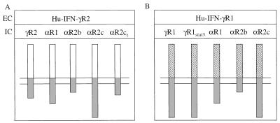
The following receptors and receptor chimeras were used in this study. The NheI site was introduced in the beginning of the transmembrane domain of the Hu-IFN-γR1/γR1 (γR1/γR1), the ligand-binding chain of the Hu-IFN-γ receptor complex (15) and the Hu-IFN-γR2/γR2 (γR2/γR2), the second chain of the Hu-IFN-γ receptor complex (16). The FLAG epitope was introduced at the NH2 terminus of the Hu-IFN-γR2 extracellular domain (FLγR2/γR2) (Fig. 1). To create chimeric receptors the extracellular domain of either Hu-IFN-γR1 or Hu-IFN-γR2 was fused to the transmembrane and intracellular domains of different subunits of the Hu-IFN-α receptor complex: the Hu-IFN-αR1 (αR1), the first chain of the Hu-IFN-α receptor complex (9); and two splice variants of the second Hu-IFN-αR2 chain of the Hu-IFN-α receptor complex: a short form, Hu-IFN-αR2b (αR2b) (10) and a long form, Hu-IFN-αR2c (11, 12). Chimeric receptors with the IFN-γR1 extracellular domain were designated γR1/αR1, γR1/αR2b, and γR1/αR2c (Fig. 1B); and chimeric receptors with the FLAG-tagged IFN-γR2 extracellular domain were designated FLγR2/αR1, FLγR2/αR2b, and FLγR2/αR2c (Fig. 1A). The FLγR2/αR2ct chimeric receptor has the IFN-αR2c intracellular domain prematurely terminated after Asp-315 (Fig. 1A). The γR1/γR1Stat3 chimeric receptor is the γR1/γR1 receptor with the Stat1α recruitment site replaced by the Stat3 recruitment site (46) (Fig. 1B).
Figure 1.
Structure of chimeric receptors. The Hu-IFN-γR1/γR1 and Hu-IFN-γR2/γR2 are the first (15) and second (16) chains of the Hu-IFN-γ receptor complex, where the NheI site was introduced in the beginning of the transmembrane domain of these receptors. The extracellular domain (EC) of either Hu-IFN-γR1 (B, hatched bars) or Hu-IFN-γR2 (A, open bars) was fused to the transmembrane and intracellular domains (IC) of different subunits of the Hu-IFN-α receptor complex (shaded bars): the Hu-IFN-αR1 (9); the Hu-IFN-αR2b (10), and the Hu-IFN-aR2c (11, 12). The FLAG epitope was introduced at the NH2 terminus of the Hu-IFN-γR2 extracellular domain. The FLγR2/αR2ct chimera has the IFN-αR2c intracellular domain prematurely terminated after Asp-315. The γR1/γR1Stat3 chimera has the Stat1α recruitment site replaced by the Stat3 recruitment site.
MHC Class I Antigen Expression and Antiviral Protection in 16-9 Cells Expressing Different Chimeric Receptors.
The chimeric receptors (Fig. 1) were used to evaluate the specific contribution of the various intracellular domains of the IFN-α receptor complex components to signaling. Because Hu-IFN-γ is highly species-specific and exhibits no activity on hamster cells, the use of the Hu-IFN-γ receptor extracellular domains allowed us to avoid the problem of variable cross-species activity of the type I IFNs and eliminate the possibility of the contribution of the endogenous hamster type I receptor chains into the Stat recruitment process.
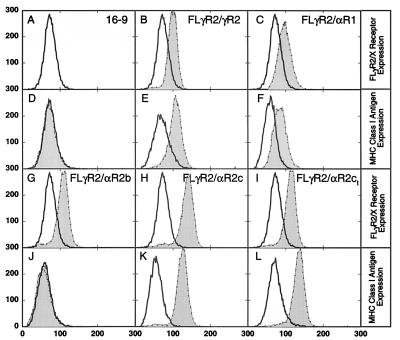
The parental 16-9 hamster cells express the Hu-IFN-γR1 chain. The chimeric receptors, FLγR2/αR1, FLγR2/αR2b, FLγR2/αR2c, and FLγR2/γR2 (Fig. 1A) were expressed in the 16-9 cells, and the transfectants obtained were designated according to the chimeric receptor expressed. After transfection clonal cell populations were obtained and the clones expressing comparable levels of the chimeric receptors as measured by flow cytometry with anti-FLAG antibody (Fig. 2) were selected and used in this study. First, the ability of Hu-IFN-γ to induce MHC class I antigen expression in these transfectants was tested (Fig. 2). Hu-IFN-γ induced MHC class I antigen expression in all cell lines except the FLγR2/αR2b cells. We then determined whether Hu-IFN-γ can induce protection against EMCV in the 16-9 cells expressing chimeric receptors with the intracellular domains of the type I IFN receptors. The FLγR2/αR1 and FLγR2/αR2b cells were not protected against EMCV in response to Hu-IFN-γ (Table 1). Only the FLγR2/αR2c cells acquired IFN-γ-induced protection against viral cytopathic effect of EMCV (Table 1).
Figure 2.
Expression of receptors on the cell surface and induction of HLA-B7 surface antigen in hamster 16-9 cells by IFN-γ. The expression of FLγR2/γR2, FLγR2/αR1, FLγR2/αR2b, FLγR2/αR2c, and FLγR2/αR2ct (B, C, G, H, and I) or induction of HLA-B7 antigen by IFN-γ (D, E, F, J, K, and L) were analyzed by flow cytometry. Ordinate, relative number of cells; abscissa, relative fluorescence. (B, C, G, H, and I) Cells were harvested and incubated with anti-FLAG monoclonal antibody (thin lines, shaded areas), the parental 16-9 cells were used as a control (A, B, C, G, H, and I, thick lines, open areas). (D, E, F, J, K, and L) Cells were treated with IFN-γ (thin lines, shaded areas) or left untreated (thick lines, open areas). Cells were the parental 16-9 cells (A and D) and clonal populations of hamster cells stably transfected with the following: FLγR2/γR2 (B and E), FLγR2/αR1 (C and F), FLγR2/αR2b (G and J), FLγR2/αR2c (H and K), and FLγR2/αR2ct (I and L).
Table 1.
Protection of cells expressing different chimeric receptors from EMCV infection by IFN-γ
| Host cell | Cell line | MHC class I antigen expression | Antiviral protection |
|---|---|---|---|
| 16-9 | − | − | |
| (Hu-IFN-γR1) | FLγR2/αR1 | + | − |
| FLγR2/αR2b | − | − | |
| FLγR2/αR2c | + | + | |
| FLγR2/αR2ct | + | − | |
| Q21 | − | − | |
| (Hu-IFN-γR2) | γR1/αR1 | − | − |
| γR1/αR2b | − | − | |
| γR1/αR2c | + | + |
Stat Activation in 16-9 Cells Expressing Different Chimeric Receptors.
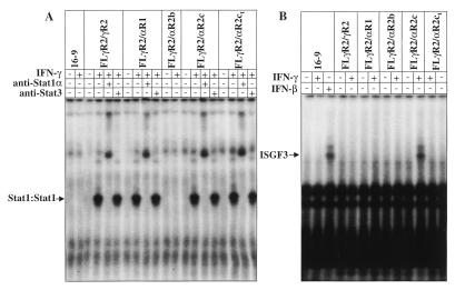
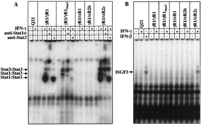
IFN-γ activates latent transcriptional factor Stat1α and induces formation of homodimeric Stat1α DNA-binding complexes which bind to the GAS element in the promoter region of the IFN-γ-inducible genes. We used a GAS probe to determine whether Stat1α was activated. The IFN-γ-induced formation of Stat1α DNA-binding complexes correlated with MHC class I antigen expression and was detected in all cell lines except the FLγR2/αR2b cells (Fig. 3A). Addition of specific anti-Stat1α antibody to the EMSA reaction caused the supershift effect, indicating that the DNA-binding complexes contained Stat1α proteins (Fig. 3A). Addition of anti-Stat3 antibody did not have any major effect (Fig. 3A), although a minor complex migrating above the Stat1 homodimeric complex was removed by anti-Stat3 antibody, indicating that a small amount of Stat3 is activated in all IFN-γ-responsive cells.
Figure 3.
EMSA in hamster 16-9 cells. Cellular lysates were prepared from untreated or IFN-γ-treated cells expressing different chimeric receptors as indicated in the figure and defined in the legends to Figs. 1 and 2. EMSAs were performed with GAS probe (A) or with ISRE probe (B). Specific anti-Stat1α and anti-Stat3 antibodies were used for supershift assays as noted. The positions of the Stat DNA-binding complexes are indicated by arrows.
Stat2 is activated in cells only by IFN-α. After IFN-α treatment, activated Stat2 and Stat1 and p48 proteins form the ISGF3 DNA-binding complex. Using the ISRE probe specific for detection of the ISGF3 complex, we tested whether IFN-γ was able to activate the ISGF3 DNA-binding complexes in cell lines expressing chimeric receptors. We detected the formation of the IFN-α-induced Stat1α/Stat2/p48 DNA-binding complexes only in the FLγR2/αR2c cells (Fig. 3B). Since Hu-IFN-β is active on hamster cells, we used Hu-IFN-β as a control for activation of the ISGF3 complex. In addition, as demonstrated above (Table 1), only the FLγR2/αR2c cells were protected against challenge with EMSV infection. Therefore, we concluded that the antiviral protection is correlated with the presence of the IFN-αR2c intracellular domain in the chimeric receptor complex and possibly with the activation of the IFN-γ-induced ISGF3 DNA-binding complexes. Thus, we decided to use the FLγR2/αR2c chain mutants to determine whether the loss of ability to activate ISGF3 will be associated with the loss of antiviral protection. We introduced a termination codon after Asp-315 of the IFN-αR2c intracellular domain, just after the putative Jak1 association site on the FLγR2/αR2c chimera (47) and expressed the truncated FLγR2/αR2ct chimeric receptor in the 16-9 cells (Fig. 2I). We still detected the IFN-γ-induced MHC class I antigen expression (Fig. 2L) and activation of Stat1α homodimeric DNA-binding complexes (Fig. 3A), but we did not detect antiviral protection in these cells (Table 1) or formation of ISGF3 complexes (Fig. 3B). In addition, these experiments narrow down the Jak1-binding region of the IFN-αR2c intracellular domain from the first 82 membrane-proximal amino acids (47) to the first 51 amino acids (Figs. 1–3).
MHC Class I Antigen Expression and Antiviral Protection in Q21 Cells Expressing Different Chimeric Receptors.
Since 16-9 cells express the Hu-IFN-γR1 chain that recruits Stat1α into the receptor complex upon IFN-γ treatment, Stat1α activation through the Hu-IFN-γR1 chain could contribute to antiviral protection in cells expressing chimeric receptors. Therefore, we focused on hamster Q21 cells (the 153B7–8 cell line) that maintained a portion of human chromosome 21 encoding the human IFNGR2 (Hu-IFN-γR2) gene (42). We created a new set of chimeric receptors by fusing the Hu-IFN-γR1 extracellular domain to the intracellular domains of all receptors that were identified to be involved in IFN-α receptor complex and signaling (Fig. 1B). The chimeric receptors γR1/αR1, γR1/αR2b, γR1/αR2c, and γR1/γR1 were expressed in Q21 cells, and the ability of Hu-IFN-γ to induce MHC class I antigen expression and antiviral protection in these transfectants was tested (Fig. 4; Table 1). Since Stat3 has been reported to be activated by type I IFNs, the chimeric receptor γR1/γR1Stat3, in which the Stat1α recruitment site was replaced by the Stat3 recruitment site, was created, expressed in Q21 cells, and served as a positive control for Stat3 activation.
Figure 4.
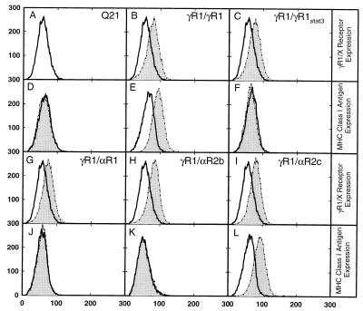
Expression of receptors on the cell surface and induction of HLA-B7 surface antigen in hamster Q21 cells by IFN-γ. The expression of γR1/γR1, γR1/γR1Stat3, γR1/αR1, γR1/αR2b, and γR1/αR2c (B, C, G, H, and I) or induction of HLA-B7 antigen by IFN-γ (D, E, F, J, K, and L) were analyzed by flow cytometry. (A, B, C, G, H, and I) Cells were harvested and incubated with anti-IFN-γR1 monoclonal antibody (thin lines, shaded areas), and the parental Q21 cells were used as a control (thick lines, open areas). (D, E, F, J, K, and L) Cells were treated with IFN-γ (thin lines, shaded areas) or left untreated (thick lines, open areas). Cells were the parental Q21 cells (A and D) and clonal populations of hamster cells stably transfected with the following: γR1/γR1 (B and E), γR1/γR1Stat3 (C and F), γR1/αR1 (G and J), γR1/αR2b (H and K), and γR1/αR2c (I and L).
As the IFN-γR2 chain does not contribute to Stat recruitment in these cells (30, 31), in response to Hu-IFN-γ the Stats can be recruited only through the intracellular domain of the transfected chimeric receptors. Both the γR1/γR1 and the γR1/αR2c cells were able to up-regulate MHC class I antigen expression after IFN-γ treatment, but the γR1/αR1 or γR1/αR2b cells were not (Fig. 4; Table 1). All tested clones had comparable levels of the cell surface expression of the chimeric receptors as demonstrated by flow cytometry with anti-Hu-IFN-γR1 antibody (Fig. 4 A, B, C, G, H, and I). These results indicate that the intracellular domains of only the IFN-γR1 and the IFN-αR2c chains are able to initiate the signal transduction cascade leading to MHC class I antigen expression, whereas the intracellular domains of the IFN-αR1 and the IFN-αR2b chains are not. When Q21 cells expressing chimeric receptors with the intracellular domains of the type I IFN receptors were challenged with EMCV, only the γR1/αR2c chimera was capable of protecting the Q21 cells against EMCV in response to Hu-IFN-γ. The other cell lines showed no antiviral protection (Table 1).
Stat Activation in Q21 Cells Expressing Different Chimeric Receptors.
We then tested whether the IFN-γ-induced up-regulation of MHC class I antigen expression and antiviral protection correlates with Stat activation. Indeed, formation of the homodimeric Stat1α DNA-binding complexes was observed in cells that were able to up-regulate MHC class I antigen expression in response to Hu-IFN-γ treatment, the γR1/γR1 and the γR1/αR2c cells (Fig. 5A). Furthermore, IFN-γ-induced activation of Stat3 as detected by EMSA with the GAS probe was observed in control the γR1/γR1Stat3 cells and in γR1/αR2c cells (Fig. 5A) indicating that the IFN-αR2c intracellular domain is able to recruit Stat3 (Fig. 5A). However, inability to detect strong Stat3 activation in 16-9 cells expressing FLγR2/αR2c (Fig. 3A) suggests that the position of the IFN-αR2c intracellular domain within the receptor complex could change the ability of the intracellular domain to recruit Stats. The presence of small amounts of Stat3 in IFN-γ-induced DNA-binding complexes was also observed in the γR1/γR1 cells (Fig. 5A).
Figure 5.
EMSA in hamster Q21 cells. Cellular lysates were prepared from untreated or IFN-γ-treated cells expressing different chimeric receptors as indicated on the figure and defined in the legends to Figs. 1 and 4. EMSAs were performed with GAS probe (A) or with ISRE probe (B). Specific anti-Stat1α and anti-Stat3 antibodies were used for supershift assays as noted. The positions of the Stat DNA-binding complexes are indicated by arrows.
Interestingly, in addition to Stat3, small amounts of Stat1 were also detected in IFNγ-induced DNA-binding complexes in Q21 cells expressing γR1/γR1Stat3 (Fig. 5A). However, IFN-γ failed to up-regulate MHC class I antigen expression in these cells (Fig. 4F). Thus, although Stat1 DNA-binding complexes can be detected in the γR1/γR1Stat3 cells (Fig. 5A), induction of MHC class I antigen on the cell surface was observed only in the γR1/γR1 and the γR1/αR2c cells (Fig. 4 E and L), and not in the γR1/γR1Stat3 cells (Fig. 4F). These observations suggest that the formation of the Stat1α DNA binding complexes in these cells might be an artifact of the EMSA, might require a minimal level of Stat1α activation to induce biological effects, or might require activation of other components in the Stat DNA-binding complexes. There appear to be two additional DNA-binding complexes; one is just above the Stat3 homodimeric complex and another one is just under the Stat1:Stat3 heterodimeric complex. The exact composition of these complexes is currently unknown.
In addition to the activation of the Stat1α and Stat3 DNA-binding complexes, IFN-γ was able to induce formation of ISGF3 complexes only in the γR1/αR2c cells, as detected by the EMSA with ISRE probe (Fig. 5B). Hu-IFN-β was used as a control for activation of the ISGF3 complex (Fig. 5B). Thus, the presence of the IFN-αR2c intracellular domain as the only Stat recruiting domain in the chimeric receptor complex enabled IFN-γ to induce activation of Stat1α, Stat3, and Stat2 proteins in the γR1/αR2c cells.
DISCUSSION
The difficulty in evaluating the contribution of each chain of the IFN-α receptor complex has resided in the lack of cell lines without the endogenous receptor chains and in the cross-species activity of the type I IFNs. To overcome these limitations we used chimeric receptors with the extracellular Hu-IFN-γ receptor chains expressed in hamster cells because Hu-IFN-γ is highly species specific and does not activate the endogenous hamster IFN-γ receptor. This strategy with the use of chimeric receptors (Fig. 1) permitted us to definitively show that the IFN-αR2c chain is necessary and sufficient for recruitment of Stat1 and Stat2.
Results of others have implicated all receptor subunits of the IFN-α receptor complex (the IFN-αR1, the IFN-αR2b, and the IFN-αR2c chains) in Stat activation (32–35). The IFN-αR1 chain was reported to bind Stat2 and Stat3 in a ligand-dependent manner through the phosphorylated Tyr-466 and the phosphorylated Tyr-527, respectively (33, 35, 37). It was originally demonstrated that the peptide containing phosphorylated Tyr-466 (pTyr-466) can inhibit type I IFN signaling in permeabilized cells and specifically interact with the SH2 domain of Stat2, but not that of Stat1 (33). Later the pTyr-466 peptide which was one amino acid longer was shown to interact with both Stat1 and Stat2 proteins (35). However, mutation of all four tyrosine residues within the IFN-αR1 intracellular domain to phenylalanine resulted in a functional receptor (48), demonstrating that phosphorylation of the IFN-αR1 chain is unnecessary for the generation of a biological response. The IFN-αR2c chain was shown to bind both Stat1 and Stat2 in a ligand-independent manner (34, 35). Because Stat1 activation by type I IFNs is Stat2-dependent (49, 50), it was proposed that Stat1 is recruited to the complex through Stat2 (35, 50). The short form, the IFN-αR2b chain, was reported to associate with Stat2 in a ligand-dependent manner (32). However, most experiments were performed in cells where the endogenous type I receptor subunits were present. It is thus likely that endogenous components interacting with the heterologous type I IFN components in the host cells contributed to the results previously reported. In our experiments, we can isolate the contributions of endogenous and exogenous components, which was not possible previously.
The chimeric receptors with the IFN-γR2 extracellular domain (Fig. 1A) were expressed in hamster 16-9 cells expressing the Hu-IFN-γR1 chain. The FLγR2/αR1 and FLγR2/αR2c chimeras rendered 16-9 cells sensitive to Hu-IFN-γ as measured by IFN-γ-induced MHC class I antigen expression and Stat1α activation, as did the intact FLγR2/γR2 (Figs. 2 and 3A). All receptors were expressed on the cell surface (Fig. 2). The FLγR2/αR2b was unable to support IFN-γ signaling because the IFN-αR2b intracellular domain does not associate with any kinase, as we previously demonstrated (30). The IFN-γ-induced activation of Stat1α DNA-binding complexes in 16-9 cells expressing chimeric receptors correlated with MHC class I antigen induction (Figs. 2 and 3A). However, formation of the IFN-γ-induced ISGF3 DNA-binding complexes was detected only in FLγR2/αR2c cells (Fig. 3B). Thus, we demonstrated that the Stat2 protein can be recruited and activated in cells expressing the chimeric receptor complex where the FLγR2/γR2 chain is replaced by the FLγR2/αR2c chimeric chain, but not by the FLγR2/αR1 or the FLγR2/αR2b chimeric chains, indicating that recruitment and activation of Stat2 occurs through the intracellular domain of the IFN-αR2c chain.
In the FLγR2/αR2c cells the presence of the Hu-IFN-γR1 chain which recruits Stat1α to the receptor complex did not permit us to answer the question whether the presence of only the IFN-αR2c intracellular domain is sufficient for recruitment and activation of Stat1α and Stat2, for formation of the ISGF3 DNA-binding complexes, and for the resultant biological activities. To further define the requirements for Stat2 recruitment, we switched to the Q21 hamster cell line. These cells express the Hu-IFN-γR2 chain, which, unlike the IFN-γR1 chain, does not recruit any Stats. Thus, the chimeric receptors with the IFN-γR1 extracellular domain (Fig. 1B) expressed in the Q21 cells were the only chains that could contribute to Stat recruitment. Therefore, MHC class I antigen induction in these cells could serve as a marker of Stat activation. The Q21 cells expressing the γR1/γR1 and γR1/αR2c chimeras demonstrated IFN-γ-induced MHC class I antigen expression, whereas the γR1/αR1 and γR1/αR2b cells did not (Fig. 4). Similarly, Stat1α DNA-binding complexes and small amounts of Stat3 DNA-binding complexes were activated in the γR1/γR1 and the γR1/αR2c cells, but not in the γR1/αR1 and the γR1/αR2b cells (Fig. 5A). However, ISGF3 DNA-binding complexes were induced only in γR1/αR2c cells (Fig. 5B). Thus, the presence of the IFN-αR2c intracellular domain as the only Stat recruiting domain in the chimeric receptor complex is sufficient for Stat1, Stat2, and Stat3 recruitment, for ISGF3 DNA-binding complex activation and for induction of MHC class I antigens.
The FLγR2/αR2c chimeric chain expressed in the 16-9 cells was able to support IFN-γ-induced protection against EMCV (Table 1). The antiviral protection correlated with induction of the ISGF3 DNA-binding complexes: the truncated FLγR2/αR2ct chain could not support antiviral activity and formation of ISGF3 complexes. The Q21 cells expressing the γR1/αR2c chimera were protected by IFN-γ treatment from EMCV infection. In none of the cells could the chimeric receptors with the αR1 or αR2b intracellular domains support antiviral activity. Thus, only when the IFN-αR2c intracellular domain was present in the chimeric receptor was IFN-γ able to induce antiviral protection in cells.
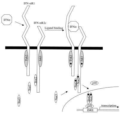
We therefore conclude that all Stats activated by type I IFNs—Stat1, Stat2, and Stat3—are activated through the IFN-αR2c intracellular domain (Fig. 6). The IFN-αR1 intracellular domain does not recruit Stats, but supports type I IFN signal transduction by bringing Tyk2 tyrosine kinase to the receptor complex. However, the IFN-αR1 intracellular domain modulates type I IFN signaling. Indeed, the deletion of the 525–544 amino acid region of the IFN-αR1 intracellular domain created a receptor that produced an enhanced response (48, 51). This same region was implicated in IFN-α-induced activation of phosphatidylinositol 3-kinase (PI 3-kinase) through association with the IFN-αR1 chain via Stat3 as an adaptor protein (52). However, it is not clear how this same region could recruit Stat3 and PI 3-kinase, which were reported to be necessary for full biological responsiveness (52, 53) but at the same time, when eliminated, produce a receptor with enhanced activity. Nevertheless, our results demonstrated that the IFN-αR2c intracellular domain was able to recruit and activate all Stats involved in type I IFN signaling, Stat1, Stat2, and Stat3, without the presence of the IFN-αR1 intracellular domain. We thus conclude that Stat recruitment by the type I IFN receptor complex is solely a function of the IFN-αR2c intracellular domain and that the IFN-αR2c chain is sufficient and necessary for recruitment of Stat1, Stat2, and Stat3.
Figure 6.
Model of type I IFN receptor complex and signaling. Ligand binding to the subunits of the type I IFN receptor complex, the IFN-αR2c and the IFN-αR1 chains, initiates the cascade of signal transduction events. All Stats involved in IFN-α signaling are activated through the intracellular domain of the IFN-αR2c chain (see text for details).
Acknowledgments
We thank James Darnell for rabbit anti-Stat1α antibody, James Ihle for rabbit anti-Stat3 antibody, Gianni Garotta for monoclonal anti-Hu-IFN-γR1 antibody, Jerry Langer for helpful discussion, and Eleanor Kells for assistance in the preparation of this manuscript. This study was supported in part by Grants RO1-CA46465 and RO1-CA52363 from the National Cancer Institute and RO1 AI36450 from the National Institute of Allergy and Infectious Diseases, and American Cancer Society Grant VM-135 to S.P, and by American Heart Association Grant AHA#9730247N to S.V.K. A special award from the Milstein Family Foundation to S.P. provided additional support for a variety of efforts in this project.
ABBREVIATIONS
- Hu-
human
- ISGF3
IFN-α-stimulated gene factor 3
- EMSA
electrophoretic mobility-shift assay
- ISRE
IFN-stimulated response element
- EMCV
encephalomyocarditis virus
- GAS
IFN-γ-activated sequence
References
- 1.Pestka S, Langer J A, Zoon K C, Samuel C E. Annu Rev Biochem. 1987;56:727–777. doi: 10.1146/annurev.bi.56.070187.003455. [DOI] [PubMed] [Google Scholar]
- 2.Pestka S. Semin Oncol. 1997;24:S9-4–S9-17. [PubMed] [Google Scholar]
- 3.Diaz M O, Pomykala H, Bohlander S K, Maltepe E, Malik K, Brownstein B, Olopade O I. Genomics. 1996;22:540–552. doi: 10.1006/geno.1994.1427. [DOI] [PubMed] [Google Scholar]
- 4.Merlin G, Falcoff E, Aguet M. J Gen Virol. 1985;66:1149–1152. doi: 10.1099/0022-1317-66-5-1149. [DOI] [PubMed] [Google Scholar]
- 5.Flores I, Mariano T M, Pestka S. J Biol Chem. 1991;266:19875–19877. [PubMed] [Google Scholar]
- 6.Cleary C M, Donnelly R J, Soh J, Mariano T M, Pestka S. J Biol Chem. 1994;269:18747–18749. [PubMed] [Google Scholar]
- 7.Cook J R, Cleary C M, Mariano T M, Izotova L, Pestka S. J Biol Chem. 1996;271:13488–13453. doi: 10.1074/jbc.271.23.13448. [DOI] [PubMed] [Google Scholar]
- 8.Cutrone E C, Langer J A. FEBS Lett. 1997;404:197–202. doi: 10.1016/s0014-5793(97)00129-4. [DOI] [PubMed] [Google Scholar]
- 9.Uzé G, Lutfalla G, Gresser I. Cell. 1990;60:225–234. doi: 10.1016/0092-8674(90)90738-z. [DOI] [PubMed] [Google Scholar]
- 10.Novick D, Cohen B, Rubinstein M. Cell. 1994;77:391–400. doi: 10.1016/0092-8674(94)90154-6. [DOI] [PubMed] [Google Scholar]
- 11.Domanski P, Witte M, Kellum M, Rubinstein M, Hackett R, Pitha P, Colamonici O R. J Biol Chem. 1995;270:21606–21611. doi: 10.1074/jbc.270.37.21606. [DOI] [PubMed] [Google Scholar]
- 12.Lutfalla G, Holland S J, Cinato E, Monneron D, Reboul J, Rogers N C, Smith J M, Stark G R, Gardiner K, Mogensen K E, Kerr I M, Uzé G. EMBO J. 1995;14:5100–5108. doi: 10.1002/j.1460-2075.1995.tb00192.x. [DOI] [PMC free article] [PubMed] [Google Scholar]
- 13.Bazan J F. Proc Natl Acad Sci USA. 1990;87:6934–6938. doi: 10.1073/pnas.87.18.6934. [DOI] [PMC free article] [PubMed] [Google Scholar]
- 14.Thoreau E, Petridou B, Kelly P A, Djiane J, Mornon J P. FEBS Lett. 1991;282:26–31. doi: 10.1016/0014-5793(91)80437-8. [DOI] [PubMed] [Google Scholar]
- 15.Aguet M, Dembic Z, Merlin G. Cell. 1998;55:273–280. doi: 10.1016/0092-8674(88)90050-5. [DOI] [PubMed] [Google Scholar]
- 16.Soh J, Donnelly R J, Kotenko S, Mariano T M, Cook J R, Wang N, Emanuel S, Schwartz B, Miki T, Pestka S. Cell. 1994;76:793–802. doi: 10.1016/0092-8674(94)90354-9. [DOI] [PubMed] [Google Scholar]
- 17.Liu Y, Wei S H-Y, Ho A S-Y, deWaal Malefut R, Moore K W. J Immunol. 1994;152:1821–1829. [PubMed] [Google Scholar]
- 18.Kotenko S V, Krause C D, Izotova L S, Pollack B P, Wu W, Pestka S. EMBO J. 1997;16:5894–5903. doi: 10.1093/emboj/16.19.5894. [DOI] [PMC free article] [PubMed] [Google Scholar]
- 19.Fu X Y, Kessler D S, Veals S A, Levy D E, Darnell J E., Jr Proc Natl Acad Sci USA. 1990;87:8555–8559. doi: 10.1073/pnas.87.21.8555. [DOI] [PMC free article] [PubMed] [Google Scholar]
- 20.Fu X Y, Schindler C, Improta T, Aebersold R, Darnell J E., Jr Proc Natl Acad Sci USA. 1992;89:7840–7843. doi: 10.1073/pnas.89.16.7840. [DOI] [PMC free article] [PubMed] [Google Scholar]
- 21.Veals S A, Schindler C, Leonard D, Fu X Y, Aebersold R, Darnell J E, Jr, Levy D E. Mol Cell Biol. 1992;12:3315–3324. doi: 10.1128/mcb.12.8.3315. [DOI] [PMC free article] [PubMed] [Google Scholar]
- 22.Velazquez L, Fellous M, Stark G R, Pellegrini S. Cell. 1992;70:313–322. [PubMed] [Google Scholar]
- 23.Schindler C, Fu X Y, Improta T, Aebersold R, Darnell J E., Jr Proc Natl Acad Sci USA. 1992;89:7836–7839. doi: 10.1073/pnas.89.16.7836. [DOI] [PMC free article] [PubMed] [Google Scholar]
- 24.Müller M, Briscoe J, Laxton C, Guschin D, Ziemiecki A, Silvennoinen O, Harpur A G, Barbieri G, Witthuhn B A, Schindler C, et al. Nature (London) 1993;366:129–135. doi: 10.1038/366129a0. [DOI] [PubMed] [Google Scholar]
- 25.Pestka S, Kotenko S V, Muthukumaran G, Izotova L S, Cook J R, Garotta G. Cytokine Growth Factor Rev. 1997;8:189–206. doi: 10.1016/s1359-6101(97)00009-9. [DOI] [PubMed] [Google Scholar]
- 26.Schindler C, Darnell J E., Jr Annu Rev Biochem. 1995;64:621–651. doi: 10.1146/annurev.bi.64.070195.003201. [DOI] [PubMed] [Google Scholar]
- 27.Greenlund A C, Farrar M A, Viviano B L, Schreiber R D. EMBO J. 1994;13:1591–1600. doi: 10.1002/j.1460-2075.1994.tb06422.x. [DOI] [PMC free article] [PubMed] [Google Scholar]
- 28.Weber-Nordt R M, Riley J K, Greenlund A C, Moore K W, Darnell J E, Schreiber R D. J Biol Chem. 1996;271:27954–27961. doi: 10.1074/jbc.271.44.27954. [DOI] [PubMed] [Google Scholar]
- 29.Kotenko S V, Izotova L S, Pollack B P, Mariano T M, Donnelly R J, Muthukumaran G, Cook J R, Garotta G, Silvennoinen O, Ihle J N, Pestka S. J Biol Chem. 1995;270:20915–20921. doi: 10.1074/jbc.270.36.20915. [DOI] [PubMed] [Google Scholar]
- 30.Kotenko S V, Izotova L S, Pollack B P, Muthukumaran G, Paukku K, Silvennoinen O, Ihle J N, Pestka S. J Biol Chem. 1996;271:17174–17182. doi: 10.1074/jbc.271.29.17174. [DOI] [PubMed] [Google Scholar]
- 31.Bach E A, Tanner J W, Marsters S, Ashkenazi A, Aguet M, Shaw A S, Schreiber R D. Mol Cell Biol. 1996;16:3214–3221. doi: 10.1128/mcb.16.6.3214. [DOI] [PMC free article] [PubMed] [Google Scholar]
- 32.Uddin S, Chamdin A, Platanias L C. J Biol Chem. 1995;270:24627–24630. doi: 10.1074/jbc.270.42.24627. [DOI] [PubMed] [Google Scholar]
- 33.Yan H, Krishnan K, Greenlund A C, Gupta S, Lim J T, Schreiber R D, Schindler C W, Krolewski J J. EMBO J. 1996;15:1064–1074. [PMC free article] [PubMed] [Google Scholar]
- 34.Domanski P, Colamonici O R. Cytokine Growth Factor Rev. 1996;7:143–151. doi: 10.1016/1359-6101(96)00017-2. [DOI] [PubMed] [Google Scholar]
- 35.Li X, Leung S, Kerr I M, Stark G R. Mol Cell Biol. 1997;17:2048–2056. doi: 10.1128/mcb.17.4.2048. [DOI] [PMC free article] [PubMed] [Google Scholar]
- 36.Hwang S Y, Hertzog P J, Holland K A, Sumarsono S H, Tymms M J, Hamilton J A, Whitty G, Bertoncello I, Kola I. Proc Natl Acad Sci USA. 1995;92:11284–11288. doi: 10.1073/pnas.92.24.11284. [DOI] [PMC free article] [PubMed] [Google Scholar]
- 37.Yang C H, Shi W, Basu L, Murti A, Constantinescu S N, Blatt L, Croze E, Mullersman J E, Pfeffer L M. J Biol Chem. 1996;271:8057–8061. doi: 10.1074/jbc.271.14.8057. [DOI] [PubMed] [Google Scholar]
- 38.Krishnan K, Yan H, Lim J T, Krolewski J J. Oncogene. 1996;13:125–133. [PubMed] [Google Scholar]
- 39.Goldman L A, Cutrone E C, Kotenko S V, Krause C D, Langer J A. BioTechniques. 1996;21:1013–1015. doi: 10.2144/96216bm10. [DOI] [PubMed] [Google Scholar]
- 40.Muthukumaran G, Kotenko S V, Donnelly R, Ihle J N, Pestka S. J Biol Chem. 1997;272:4993–4999. doi: 10.1074/jbc.272.8.4993. [DOI] [PubMed] [Google Scholar]
- 41.Soh J, Donnelly R J, Mariano T M, Cook J R, Schwartz B, Pestka S. Proc Natl Acad Sci USA. 1993;90:8737–8741. doi: 10.1073/pnas.90.18.8737. [DOI] [PMC free article] [PubMed] [Google Scholar]
- 42.Jung V, Jones C, Kumar C S, Stefanos S, O’Connell S, Pestka S. J Biol Chem. 1990;265:1827–1830. [PubMed] [Google Scholar]
- 43.Campbell M J. BioTechniques. 1995;18:1027–1032. [PubMed] [Google Scholar]
- 44.Barnstable C J, Bodmer W F, Brown G, Galfre G, Milstein C, Williams A F, Ziegler A. Cell. 1978;14:9–20. doi: 10.1016/0092-8674(78)90296-9. [DOI] [PubMed] [Google Scholar]
- 45.Familletti P C, Rubinstein S, Pestka S. Methods Enzymol. 1981;78:387–394. doi: 10.1016/0076-6879(81)78146-1. [DOI] [PubMed] [Google Scholar]
- 46.Stahl N, Farruggella T J, Boulton T G, Zhong Z, Darnell J E, Jr, Yancopoulos G D. Science. 1995;267:1349–1353. doi: 10.1126/science.7871433. [DOI] [PubMed] [Google Scholar]
- 47.Domanski P, Fish E, Nadeau O W, Witte M, Platanias L C, Yan H, Krolewski J, Pitha P, Colamonici O R. J Biol Chem. 1997;272:26388–26393. doi: 10.1074/jbc.272.42.26388. [DOI] [PubMed] [Google Scholar]
- 48.Gibbs V C, Takahashi M, Aguet M, Chuntharapai A. J Biol Chem. 1996;271:28710–28716. doi: 10.1074/jbc.271.45.28710. [DOI] [PubMed] [Google Scholar]
- 49.Improta T, Schindler C, Horvath C M, Kerr I M, Stark G R, Darnell J E., Jr Proc Natl Acad Sci USA. 1994;91:4776–4780. doi: 10.1073/pnas.91.11.4776. [DOI] [PMC free article] [PubMed] [Google Scholar]
- 50.Leung S, Qureshi S A, Kerr I M, Darnell J E, Jr, Stark G R. Mol Cell Biol. 1995;15:1312–1317. doi: 10.1128/mcb.15.3.1312. [DOI] [PMC free article] [PubMed] [Google Scholar]
- 51.Basu L, Yang C H, Murti A, Garcia J V, Croze E, Constantinescu S N, Mullersman J E, Pfeffer L M. Virology. 1998;242:14–21. doi: 10.1006/viro.1997.9002. [DOI] [PubMed] [Google Scholar]
- 52.Pfeffer L M, Mullersman J E, Pfeffer S R, Murti A, Yang C H. Science. 1997;276:1418–1420. doi: 10.1126/science.276.5317.1418. [DOI] [PubMed] [Google Scholar]
- 53.Yang C H, Murti A, Pfeffer L M. Proc Natl Acad Sci USA. 1998;95:5568–5572. doi: 10.1073/pnas.95.10.5568. [DOI] [PMC free article] [PubMed] [Google Scholar]


