

Abstract
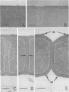
The changes in cell morphology of Bacillus subtilis rodB during a temperature shift from 20 to 42 degrees C, in the absence of added anions, are described. At 20 degrees C the organisms grow as rods but gradually become spherical in shape when placed at 42 degrees C. The shape change is initiated by an increase in diameter at the cell equator, resulting in a bulged morphology, which is further modified to the morphology of a coccus. This change may involve a modification of the pattern of normal cylindrical extension such that incorporation of newly synthesized wall leads only to increase in diameter, perhaps from a growth zone of limited extent. The pattern of surface growth was followed by reconstructing the sequence of cross wall formation and pole construction in rods grown at 20 degrees C and in organisms incubated at 42 degrees C for 75 and 150 min. In thin section, wall forming the septum and nascent poles can be distinguished from the surface distal to the division site by the presence of raised tears, perhaps analogous to the wall bands of streptococci. By using an analog rotation technique involving the three-dimensional reconstruction of cells by mathematical rotation of axial thin sections about their longitudinal axis, it is shown that the proportion of septal wall increases during the shape change. In the coccal forms, all surface growth may arise from septal growth sites.
Full text
PDF


Images in this article


Selected References
These references are in PubMed. This may not be the complete list of references from this article.
- Archibald A. R., Coapes H. E. Bacteriophage SP50 as a marker for cell wall growth in Bacillus subtilis. J Bacteriol. 1976 Mar;125(3):1195–1206. doi: 10.1128/jb.125.3.1195-1206.1976. [DOI] [PMC free article] [PubMed] [Google Scholar]
- Boylan R. J., Mendelson N. H. Initial characterization of a temperature-sensitive rod--mutant of Bacillus subtilis. J Bacteriol. 1969 Dec;100(3):1316–1321. doi: 10.1128/jb.100.3.1316-1321.1969. [DOI] [PMC free article] [PubMed] [Google Scholar]
- Burdett I. D., Higgins M. L. Study of pole assembly in Bacillus subtilis by computer reconstruction of septal growth zones seen in central, longitudinal thin sections of cells. J Bacteriol. 1978 Feb;133(2):959–971. doi: 10.1128/jb.133.2.959-971.1978. [DOI] [PMC free article] [PubMed] [Google Scholar]
- Cole R. M., Popkin T. J., Boylan R. J., Mendelson N. H. Ultrastructure of a temperature-sensitive rod- mutant of Bacillus subtilis. J Bacteriol. 1970 Sep;103(3):793–810. doi: 10.1128/jb.103.3.793-810.1970. [DOI] [PMC free article] [PubMed] [Google Scholar]
- Higgins M. L., Pooley H. M., Shockman G. D. Reinitiation of cell wall growth after threonine starvation of Streptococcus faecalis. J Bacteriol. 1971 Mar;105(3):1175–1183. doi: 10.1128/jb.105.3.1175-1183.1971. [DOI] [PMC free article] [PubMed] [Google Scholar]
- Higgins M. L., Shockman G. D. Model for cell wall growth of Streptococcus faecalis. J Bacteriol. 1970 Feb;101(2):643–648. doi: 10.1128/jb.101.2.643-648.1970. [DOI] [PMC free article] [PubMed] [Google Scholar]
- Higgins M. L., Shockman G. D. Study of cycle of cell wall assembly in Streptococcus faecalis by three-dimensional reconstructions of thin sections of cells. J Bacteriol. 1976 Sep;127(3):1346–1358. doi: 10.1128/jb.127.3.1346-1358.1976. [DOI] [PMC free article] [PubMed] [Google Scholar]
- Higgins M. L. Three-dimensional reconstruction of whole cells of Streptococcus faecalis from thin sections of cells. J Bacteriol. 1976 Sep;127(3):1337–1345. doi: 10.1128/jb.127.3.1337-1345.1976. [DOI] [PMC free article] [PubMed] [Google Scholar]
- Highton P. J., Hobbs D. G. Penicillin and cell wall synthesis: a study of Bacillus cereus by electron microscopy. J Bacteriol. 1972 Mar;109(3):1181–1190. doi: 10.1128/jb.109.3.1181-1190.1972. [DOI] [PMC free article] [PubMed] [Google Scholar]
- Hughes R. C., Stokes E. Cell wall growth in Bacillus licheniformis followed by immunofluorescence with mucopeptide-specific antiserum. J Bacteriol. 1971 May;106(2):694–696. doi: 10.1128/jb.106.2.694-696.1971. [DOI] [PMC free article] [PubMed] [Google Scholar]
- KELLENBERGER E., ARBER W. Electron microscopical studies of phage multiplication. I. A method for quantitative analysis of particle suspensions. Virology. 1957 Apr;3(2):245–255. doi: 10.1016/0042-6822(57)90091-0. [DOI] [PubMed] [Google Scholar]
- KELLENBERGER E., RYTER A., SECHAUD J. Electron microscope study of DNA-containing plasms. II. Vegetative and mature phage DNA as compared with normal bacterial nucleoids in different physiological states. J Biophys Biochem Cytol. 1958 Nov 25;4(6):671–678. doi: 10.1083/jcb.4.6.671. [DOI] [PMC free article] [PubMed] [Google Scholar]
- Karamata D., McConnell M., Rogers H. J. Mapping of rod mutants of Bacillus subtilis. J Bacteriol. 1972 Jul;111(1):73–79. doi: 10.1128/jb.111.1.73-79.1972. [DOI] [PMC free article] [PubMed] [Google Scholar]
- Marr A. G., Harvey R. J., Trentini W. C. Growth and division of Escherichia coli. J Bacteriol. 1966 Jun;91(6):2388–2389. doi: 10.1128/jb.91.6.2388-2389.1966. [DOI] [PMC free article] [PubMed] [Google Scholar]
- Mendelson N. H., Reeve J. N. Growth of the Bacillus subtilis cell surface. Nat New Biol. 1973 May 9;243(123):62–64. [PubMed] [Google Scholar]
- Polley H. M., Schlaeppi J. M., Karamata D. Localised insertion of new cell wall in Bacillus subtilis. Nature. 1978 Jul 20;274(5668):264–266. doi: 10.1038/274264a0. [DOI] [PubMed] [Google Scholar]
- Pooley H. M. Layered distribution, according to age, within the cell wall of bacillus subtilis. J Bacteriol. 1976 Mar;125(3):1139–1147. doi: 10.1128/jb.125.3.1139-1147.1976. [DOI] [PMC free article] [PubMed] [Google Scholar]
- Pooley H. M. Turnover and spreading of old wall during surface growth of Bacillus subtilis. J Bacteriol. 1976 Mar;125(3):1127–1138. doi: 10.1128/jb.125.3.1127-1138.1976. [DOI] [PMC free article] [PubMed] [Google Scholar]
- Reeve J. N., Mendelson N. H. Cell morphology of Bacillus subtilis: the effect of genetic background on the expression of a rod - gene. Mol Gen Genet. 1972;119(1):11–26. doi: 10.1007/BF00270440. [DOI] [PubMed] [Google Scholar]
- Rogers H. J., McConnell M., Burdett I. D. Cell wall or membrane mutants of Bacillus subtilis and Bacillus licheniformis with grossly deformed morphology. Nature. 1968 Jul 20;219(5151):285–288. doi: 10.1038/219285a0. [DOI] [PubMed] [Google Scholar]
- Rogers H. J., McConnell M., Burdett I. D. The isolation and characterization of mutants of Bacillus subtilis and Bacillus licheniformis with disturbed morphology and cell division. J Gen Microbiol. 1970 May;61(2):155–171. doi: 10.1099/00221287-61-2-155. [DOI] [PubMed] [Google Scholar]
- Rogers H. J., Taylor C. Autolysins and shape change in rodA mutants of Bacillus subtilis. J Bacteriol. 1978 Sep;135(3):1032–1042. doi: 10.1128/jb.135.3.1032-1042.1978. [DOI] [PMC free article] [PubMed] [Google Scholar]
- Rogers H. J., Thurman P. F., Buxton R. S. Magnesium and anion requirements of rodB mutants of Bacillus subtilis. J Bacteriol. 1976 Feb;125(2):556–564. doi: 10.1128/jb.125.2.556-564.1976. [DOI] [PMC free article] [PubMed] [Google Scholar]
- Rogers H. J., Thurman P. F. Temperature-sensitive nature of the rodB maturation in Bacillus subtilis. J Bacteriol. 1978 Jan;133(1):298–305. doi: 10.1128/jb.133.1.298-305.1978. [DOI] [PMC free article] [PubMed] [Google Scholar]
- Shiflett M. A., Brooks D., Young F. E. Cell wall and morphological changes induced by temperature shift in Bacillus subtilis cell wall mutants. J Bacteriol. 1977 Nov;132(2):681–690. doi: 10.1128/jb.132.2.681-690.1977. [DOI] [PMC free article] [PubMed] [Google Scholar]
- de Chastellier C., Hellio R., Ryter A. Study of cell wall growth in Bacillus megaterium by high-resolution autoradiography. J Bacteriol. 1975 Sep;123(3):1184–1196. doi: 10.1128/jb.123.3.1184-1196.1975. [DOI] [PMC free article] [PubMed] [Google Scholar]