

Abstract
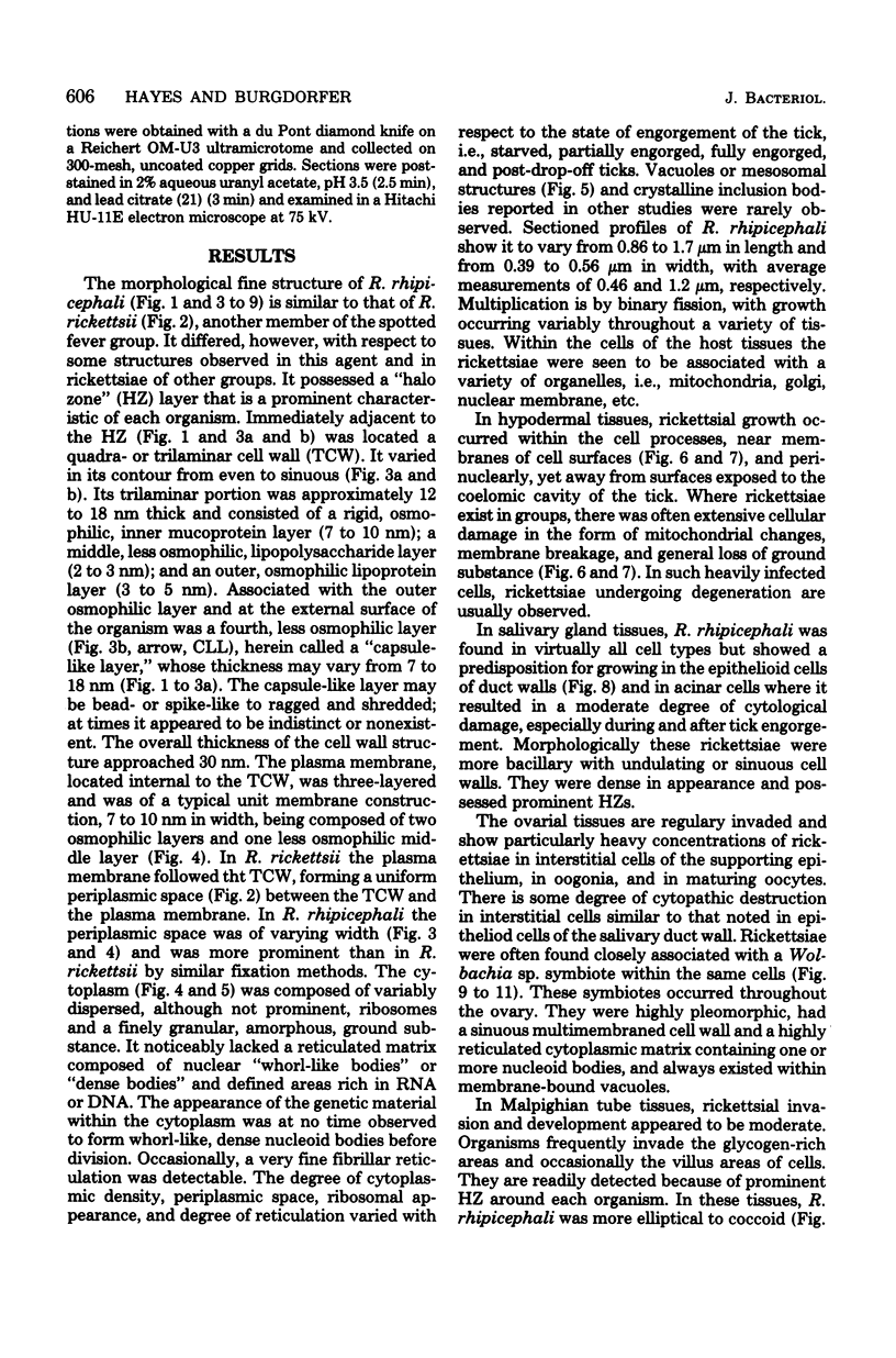
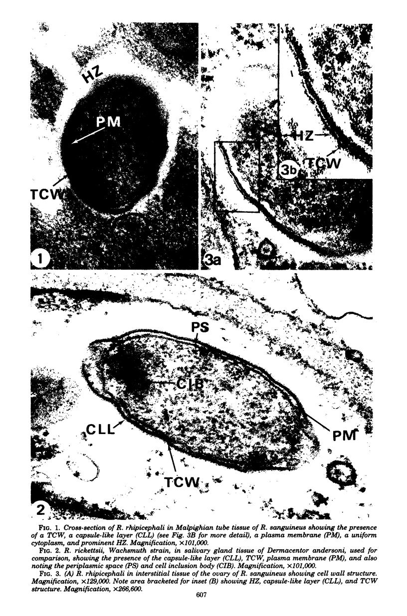
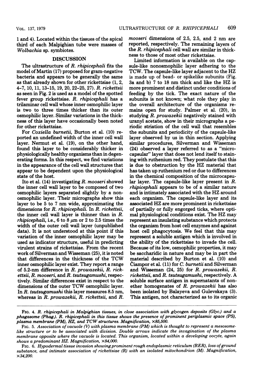
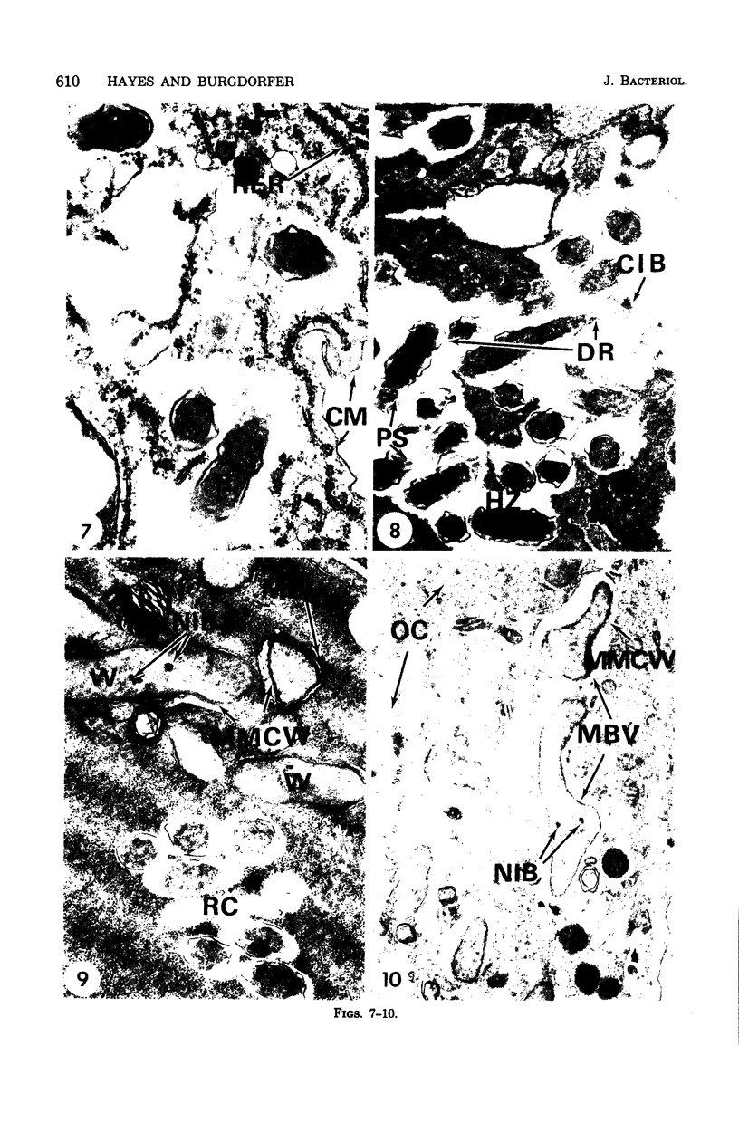
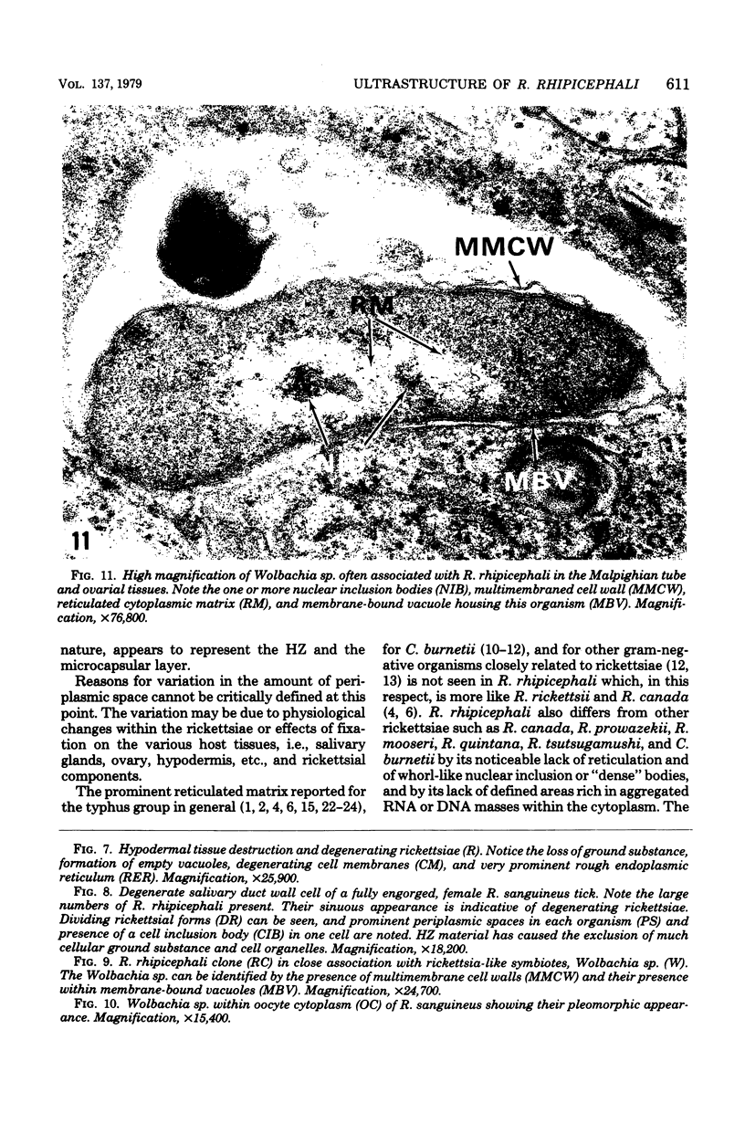
Rickettsia rhipicephali is similar in ultrastructure to R. rickettsii while differing from other rickettsiae of the typhus group and of Q fever and others by its lack of a prominently reticulated cytoplasmic matrix and in the thickness of the inner osmophilic layer of the cell wall. In tissues of the tick vector Rhipicephalus sanguineus, R. rhipicephali had a mean length and width of 1.2 and 0.46 micrometer, respectively. It possessed a trilaminar cell wall with an adhering capsule-like layer. The trilaminar cell wall was approximately 12 to 18 nm thick; its inner osmophilic layer was thicker than that previously reported for other rickettsiae. The capsule-like layer varied from 7 to 18 nm thick. The plasma membrane was similar in structure, measurement, and appearance to that of other reported rickettsiae. The cytoplasm appeared to be composed of a finely granular, amorphous, ground substance and randomly dispersed ribosomes and lacked a reticular matrix or nuclear fibrils. In massively infected salivary glands and ovarial tissues of its tick vector, R. rhipicephali produced a low degree of histopathology which does not appear to affect the engorgement and egg-laying process of the ticks.
Full text
PDF
Images in this article
Selected References
These references are in PubMed. This may not be the complete list of references from this article.
- Anacker R. L., Pickens E. G., Lackman D. B. Details of the ultrastructure of Rickettsia prowazekii grown in the chick yolk sac. J Bacteriol. 1967 Jul;94(1):260–262. doi: 10.1128/jb.94.1.260-262.1967. [DOI] [PMC free article] [PubMed] [Google Scholar]
- Anderson D. R., Hopps H. E., Barile M. F., Bernheim B. C. Comparison of the ultrastructure of several rickettsiae, ornithosis virus, and Mycoplasma in tissue culture. J Bacteriol. 1965 Nov;90(5):1387–1404. doi: 10.1128/jb.90.5.1387-1404.1965. [DOI] [PMC free article] [PubMed] [Google Scholar]
- Balayeva N. M., Gulevskaya S. A. Comparative characteristics of "ether" and "non-ether" soluble ricketssia prowazekii antigens and electron microscopy findings on the morphology of the rickettsiae during isolation of the antigens. J Hyg Epidemiol Microbiol Immunol. 1972;16(1):92–100. [PubMed] [Google Scholar]
- Brinton L. P., Burgdorfer W. Fine structure of Rickettsia canada in tissues of Dermacentor andersoni Stiles. J Bacteriol. 1971 Mar;105(3):1149–1159. doi: 10.1128/jb.105.3.1149-1159.1971. [DOI] [PMC free article] [PubMed] [Google Scholar]
- Burgdorfer W., Anacker R. L., Bird R. G., Bertram D. S. Intranuclear growth of Rickettsia rickettsii. J Bacteriol. 1968 Oct;96(4):1415–1418. doi: 10.1128/jb.96.4.1415-1418.1968. [DOI] [PMC free article] [PubMed] [Google Scholar]
- Burgdorfer W., Brinton L. P., Hughes L. E. Isolation and characterization of symbiotes from the Rocky Mountain wood tick, Dermacentor andersoni. J Invertebr Pathol. 1973 Nov;22(3):424–434. doi: 10.1016/0022-2011(73)90173-0. [DOI] [PubMed] [Google Scholar]
- Burgdorfer W., Brinton L. P. Intranuclear growth of rickettsia Canada, a member of the typhus group. Infect Immun. 1970 Jul;2(1):112–114. doi: 10.1128/iai.2.1.112-114.1970. [DOI] [PMC free article] [PubMed] [Google Scholar]
- Burgdorfer W., Sexton D. J., Gerloff R. K., Anacker R. L., Philip R. N., Thomas L. A. Rhipicephalus sanguineus: vector of a new spotted fever group rickettsia in the United States. Infect Immun. 1975 Jul;12(1):205–210. doi: 10.1128/iai.12.1.205-210.1975. [DOI] [PMC free article] [PubMed] [Google Scholar]
- Burton P. R., Stueckemann J., Paretsky D. Electron microscopy studies of the limiting layers of the rickettsia Coxiella burneti. J Bacteriol. 1975 Apr;122(1):316–324. doi: 10.1128/jb.122.1.316-324.1975. [DOI] [PMC free article] [PubMed] [Google Scholar]
- Ciampor F., Schramek S., Brezina R. Electron microscopy of ruthenium red-stained phase I and II Coxiella burneti. Acta Virol. 1972 Nov;16(6):503–506. [PubMed] [Google Scholar]
- Higashi N. Recent advances in electron microscope studies on ultrastructure of rickettsiae. Zentralbl Bakteriol Orig. 1968 Apr;206(3):277–283. [PubMed] [Google Scholar]
- ITO S., VINSON J. W. FINE STRUCTURE OF RICKETTSIA QUINTANA CULTIVATED IN VITRO AND IN THE LOUSE. J Bacteriol. 1965 Feb;89:481–495. doi: 10.1128/jb.89.2.481-495.1965. [DOI] [PMC free article] [PubMed] [Google Scholar]
- Ito S., Vinson J. W., McGuire T. J., Jr Murine typhus Rickettsiae in the Oriental rat flea. Ann N Y Acad Sci. 1975;266:35–60. doi: 10.1111/j.1749-6632.1975.tb35087.x. [DOI] [PubMed] [Google Scholar]
- Jadin J., Creemers J., Jadin J. M., Giroud P. Ultrastructure of Rickettsia prowazeki. Acta Virol. 1968 Jan;12(1):7–10. [PubMed] [Google Scholar]
- LUFT J. H. Improvements in epoxy resin embedding methods. J Biophys Biochem Cytol. 1961 Feb;9:409–414. doi: 10.1083/jcb.9.2.409. [DOI] [PMC free article] [PubMed] [Google Scholar]
- Martin H. H. Bacterial protoplasts--a review. J Theor Biol. 1963 Jul;5(1):1–34. doi: 10.1016/0022-5193(63)90034-1. [DOI] [PubMed] [Google Scholar]
- Morgan C., Rosenkranz H. S. Ultrastructure of Escherichia coli depleted of an amino acid. J Bacteriol. 1970 May;102(2):584–587. doi: 10.1128/jb.102.2.584-587.1970. [DOI] [PMC free article] [PubMed] [Google Scholar]
- Nermut M. V., Schramek S., Brezina R. Electron microscopy of Coxiella burneti phase I and II. Acta Virol. 1968 Sep;12(5):446–452. [PubMed] [Google Scholar]
- Palmer E. L., Mallavia L. P., Tzianabos T., Obijeski J. F. Electron microscopy of the cell wall of Rickettsia prowazeki. J Bacteriol. 1974 Jun;118(3):1158–1166. doi: 10.1128/jb.118.3.1158-1166.1974. [DOI] [PMC free article] [PubMed] [Google Scholar]
- REYNOLDS E. S. The use of lead citrate at high pH as an electron-opaque stain in electron microscopy. J Cell Biol. 1963 Apr;17:208–212. doi: 10.1083/jcb.17.1.208. [DOI] [PMC free article] [PubMed] [Google Scholar]
- SCHAECHTER M., TOUSIMIS A. J., COHN Z. A., ROSEN H., CAMPBELL J., HAHN F. E. Morphological, chemical, and serological studies of the cell walls of Rickettsia mooseri. J Bacteriol. 1957 Dec;74(6):822–829. doi: 10.1128/jb.74.6.822-829.1957. [DOI] [PMC free article] [PubMed] [Google Scholar]
- Shkolnik L. Y., Zatulovsky B. G., Shestopalova N. M. Ultrastructure of Rickettsia prow azeki. An electron microscope study of ultrathin sections from infected louse guts and chick embryo yolk sacs. Acta Virol. 1966 May;10(3):260–265. [PubMed] [Google Scholar]
- Silverman D. J., Wisseman C. L., Jr Comparative ultrastructural study on the cell envelopes of Rickettsia prowazekii, Rickettsia rickettsii, and Rickettsia tsutsugamushi. Infect Immun. 1978 Sep;21(3):1020–1023. doi: 10.1128/iai.21.3.1020-1023.1978. [DOI] [PMC free article] [PubMed] [Google Scholar]
- Spendlove R. S., Crosbie R. B., Hayes S. F., Keeler R. F. TRICINE-buffered tissue culture media for control of mycoplasma contaminants. Proc Soc Exp Biol Med. 1971 May;137(1):258–263. doi: 10.3181/00379727-137-35556. [DOI] [PubMed] [Google Scholar]
- Walker D. H., Harrison A., Henderson F., Murphy F. A. Identification of Rickettsia rickettsii in a guinea pig model by immunofluorescent and electron microscopic techniques. Am J Pathol. 1977 Feb;86(2):343–358. [PMC free article] [PubMed] [Google Scholar]