Abstract
Sorted CD4+ and CD8+ T cells from the peripheral blood or bone marrow of donor C57BL/6 (H-2b) mice were tested for their capacity to induce graft-versus-host disease (GVHD) by injecting the cells, along with stringently T cell–depleted donor marrow cells, into lethally irradiated BALB/c (H-2d) host mice. The peripheral blood T cells were at least 30 times more potent than the marrow T cells in inducing lethal GVHD. As NK1.1+ T cells represented <1% of all T cells in the blood and ∼30% of T cells in the marrow, the capacity of sorted marrow NK1.1− CD4+ and CD8+ T cells to induce GVHD was tested. The latter cells had markedly increased potency, and adding back marrow NK1.1+ T cells suppressed GVHD. The marrow NK1.1+ T cells secreted high levels of both interferon γ (IFN-γ) and interleukin 4 (IL-4), and the NK1.1− T cells secreted high levels of IFN-γ with little IL-4. Marrow NK1.1+ T cells obtained from IL-4−/− rather than wild-type C57BL/6 donors not only failed to prevent GVHD but actually increased its severity. Together, these results demonstrate that GVHD is reciprocally regulated by the NK1.1− and NK1.1+ T cell subsets via their differential production of cytokines.
Keywords: bone marrow transplantation, immune regulation, regulatory T cells, cytokines, interleukin 4
Allogeneic GVHD is a major barrier to the widespread use of bone marrow transplantation in clinical medicine (1–4). Acute GVHD is mediated by donor T cells expressing α/β+ TCRs (α/β+ T cells) that recognize molecules of the MHC (referred to as the H-2 complex in mice) and their associated peptides (5–7). The central role of donor T cells has been documented by the markedly reduced frequency and severity of GVHD in hosts given bone marrow transplants stringently depleted of T cells (8, 9). In humans undergoing bone marrow transplantation, the major potential sources of GVHD-inducing donor α/β+ T cells come from the bone marrow itself or from the peripheral blood that contaminates the bone marrow during its harvest (10–12). In humans, GVHD is severe even in MHC-matched siblings with only minor histocompatibility antigens mismatched, unless anti-GVHD prophylactic drugs are used (1–4, 8, 9). In contrast, GVHD in mice after transplantation of both MHC- and minor antigen–mismatched bone marrow is usually mild, even when T cells are not depleted and prophylactic drugs are not used (13–15). In most murine GVHD models, α/β+ T cells from other tissue sources, such as the spleen or lymph nodes, are added to the bone marrow transplant for disease induction (16, 17). This suggests either that bone marrow T cells may be intrinsically ineffective in causing GVHD in mice or that cellular mechanisms may act to inhibit the GVHD-inducing potential of bone marrow T cells.
The makeup of T cell subsets in the bone marrow differs from that in the periphery, and the marrow T cells contain an unusually high proportion of NK1.1+ T cells and CD4− CD8− (double negative) T cells (18–21). NK1.1+ T cells have been reported to ameliorate autoimmune diseases (22– 24), and CD4−CD8− T cells have been reported to ameliorate GVHD (25–27). The object of the current report was to study the abilities of purified NK1.1−, NK1.1+, and CD4− CD8− T cells in the marrow to induce or regulate GVHD. The experimental results showed that sorted NK1.1− CD4+ and CD8+ T cells from the blood or marrow induced acute lethal GVHD, and NK1.1+ T cells suppressed GVHD, regardless of whether they were found amongst the CD4− CD8− or CD4+ and CD8+ T cells in the marrow. Almost all CD4−CD8− marrow T cells were NK1.1+, but only a minority of CD4+ and CD8+ T cells were NK1.1+. The suppressive activity of the NK1.1+ T cells was dependent on their secretion of IL-4, as NK1.1+ T cells from IL-4−/− donor mice failed to ameliorate and even worsened GVHD as compared to those from wild-type mice.
Materials and Methods
Mice and Monitoring of GVHD.
C57BL/6 (H-2b) wild-type, C57BL/6 IL-4−/−, and BALB/c (H-2d) wild-type mice were obtained from the Department of Comparative Medicine, Stanford University breeding facility. C57BL/6 IL-4−/− mice have been described in detail previously (28, 29). Only male mice were used at 8–12 wk of age. Care of all experimental animals was in accordance with institutional guidelines. Host mice were given 800 cGy whole body irradiation from a 250 kV x-ray source and injected via the tail vein within 12 h. Survival and appearance of mice was monitored daily, and body weight was measured weekly. Mean body weights of surviving mice in each group were determined at day 100. Chimerism in the peripheral blood of hosts at day 100 was measured by staining PBMC from Ficoll-hypaque gradients with fluorochrome-conjugated anti–H-2b mAbs (PharMingen) and analysis by one-color flow cytometry.
Monoclonal Antibodies, Immunofluorescent Staining, and Flow Cytometric Analysis.
Bone marrow cells were obtained from the femur and tibia and stained with mAbs as described previously (21, 30). Stainings were performed in the presence of anti-CD16/32 (2.4G2; PharMingen) at saturation to block FcRII/III receptors, and propidium iodide (Sigma Chemical Co.) was added to staining reagents to exclude dead cells. Three-color FACS® analysis was performed using a modified dual laser FACS Vantage™ (Becton Dickinson), and data was analyzed using FACS®/Desk software (Becton Dickinson; 21, 30). The following conjugated antibodies were used for staining: FITC–anti-CD8 (CT-CD8α) purchased from Caltag, Inc., and allophycocyanin–anti-TCRαβ (H57-597), PE–anti-NK1.1 (PK136), and FITC–anti-CD4 (RM4-5) purchased from PharMingen.
Sorting of Peripheral Blood and Bone Marrow T Cells.
PBMC or marrow cells from wild-type C57BL/6 mice were stained with anti-CD4, anti-CD8, and anti-TCRαβ mAbs and sorted CD4+ and CD8+ α/β+ T cells (CD4+/CD8+) from the blood or stringently T cell–depleted (TCD)1 marrow cells (CD4−CD8−α/β−) were obtained by flow cytometry using a FACStar™ (Becton Dickinson) as described previously (21, 30). Sorted CD4+/CD8+ T cells from the marrow were obtained by flow cytometry after enrichment of bone marrow T cells on immunomagnetic bead columns (Miltenyi Biotec). Marrow cells were first incubated with biotinylated anti–Thy-1 mAb (Caltag, Inc.) and then incubated with streptavidin magnetic beads. Thy-1+ cells were positively selected by retention on the magnetic columns and subsequent release.
In Vitro Secretion and Measurement of Cytokines.
Sorted T cells (105) from the peripheral blood or bone marrow from wild-type or IL-4−/− mice were stimulated in vitro with 20 ng/ml PMA (Sigma Chemical Co.) and 1 μM Ionomycin (Calbiochem Corp.) in 10% FBS and RPMI complete medium in 96-well round-bottom plates and harvested at the peak time point (48 h) as described previously (21). Supernatants were assayed for the secretion of IFN-γ and IL-4 using commercial ELISA kits (Biosource International). Assays were developed with avidin-peroxidase and substrate, and plates were read at 450 nm using a microplate reader (21).
Histopathology of Skin and Large Intestine.
Histopathological specimens from the skin and large intestines of hosts were obtained 40–60 d after transplantation and fixed in formalin before embedding in paraffin blocks. Tissue sections were stained with hematoxylin and eosin and examined at 400×.
Results
Comparing Abilities of CD4+ and CD8+ T Cells from Blood or Bone Marrow to Induce GVHD.
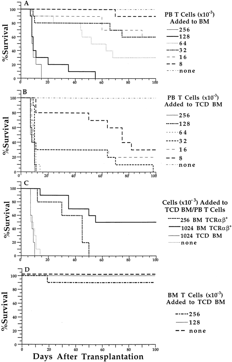
To test the ability of T cells from murine peripheral blood to cause GVHD, we stained PBMC from C57BL/6 mice and isolated CD4+ and CD8+ T cells as a single pool by flow cytometry (hereafter referred to as CD4+/CD8+ T cells). Graded numbers of these purified T cells were mixed with a constant number of unfractionated (1.5 × 10⁶) C57BL/6 bone marrow cells and injected intravenously into lethally irradiated BALB/c mice. Injection of complete H-2–mismatched unfractionated bone marrow from C57BL/6 mice into lethally irradiated BALB/c mice allowed 100% of mice to survive for at least 100 d (Fig. 1 A). Control irradiated hosts given no donor cells all died by day 14, whereas those reconstituted with the unfractionated bone marrow cells alone were complete chimeras with donor-origin (H-2b) blood mononuclear cells at day 100, based on staining for surface H-2b expression and flow cytometric analysis (data not shown). The addition of graded numbers of purified CD4+/CD8+ peripheral blood T cells to the transplant resulted in dose-dependent mortality, with the addition of as few as 1.6 × 10⁴ of these cells causing a significant decrease (P < 0.05 by the log rank test) in survival compared to transplantation of unfractionated bone marrow alone (Fig. 1 A).
Figure 1.

Ability of peripheral blood (PB) and bone marrow (BM) CD4+ and CD8+ T cells to induce lethal GVHD. Graded numbers of sorted CD4+/CD8+ T cells were added to a constant number (1.5 × 10⁶) of unfractionated or TCD C57BL/6 marrow cells and injected intravenously into lethally irradiated (800 cGy) BALB/c hosts. Survival over a 100-d observation period is shown in groups of 10 mice. (A) Graded numbers of sorted peripheral blood CD4+/CD8+ T cells were added to unfractionated marrow cells. (B) Graded number of sorted peripheral blood CD4+/CD8+ T cells were added to TCD marrow cells. (C) 6.4 × 10⁴ peripheral blood CD4+/CD8+ T cells and 1.5 × 10⁶ TCD marrow cells were or were not injected with sorted TCRα/β+ marrow T cells or with TCD marrow cells. (D) Sorted CD4+/CD8+ T cells from the marrow were injected with 1.5 × 10⁶ TCD marrow cells.
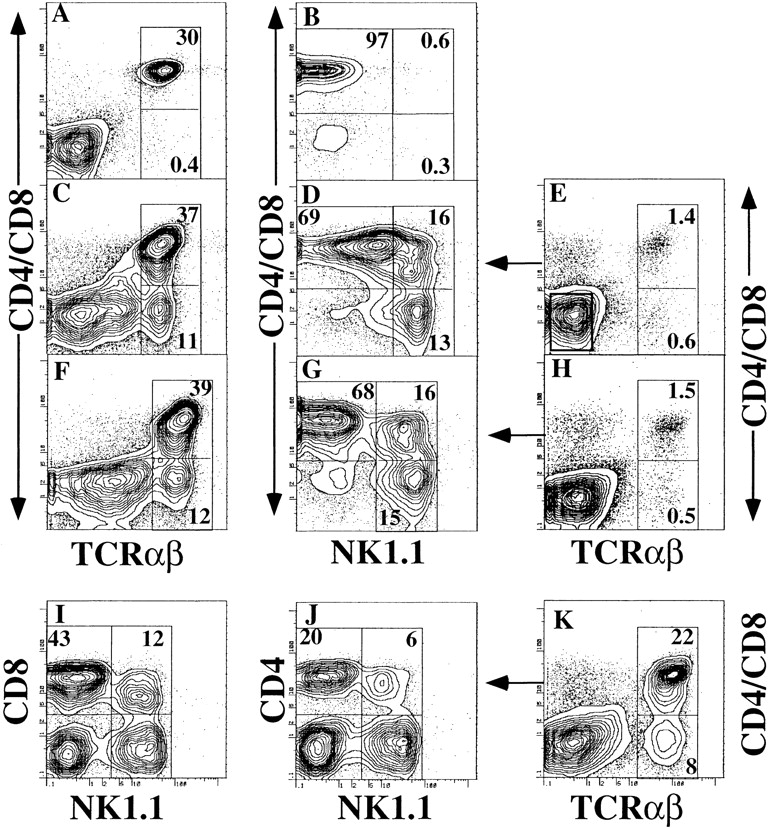
Because ∼2% of nucleated bone marrow cells were brightly staining α/β+ T cells (Fig. 2 E), we next determined whether stringent depletion of T cells (Fig. 2 E, left box) prior to transplant would further decrease the mortality from the addition of peripheral blood CD4+/CD8+ T cells. Surprisingly, T cell depletion of the bone marrow actually increased rather than decreased mortality compared to transplantation with unfractionated bone marrow, and this effect was observed over a large range of doses of peripheral blood CD4+/CD8+ T cells (Fig. 1 B). Strikingly, the addition of as few as 8 × 10³ CD4+/CD8+ T cells from the peripheral blood to TCD bone marrow cells resulted in significantly decreased host survival as compared to either transplantation of TCD bone marrow cells alone or to hosts given 8 × 10³ CD4+/CD8+ blood T cells plus unfractionated bone marrow cells (Fig. 1, A and B; P < 0.01 and 0.001, respectively). Only ∼30% of the hosts given the combination with TCD marrow cells survived more than 100 d, and those that died showed typical signs of GVHD including weight loss, hunched back, ruffled fur, diarrhea, and facial swelling. Necropsies of moribund hosts with severe clinical signs after day 40 showed typical GVHD histopathological lesions of the large intestines and skin (see below). Increasing the dose of CD4+/CD8+ peripheral blood T cells accelerated the rapidity of death of the hosts until a plateau was reached between 6.4 × 10⁴ and 2.56 × 10⁵ T cells. Hosts given the latter cell doses all died by day 14 after cell transplantation (Fig. 1 B).
Figure 2.

Two-color flow cytometric analysis of α/β+ T cells in the peripheral blood, bone marrow, and T cell–enriched bone marrow of C57BL/6 wild-type or C57BL IL-4−/− mice. A and B show staining of wild-type PBMC. (A) CD4 and CD8 (using same fluorochrome) versus TCRαβ markers; the two boxes enclose CD4+/CD8+ (upper) and CD4−CD8− (lower) TCRαβhi cells. (B) Analysis of the gated TCRαβhi cells for CD4 and CD8 versus NK1.1. Left upper box encloses NK1.1− CD4+/CD8+ cells, right upper box encloses NK1.1+ CD4+/CD8+ cells, and right lower box encloses NK1.1+ CD4−CD8− cells. C and D show similar analyses for immunomagnetic bead–enriched wild-type bone marrow T cells. F and G show analyses of enriched IL-4−/− bone marrow T cells. E shows analysis of whole wild-type bone marrow cells before enrichment of T cells and thresholds for gating TCD bone marrow cells (E, left box). Sorted TCRαβ+ bone marrow cells were all cells to the right of the TCRαβ gating threshold. H shows analysis of whole bone marrow cells before enrichment from IL-4−/− mice. K shows analysis of enriched bone marrow cells from wild-type mice; box encloses TCRαβhi cells. I and J show the analyses of the gated TCRαβhi cells for CD8 or CD4 versus NK1.1, respectively. Percentages of cell subsets enclosed in boxes are shown.
To test the notion that α/β+ T cells in the bone marrow were responsible for protection from GVHD, these cells were purified from the bone marrow (Fig. 2 E) and added back to the TCD bone marrow with 6.4 × 10⁴ peripheral blood CD4+/CD8+ T cells, a dose that resulted in 100% mortality by day 14 posttransplantation (Fig. 1 B). Under these conditions, the inclusion of 2.56 × 10⁵ α/β+ T cells significantly delayed mortality (P < 0.05), and the inclusion of 1.024 × 10⁶ of these cells resulted in the long-term survival of 50% of the transplant recipients (Fig. 1 C). This inhibitory effect on GVHD was specific for α/β+ T cells of the bone marrow, in that the addition of 1.024 × 10⁶ TCD bone marrow cells provided no protection from mortality (Fig. 1 C).
Because bone marrow α/β+ T cells enriched on immunomagnetic bead columns include populations that express CD4 or CD8 markers or neither marker (CD4−CD8−; Fig. 2 C), we next directly assessed the capacity of purified CD4+/CD8+ α/β+ T cells from the enriched bone marrow to mediate GVHD. When these were added at a ratio of 1.28 × 10⁵ or 2.56 × 10⁵ donor T cells/1.5 × 106 TCD marrow cells, 100 and 90% of the hosts, respectively, survived for more than 100 d (Fig. 1 D). The surviving hosts did not show signs of GVHD, and their mean body weight at day 100 posttransplantation did not differ significantly from that of mice that had received TCD bone marrow alone (data not shown). Bone marrow CD4+/CD8+ T cells were therefore strikingly ineffective in inducing lethal GVHD. In control experiments, PBMC were applied to immunomagnetic bead columns, and sorted CD4+/CD8+ T cells were obtained thereafter by flow cytometry. These T cells (3.2 or 6.4 × 10⁴) were injected with TCD marrow cells into groups of 10 BALB/c hosts, and survival was compared to that of hosts given the same number of sorted peripheral blood CD4+/CD8+ T cells without column enrichment. The column-enriched cells remained as effective as an equivalent number of these T cells obtained without column enrichment in mediating lethal GVHD (data not shown).
NK1.1+ T Cells Regulate GVHD.
Previous studies have shown that bone marrow α/β+ T cells from C57BL/6 mice, as well as several other strains, have a substantially greater proportion of cells with surface expression of NK1.1 than peripheral blood α/β+ T cells (18, 21). As there is recent evidence that NK1.1+ α/β+ T cells may act as negative regulators of T cell–dependent autoimmune diseases (22– 24), it was plausible that a bone marrow NK1.1+ T cell population might act in a similar negative regulatory fashion in GVHD. Therefore, we analyzed bone marrow and peripheral blood α/β+ T cells for their surface expression of CD4 plus CD8 versus NK1.1. This revealed that ∼30% of C57BL/6 PBMC were α/β+ T cells that expressed either CD4 or CD8 (Fig. 2 A). CD4 and CD8 are expressed in a mutually exclusive pattern (21). Less than 1% of the peripheral blood α/β+ T cells expressed NK1.1 (Fig. 2 B), in agreement with previous reports (19, 20). In contrast, ∼20% of bone marrow α/β+ T cells were CD4−CD8− (Fig. 2 C, lower box), and most of the CD4−CD8− T cells were NK1.1+ (Fig. 2 D, lower box). 16% of bone marrow α/β+ T cells were NK1.1+ and either CD4+ and/or CD8+ (upper box). Further analysis of gated α/β+ T cells from the bone marrow (Fig. 2 K) showed that the percentage of NK1.1+CD8+ T cells exceeded that of NK1.1+CD4+ T cells (Fig. 2, compare I and J). Although NK1.1+CD8+ T cells were easily identified in the bone marrow, this subset was not detected in the peripheral blood, thymi, or spleens of the donor mice (data not shown).
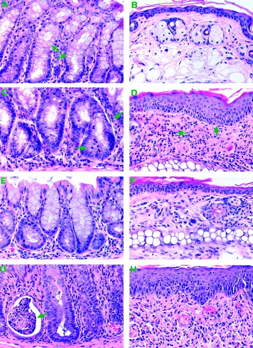
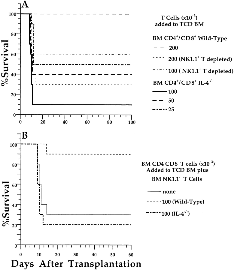
Because the CD4+/CD8+ T cell subset of bone marrow cells contained a substantial proportion of NK1.1+ cells but the peripheral blood did not, we tested the ability of purified CD4+/CD8+ T cells depleted of NK1.1+ T cells to induce GVHD. As before, the addition of 2.00 × 10⁵ CD4+/CD8+ T cells (including the NK1.1+ population) to the TCD bone marrow transplant induced minimal clinical signs of GVHD in irradiated BALB/c hosts, and no deaths occurred during a 100-d observation period (Fig. 3 A). In contrast, an equal number of NK1.1-depleted CD4+/ CD8+ T cells added to TCD bone marrow resulted in the death of ∼70% of hosts within 20 d and was associated with clinical signs of GVHD including facial swelling, hair loss, hunched back, and weight loss (Fig. 3 A). Reduction of the NK1.1-depleted CD4+/CD8+ T cell dose to 1.00 × 10⁵ cells still resulted in the death of ∼40% of hosts by day 20. This was a significant reduction in survival (P < 0.01, log rank test) as compared to the group given sorted CD4+/ CD8+ T cells. Histopathological examination of the large intestines and skin of additional mice given 2.00 × 10⁵ NK1.1− T cells showed lesions of GVHD, including inflammation in the intestinal crypts and hyperplasia and apoptosis of crypt epithelial cells, inflammation of the dermis, and epidermal hyperplasia as compared to mice given TCD bone marrow alone (Fig. 4, A–D).
Figure 3.
Different subsets of T cells induce or suppress GVHD, depending on their cytokine secretion profiles. (A) Sorted C57BL/6 wild-type CD4+ and CD8+ bone marrow (BM) T cells with or without depletion of NK1.1+ T cells were added to C57BL/6 wild-type TCD bone marrow cells (1.5 × 10⁶) and injected into irradiated BALB/c hosts. In addition, sorted C57BL IL-4−/− CD4+ and CD8+ bone marrow T cells without NK1.1+ T cell depletion were added to wild-type TCD bone marrow cells and injected into BALB/c hosts. (B) Sorted CD4−CD8− T cells from the bone marrow of wild-type or IL-4−/− C57BL/6 donor mice were added to 2.00 × 10⁵ sorted wild-type NK1.1− CD4+/CD8+ bone marrow T cells and 1.5 × 10⁶ wild-type TCD bone marrow cells. Control mice received sorted wild-type NK1.1− CD4+/CD8+ bone marrow T cells and TCD bone marrow cells without added-back CD4−CD8+ bone marrow T cells (none). There were 10 mice in each group.
Figure 4.

Different subsets of NK1.1− and NK1.1+ T cells induce or suppress histopathological lesions of GVHD in the large intestine and skin. Sections of intestine (A) and skin (B) from irradiated BALB/c hosts injected 60 d earlier with only TCD bone marrow from wild-type C57BL/6 donor mice. Plump, mucin-containing epithelial cells are seen in the intestine (arrows) with little or no inflammation. C and D show BALB/c hosts injected with wild-type TCD bone marrow and sorted wild-type NK1.1− CD4+/ CD8+ bone marrow T cells. An inflammatory infiltrate between intestinal crypts and hyperplasia of crypt epithelial cells with reduced mucin characteristic of GVHD is seen (arrow). An inflammatory infiltrate in the dermis and epidermal hyperplasia characteristic of GVHD is observed in the skin (arrows). E and F show hosts injected with wild-type TCD bone marrow, sorted wild-type NK1.1− CD4+/CD8+ bone marrow T cells, and sorted wild-type CD4−CD8− (NK1.1+) bone marrow T cells. Lesions of GVHD are markedly reduced. G and H show hosts injected with wild-type TCD bone marrow, sorted wild-type NK1.1− CD4+/CD8+ bone marrow T cells, and sorted IL-4−/− CD4−CD8− (NK1.1+) bone marrow T cells. Severe lesions of GVHD are seen, including an abscess in an intestinal crypt (arrow).
Regulation of GVHD by NK1.1+ T Cells is IL-4 Dependent.
Unlike conventional α/β+ T cells, NK1.1+ T-lineage cells of the thymus and spleen rapidly secrete large amounts of IL-4 and IFN-γ after engagement of the TCR–CD3 complex or after stimulation with calcium ionophore and phorbol PMA, without previous exposure to antigens or polyclonal T cell activators (31–33). IFN-γ and IL-4 are important in helping to polarize T cell responses toward Th-1 or Th-2 patterns of cytokine production that have been implicated in the induction and amelioration of GVHD, respectively (34–36). Therefore, we characterized the capacity of bone marrow T cells, including the NK1.1+ population, to produce these cytokines after in vitro stimulation with calcium ionophore and PMA for 48 h, a time point at which cytokine levels were maximal (21). Depletion of NK1.1+ T cells from the bone marrow CD4+/CD8+ T cell population resulted in an eightfold reduction of IL-4 production (Table I) that was highly significant (P < 0.01, two-tailed Student's t test). In contrast, secretion of IFN-γ was similar in unfractionated versus NK1.1-depleted bone marrow CD4+/CD8+ T cells. In comparison, sorted peripheral blood CD4+/CD8+ T cells secreted high levels of IFN-γ and a low level of IL-4 (Table I). Therefore, a high ratio of IFN-γ/IL-4 production correlated with the capacity to induce severe GVHD.
Table I.
Cytokine Production by Bone Marrow (BM) and Peripheral Blood (PB) T Cells
| Cells (1.00 × 10⁵) | IL-4* | IFN-γ* | IFN-γ/IL-4 ratio | |||
|---|---|---|---|---|---|---|
| pg/ml | pg/ml | |||||
| PB CD4+/CD8+ T (wt, n = 6) | 18 ± 4 | 566 ± 75 | 32 | |||
| BM CD4+/CD8+ T (wt, n = 6) | 528 ± 67 | 1,216 ± 108 | 2.3 | |||
| BM CD4+/CD8+ T NK1.1− (wt, n = 6) | 73 ± 13 | 992 ± 52 | 14 | |||
| BM CD4+/CD8+ T (IL-4−/−, n = 6) | 0 ± 0 | 1,657 ± 267 | — | |||
| BM CD4−/CD8− T (wt, n = 6) | 829 ± 58 | 1,492 ± 100 | 1.8 | |||
| BM CD4−/CD8− T (IL-4−/−, n = 4) | 0 ± 0 | 1,024 ± 56 | — | |||
| BM CD4+/CD8+ T (wt, anti-NK1.1, n = 4) | 281 ± 19 | 1,419 ± 28 | 5 | |||
| BM CD4−/CD8− T (wt, anti-NK1.1, n = 4) | 247 ± 8 | 1,305 ± 114 | 5.3 |
Values are the means ± SE of replicate experiments from individual donor mice. In some experiments, BM cells were stained with anti-NK1.1 monoclonal antibodies as well as with the anti-CD4, anti-CD8, and anti-TCRαβ antibodies used for sorting. Cytokine patterns of sorted cells in which anti-NK1.1 antibodies were included in the staining mixture are also shown (anti-NK1.1). wt, wild type.
To determine whether the high capacity of NK1.1+ bone marrow T cells to produce IL-4 was important for the negative regulation of GVHD by this cell population, bone marrow CD4+/CD8+ T cells from wild-type and IL-4−/− mice of the C57BL/6 background were used to induce GVHD in the irradiated BALB/c hosts. The surface phenotype and percentages of bone marrow α/β+ T cells, particularly the NK1.1+ and CD4−CD8− T cell subsets, were similar in IL-4−/− (Fig. 2, F–H) and wild-type mice (Fig. 2, C–E), indicating that IL-4 is not required for NK1.1+ T cell development. The percentage of CD4+ and CD8+ NK1.1+ T cells in the IL-4−/− mice was also similar to that in the wild type (data not shown). Whereas 2.00 × 10⁵ of wild-type CD4+/CD8+ bone marrow T cells failed to induce lethal GVHD (Fig. 3 A), the addition of 1.00 × 10⁵ of these bone marrow T cells from IL-4−/− mice to the transplant induced lethal GVHD in 90% of irradiated BALB/c hosts by day 11 (Fig. 3 A). Reduction of the dose of IL-4−/− CD4+/CD8+ T cells to 5.0 and 2.5 × 10⁴ resulted in the reduction of lethal GVHD to 60 and 50%, respectively (Fig. 3 A). Examination of surviving hosts at day 40 again showed histopathological evidence of GVHD of the large intestine (data not shown). Purified CD4+ and CD8+ bone marrow T cells from IL-4−/− donors secreted no detectable IL-4, and high levels of IFN-γ that were not significantly different than that of cells from wild-type C57BL/6 mice (Table I). Taken together, these results indicate that the NK1.1+ subset of CD4+/CD8+ bone marrow T cells inhibited the induction of GVHD by NK1.1− subset and that this inhibition was IL-4 dependent.
We attempted to directly test the suppressive capacity of purified NK1.1+ CD4+/CD8+ T cells on GVHD. However, as immunofluorescent staining of bone marrow T cells with anti-NK1.1 mAb, a step required for cell purification by positive selection, substantially reduced the capacity of CD4+/CD8+ T cells to secrete IL-4 in vitro (Table I), this approach was not pursued. As an alternative approach, we performed experiments in which purified CD4−CD8− α/β+ T cells from the bone marrow, which are almost all NK1.1+ (Fig. 2 D), were combined with NK1.1− CD4+/CD8+ bone marrow T cells and transplanted with TCD bone marrow. These purified bone marrow CD4−CD8− α/β+ T cells secreted levels of IL-4 (mean 829 pg/ml) and IFN-γ (mean 1,216 pg/ml) that lacked a statistically significant difference (P > 0.1) from that of NK1.1+-containing bone marrow CD4+/CD8+ T cells (Table I). Addition of 1.00 × 10⁵ CD4−CD8− bone marrow T cells and 2.00 × 10⁵ NK1.1− CD4+/CD8+ bone marrow T cells to the transplant significantly increased the survival of the irradiated hosts (P < 0.01, log rank test) as compared to the injection of only NK1.1− CD4+/CD8+ bone marrow T cells and TCD bone marrow (Fig. 3 B). Only 10% of host mice died by day 60 in the group that received CD4−CD8− T cells. In contrast, the addition of 1.00 × 10⁵ sorted CD4−CD8− bone marrow T cells from IL-4−/− mice resulted in reduced survival, and 80% of hosts died by day 12 posttransplantation. Surviving hosts given CD4−CD8− bone marrow T cells from wild-type or IL-4−/− donors were killed on day 60. Interestingly, the large intestines and skin of hosts given IL-4−/− CD4−CD8− T cells showed more severe histopathological lesions of GVHD compared not only to recipients of wild-type CD4−CD8− T cells but also to hosts that received only NK1.1− CD4+/CD8+ T cells with TCD bone marrow (Fig. 4, E–H). The sorted CD4−CD8− bone marrow T cells from the IL-4−/− donors secreted a high level of IFN-γ (mean 1,024 pg/ml) that was similar to the level secreted by CD4−CD8− bone marrow T cells (Table I). This indicates that the secretion of IFN-γ by NK1.1+ T cells, when it is unopposed by IL-4 secretion, exacerbates rather than inhibits GVHD.
Discussion
We directly compared the ability of sorted CD4 and CD8 T cells obtained from the peripheral blood or bone marrow of C57BL/6 mice to induce acute GVHD in lethally irradiated BALB/c hosts coinjected with stringently TCD C57BL/6 marrow cells. The peripheral blood T cells were at least 30-fold more potent than the marrow T cells on a per-cell basis as judged by mortality of the hosts during a 100-d observation period. In fact, no significant mortality was induced by the sorted marrow T cells in the dose range tested unless the NK1.1+ T cells were removed or the sorted marrow T cells were obtained from IL-4−/− donors. Surprisingly, the bone marrow NK1.1+ T cells contained CD4+, CD8+, and CD4−CD8− subsets, whereas only the CD4+ and CD4−CD8− subsets could be detected in the thymus and spleen as reported previously (20). CD4+ and CD8+ NK1.1+ T cells in the marrow show similar cytokine secretion profiles (Zeng, D., and S. Strober, manuscript in preparation). Sorted wild-type NK1.1+ T cells (CD4−CD8−) from the marrow that secreted high levels of both IFN-γ and IL-4 suppressed GVHD as judged by mortality and by histopathological changes in the skin and large intestines. In contrast, sorted IL-4−/− NK1.1+ T cells that secreted high levels of IFN-γ without IL-4 exacerbated mortality and the histopathological changes of GVHD.
The experimental results show that peripheral blood or bone marrow NK1.1− α/β+ T cells induce and NK1.1+ α/β+ T cells potently suppress acute lethal GVHD and that this negative regulatory effect requires IL-4. These results agree with and extend previous studies showing that CD4− CD8− T cells (“natural suppressor” cells) inhibit GVHD (25–27), and CD4+ or CD8+ T cells that secrete a Th1-type cytokine pattern (e.g., IFN-γ and IL-2, but not IL-4) induce GVHD and those that secrete a Th2-type pattern (e.g., IL-4, but not IFN-γ and IL-2) are protective (34–36). However, two recent studies concluded that secretion of IFN-γ by allogeneic donor cells is not required for the induction of lethal GVHD in irradiated hosts, as donor cells from IFN-γ−/− mice induced lethal disease in either wild-type or IFN-γ−/− hosts (37, 38). In some studies, injection of IFN-γ or IL-12 into allogeneic bone marrow transplant hosts protected against GVHD when given at the same time or shortly after the injection of the donor cells (39, 40). The current study did not determine whether the induction of GVHD was dependent upon IFN-γ secretion by donor NK1.1− T cells, and it is possible that alloantigen-induced secretion of other cytokines such as IL-2 is critical for the development of the disease (37). The current results suggest that NK1.1+ T cell secretion of IFN-γ in the absence of IL-4 can worsen GVHD when it is induced by donor NK1.1− T cells, perhaps due to the specific kinetics and localization of IFN-γ secretion by the NK1.1+ T cells.
GVHD induced by donor spleen cells added to TCD donor bone marrow cells has been reported to be less vigorous when IL-4−/− rather than wild-type donor mice are used (37). The latter result appears contradictory to the increase in severity of GVHD induced by bone marrow CD4+/ CD8+ T cells from IL-4−/− donors as compared to wild-type donors in the current study. Because the percentage of NK1.1+ T cells in the spleen is quite low and comparable to that in the peripheral blood, changes in the ability of splenic T cells from wild-type versus cytokine-deficient donors to induce GVHD are likely to reflect changes mainly in the function of NK1.1− T cells, which recognize predominantly polymorphic MHC class I and II antigens. However, changes in the ability of bone marrow T cells from IL-4−/− donor mice to induce GVHD are due mainly to changes in the function of NK1.1+ T cells, which recognize the nonpolymorphic CD1 antigen (20). A simple Th1/ Th2 paradigm cannot explain the capacity of all T cell subsets to induce or protect against GVHD in a variety of animal models of bone marrow transplantation, as a given cytokine such as IFN-γ can ameliorate or worsen disease depending on the preparatory regimen (lethal versus sublethal or no irradiation) used to treat the host (37, 41).
Decreased production of NK1.1+ α/β thymocytes and a resulting decrease in NK1.1+ α/β T-lineage cell–mediated IL-4 production appears to play a key role in the development of diabetes mellitus in nonobese diabetic mice, a disease which depends on NK1.1− α/β+ T cells for its development (22–24). Taken together with our observations, this suggests that the NK1.1+ α/β+ T cell population is an important negative regulator of other α/β+ T cell populations. A human CD4−CD8− α/β+ T cell subset (Vα24-JαQ) is found in most healthy individuals and has striking similarities to the murine NK1.1+ α/β+ T cell population (42, 43). These similarities include a highly limited αβ-TCR repertoire that is restricted, at least in part, by the CD1d molecule (43). In addition, this subset expresses high levels of NKR-P1A, the human homologue of murine NK1.1 molecule, and has a high capacity to produce IL-4 and IFN-γ (43). Therefore, given our results, it will be of interest to determine if inclusion rather than depletion of donor Vα24-JαQ CD4−CD8− T cells in allogeneic bone marrow transplantation reduces GVHD in humans. Our results also raise the possibility that IL-4 produced by this CD4−CD8− T cell population or administered therapeutically might prevent or ameliorate GVHD in humans.
Acknowledgments
We thank A. Mukhopadhyay, A. Van Vollenhoven, and P. Stepak-Biek for excellent technical assistance and V. Cleaver for preparation of the manuscript.
This work was supported by National Institutes of Health grants R01 HL57443, R01 HL58250, R01 AI40093, and R01 AI26940. D. Zeng was the recipient of a postdoctoral fellowship award from Dendreon Corporation, Mountain View, CA.
Abbreviation used in this paper
- TCD
T cell–depleted
References
- 1.Sullivan, K.M. 1994. Graft-versus-host disease. In Bone Marrow Transplantation. S.J. Forman, K.G. Blume, and E.D. Thomas, editors. Blackwell Scientific Publications, Boston. xxiii, 942.
- 2.Weisdorf D, Hakke R, Blazar B, Miller W, McGlave P, Ramsay N, Kersey J, Filipovich A. Risk factors for acute graft-versus-host disease in histocompatible donor bone marrow transplantation. Transplantation. 1991;51:1197–1203. doi: 10.1097/00007890-199106000-00010. [DOI] [PubMed] [Google Scholar]
- 3.Bross DS, Tutschka PJ, Farmer ER, Beschorner WE, Braine HG, Mellits ED, Bias WB, Santos GW. Predictive factors for acute graft-versus-host disease in patients transplanted with HLA-identical bone marrow. Blood. 1984;63:1265–1270. [PubMed] [Google Scholar]
- 4.Ferrara JL, Cooke KR, Pan L, Krenger W. The immunopathophysiology of acute graft-versus-host-disease. Stem Cells. 1996;14:473–489. doi: 10.1002/stem.140473. [DOI] [PubMed] [Google Scholar]
- 5.Korngold R, Sprent J. Surface markers of T cells causing lethal graft-vs-host disease to class I vs class II H-2 differences. J Immunol. 1985;135:3004–3010. [PubMed] [Google Scholar]
- 6.Korngold R, Sprent J. T cell subsets and graft-versus-host disease. Transplantation. 1987;44:335–339. doi: 10.1097/00007890-198709000-00002. [DOI] [PubMed] [Google Scholar]
- 7.Korngold R, Sprent J. Graft-versus-host disease in experimental allogeneic bone marrow transplantation. Proc Soc Exp Biol Med. 1991;197:12–18. doi: 10.3181/00379727-197-43217a. [DOI] [PubMed] [Google Scholar]
- 8.Martin, P.J., and N. Kernan. 1990. T cell depletion for GVHD prevention in humans. In Graft Versus Host Disease: Research and Clinical Management. S. Burakoff, H.J. Keeg, J. Ferrara, and K. Atkinson, editors. Marcel Dekker, Inc., New York. 371–387.
- 9.Burnett AK, Hann IM, Robertson AG, Alcorn M, Gibson B, McVicar I, Niven L, Mackinnon S, Hambley H, Morrison A, et al. Prevention of graft-versus-host disease by ex vivo T cell depletion: reduction in graft failure with augmented total body irradiation. Leukemia. 1988;2:300–303. [PubMed] [Google Scholar]
- 10.Fauci AS. Human bone marrow lymphocytes. I. Distribution of lymphocyte subpopulations in the bone marrow of normal individuals. J Clin Invest. 1975;56:98–110. doi: 10.1172/JCI108085. [DOI] [PMC free article] [PubMed] [Google Scholar]
- 11.Gale RP, Opelz G, Kiuchi OM, Golde DW. Thymus-dependent lymphocytes in human bone marrow. J Clin Invest. 1975;56:1491–1498. doi: 10.1172/JCI108230. [DOI] [PMC free article] [PubMed] [Google Scholar]
- 12.Batinic D, Marusic M, Pavletic Z, Bogdanic V, Uzarevic B, Nemet D, Labar B. Relationship between differing volumes of bone marrow aspirates and their cellular composition. Bone Marrow Transplant. 1990;6:103–107. [PubMed] [Google Scholar]
- 13.Almaraz R, Ballinger W, Sachs DH, Rosenberg SA. The effect of peripheral lymphoid cells on the incidence of lethal graft versus host disease following allogeneic mouse bone marrow transplantation. J Surg Res. 1983;34:133–144. doi: 10.1016/0022-4804(83)90052-5. [DOI] [PubMed] [Google Scholar]
- 14.Palathumpat V, Dejbakhsh-Jones S, Holm B, Strober S. Different subsets of T cells in the adult mouse bone marrow and spleen induce or suppress acute graft-versus-host disease. J Immunol. 1992;149:808–817. [PubMed] [Google Scholar]
- 15.Palathumpat V, Dejbakhsh-Jones S, Strober S. The role of purified CD8+T cells in graft-versus-leukemia activity and engraftment after allogeneic bone marrow transplantation. Transplantation. 1995;60:355–361. doi: 10.1097/00007890-199508270-00010. [DOI] [PubMed] [Google Scholar]
- 16.Korngold B, Sprent J. Lethal graft-versus-host disease after bone marrow transplantation across minor histocompatibility barriers in mice. Prevention by removing mature T cells from marrow. J Exp Med. 1978;148:1687–1698. doi: 10.1084/jem.148.6.1687. [DOI] [PMC free article] [PubMed] [Google Scholar]
- 17.Vallera DA, Soderling CC, Kersey JH. Bone marrow transplantation across major histocompatability barriers in mice. III. Treatment of donor grafts with monoclonal antibodies directed against Lyt determinants. J Immunol. 1982;128:871–875. [PubMed] [Google Scholar]
- 18.Sykes M. Unusual T cell populations in adult murine bone marrow. Prevalence of CD3+CD4−CD8− and alpha beta TCR+NK1.1+cells. J Immunol. 1990;145:3209–3215. [PubMed] [Google Scholar]
- 19.Makino Y, Kanno R, Ito T, Higashino K, Taniguchi M. Predominant expression of invariant V alpha 14+ TCR alpha chain in NK1.1+T cell populations. Int Immunol. 1995;7:1157–1161. doi: 10.1093/intimm/7.7.1157. [DOI] [PubMed] [Google Scholar]
- 20.Bendelac A, Rivera MN, Park SH, Roark JH. Mouse CD1-specific NK1 T cells: development, specificity, and function. Annu Rev Immunol. 1997;15:535–562. doi: 10.1146/annurev.immunol.15.1.535. [DOI] [PubMed] [Google Scholar]
- 21.Zeng D, Dejbakhsh-Jones S, Strober S. Granulocyte colony-stimulating factor reduces the capacity of blood mononuclear cells to induce graft-versus-host disease: impact on blood progenitor cell transplantation. Blood. 1997;90:453–463. [PubMed] [Google Scholar]
- 22.Gombert JM, Herbelin A, Tancrede-Bohin E, Dy M, Carnaud C, Bach JF. Early quantitative and functional deficiency of NK1+-like thymocytes in the NOD mouse. Eur J Immunol. 1996;26:2989–2998. doi: 10.1002/eji.1830261226. [DOI] [PubMed] [Google Scholar]
- 23.Baxter AG, Kinder SJ, Hammond KJ, Scollay R, Godfrey DI. Association between alpha beta TCR+CD4−CD8−T-cell deficiency and IDDM in NOD/ Lt mice. Diabetes. 1997;46:572–582. doi: 10.2337/diab.46.4.572. [DOI] [PubMed] [Google Scholar]
- 24.Hammond KJL, Poulton LD, Palmisano LJ, Silveira PA, Godfrey DI, Baxter AG. α/β–T cell receptor (TCR)+CD4−CD8−(NKT) thymocytes prevent insulin-dependent diabetes mellitus in nonobese diabetic (NOD)/Lt mice by the influence of interleukin (IL)-4 and/ or IL-10. J Exp Med. 1998;187:1047–1056. doi: 10.1084/jem.187.7.1047. [DOI] [PMC free article] [PubMed] [Google Scholar]
- 25.Palathumpat V, Dejbakhsh-Jones S, Holm B, Wang H, Liang O, Strober S. Studies of CD4− CD8−alpha beta bone marrow T cells with suppressor activity. J Immunol. 1992;148:373–380. [PubMed] [Google Scholar]
- 26.Sykes M, Hoyles KA, Romick ML, Sachs DH. In vitro and in vivo analysis of bone marrow-derived CD3+, CD4−, CD8−, NK1.1+cell lines. Cell Immunol. 1990;129:478–493. doi: 10.1016/0008-8749(90)90222-d. [DOI] [PubMed] [Google Scholar]
- 27.Strober S, Cheng L, Zeng D, Palathumpat R, Dejbakhsh-Jones S, Huie P, Sibley R. Double negative (CD4−CD8− αβ+) T cells which promote tolerance induction and regulate autoimmunity. Immunol Rev. 1996;149:217–230. doi: 10.1111/j.1600-065x.1996.tb00906.x. [DOI] [PubMed] [Google Scholar]
- 28.Kuhn R, Rajewsky K, Muller W. Generation and analysis of interleukin-4 deficient mice. Science. 1991;254:707–710. doi: 10.1126/science.1948049. [DOI] [PubMed] [Google Scholar]
- 29.Zhang Y, Rogers KH, Lewis DB. β2-microglobulin–dependent T cells are dispensable for allergen- induced T helper 2 responses. J Exp Med. 1996;184:1507–1512. doi: 10.1084/jem.184.4.1507. [DOI] [PMC free article] [PubMed] [Google Scholar]
- 30.Garcia-Ojeda ME, Dejbakhsh-Jones S, Weissman IL, Strober S. An alternate pathway for T cell development supported by the bone marrow microenvironment: recapitulation of thymic maturation. J Exp Med. 1998;187:1813–1823. doi: 10.1084/jem.187.11.1813. [DOI] [PMC free article] [PubMed] [Google Scholar]
- 31.Yoshimoto T, Paul WE. CD4pos, NK1.1pos T cells promptly produce interleukin 4 in response to in vivo challenge with anti-CD3. J Exp Med. 1994;179:1285–1295. doi: 10.1084/jem.179.4.1285. [DOI] [PMC free article] [PubMed] [Google Scholar]
- 32.Arase H, Arase N, Nakagawa K, Good RA, Onoe K. NK1.1+ CD4+ CD8−thymocytes with specific lymphokine secretion. Eur J Immunol. 1993;23:307–310. doi: 10.1002/eji.1830230151. [DOI] [PubMed] [Google Scholar]
- 33.Zlotnik A, Godfrey DI, Fischer M, Suda T. Cytokine production by mature and immature CD4−CD8− T cells. Alpha beta− T cell receptor+ CD4−CD8−T cells produce IL-4. J Immunol. 1992;149:1211–1215. [PubMed] [Google Scholar]
- 34.Fowler DH, Kurasawa K, Husebekk A, Cohen PA, Gress RE. Cells of Th2 cytokine phenotype prevent LPS-induced lethality during murine graft-versus-host reaction. Regulation of cytokines and CD8+lymphoid engraftment. J Immunol. 1994;152:1004–1013. [PubMed] [Google Scholar]
- 35.Krenger W, Snyder KM, Byon JC, Falzarano G, Ferrara JL. Polarized type 2 alloreactive CD4+ and CD8+donor T cells fail to induce experimental acute graft-versus-host disease. J Immunol. 1995;155:585–593. [PubMed] [Google Scholar]
- 36.Fowler DH, Breglio J, Nagel G, Eckhaus MA, Gress RE. Allospecific CD8+Tc1 and Tc2 populations in graft-versus-leukemia effect and graft-versus-host disease. J Immunol. 1996;157:4811–4821. [PubMed] [Google Scholar]
- 37.Murphy WJ, Welniak LA, Taub DD, Wiltrout RH, Taylor PA, Vallera DA, Kopf M, Young H, Longo DL, Blazar BR. Differential effects of the absence of interferon-gamma and IL-4 in acute graft-versus-host disease after allogeneic bone marrow transplantation in mice. J Clin Invest. 1998;102:1742–1748. doi: 10.1172/JCI3906. [DOI] [PMC free article] [PubMed] [Google Scholar]
- 38.Yang YG, Dey BR, Sergio JJ, Pearson DA, Sykes M. Donor-derived interferon gamma is required for inhibition of acute graft-versus-host disease by interleukin 12. J Clin Invest. 1998;102:2126–2135. doi: 10.1172/JCI4992. [DOI] [PMC free article] [PubMed] [Google Scholar]
- 39.Brok HP, Heidt PJ, van der Meide PH, Zurcher C, Vossen JM. Interferon-gamma prevents graft-versus-host disease after allogeneic bone marrow transplantation in mice. J Immunol. 1993;151:6451–6459. [PubMed] [Google Scholar]
- 40.Sykes M, Szot GL, Nguyen PL, Pearson DA. Interleukin-12 inhibits murine graft-versus-host disease. Blood. 1995;86:2429–2438. [PubMed] [Google Scholar]
- 41.Ellison CA, Fischer JM, HayGlass KT, Gartner JG. Murine graft-versus-host disease in an F1-hybrid model using IFN-gamma gene knockout donors. J Immunol. 1998;161:631–640. [PubMed] [Google Scholar]
- 42.Lantz O, Bendelac A. An invariant T cell receptor alpha chain is used by a unique subset of major histocompatibility complex class I–specific CD4+ and CD4−8−T cells in mice and humans. J Exp Med. 1994;180:1097–1106. doi: 10.1084/jem.180.3.1097. [DOI] [PMC free article] [PubMed] [Google Scholar]
- 43.Exley M, Garcia J, Balk SP, Porcelli S. Requirements for CD1d recognition by human invariant Valpha24+ CD4−CD8−T cells. J Exp Med. 1997;186:109–120. doi: 10.1084/jem.186.1.109. [DOI] [PMC free article] [PubMed] [Google Scholar]


