

Abstract
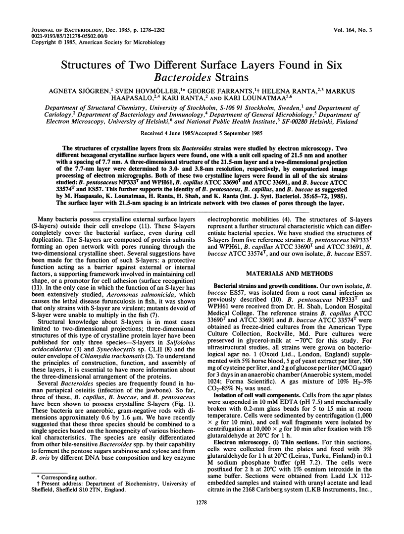
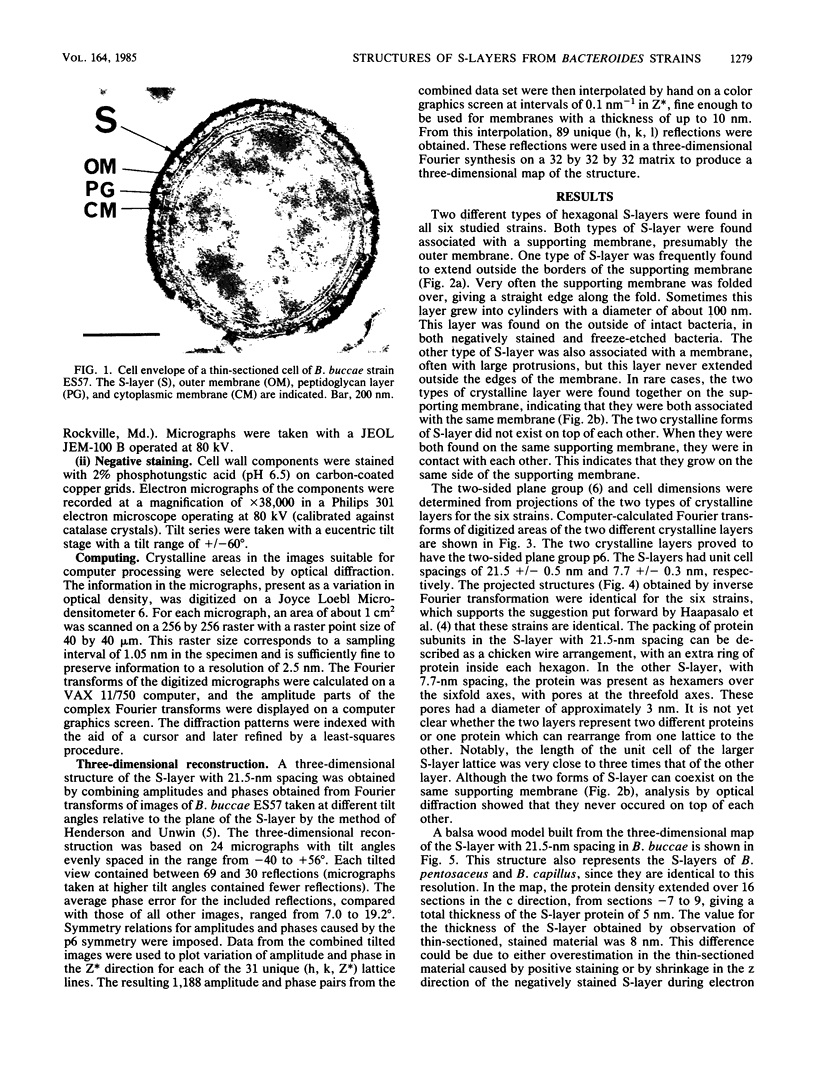
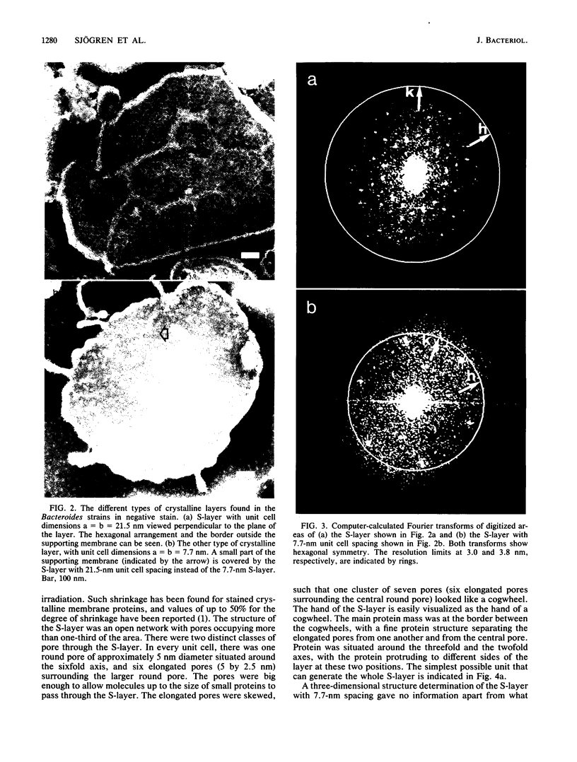
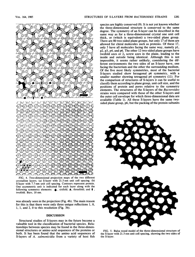
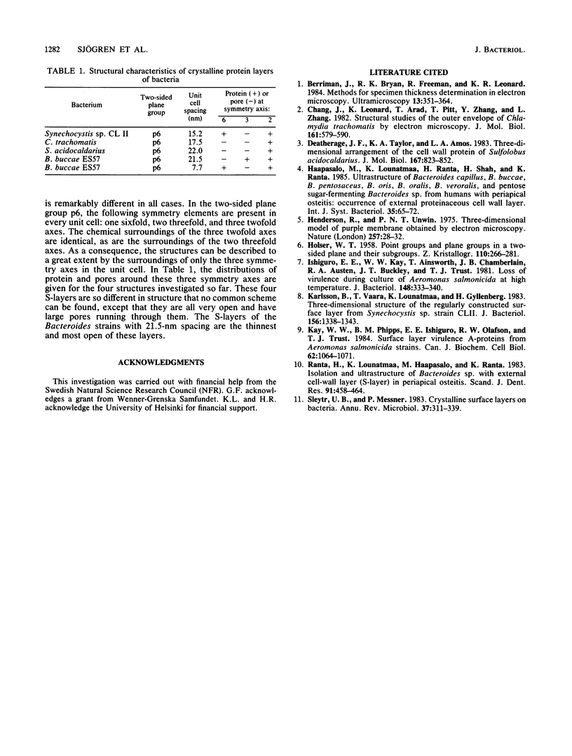
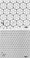
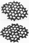
The structures of crystalline layers from six Bacteroides strains were studied by electron microscopy. Two different hexagonal crystalline surface layers were found, one with a unit cell spacing of 21.5 nm and another with a spacing of 7.7 nm. A three-dimensional structure of the 21.5-nm layer and a two-dimensional projection of the 7.7-nm layer were determined to 3.0- and 3.8-nm resolution, respectively, by computerized image processing of electron micrographs. Both of these two crystalline layers were found in all six strains studied: B. pentosaceus NP333T and WPH61, B. capillus ATCC 33690T and ATCC 33691, and B. buccae ATCC 33574T and ES57. This further supports the identity of B. pentosaceus, B. capillus, and B. buccae as suggested by M. Haapasalo, K. Lounatmaa, H. Ranta, H. Shah, and K. Ranta (Int. J. Syst. Bacteriol. 35:65-72, 1985). The surface layer with 21.5-nm spacing is an intricate network with two classes of pores through the layer.
Full text
PDF
Images in this article
Selected References
These references are in PubMed. This may not be the complete list of references from this article.
- Berriman J., Bryan R. K., Freeman R., Leonard K. R. Methods for specimen thickness determination in electron microscopy. Ultramicroscopy. 1984;13(4):351–364. doi: 10.1016/0304-3991(84)90001-9. [DOI] [PubMed] [Google Scholar]
- Chang J. J., Leonard K., Arad T., Pitt T., Zhang Y. X., Zhang L. H. Structural studies of the outer envelope of Chlamydia trachomatis by electron microscopy. J Mol Biol. 1982 Nov 15;161(4):579–590. doi: 10.1016/0022-2836(82)90409-0. [DOI] [PubMed] [Google Scholar]
- Deatherage J. F., Taylor K. A., Amos L. A. Three-dimensional arrangement of the cell wall protein of Sulfolobus acidocaldarius. J Mol Biol. 1983 Jul 15;167(4):823–848. doi: 10.1016/s0022-2836(83)80113-2. [DOI] [PubMed] [Google Scholar]
- Henderson R., Unwin P. N. Three-dimensional model of purple membrane obtained by electron microscopy. Nature. 1975 Sep 4;257(5521):28–32. doi: 10.1038/257028a0. [DOI] [PubMed] [Google Scholar]
- Ishiguro E. E., Kay W. W., Ainsworth T., Chamberlain J. B., Austen R. A., Buckley J. T., Trust T. J. Loss of virulence during culture of Aeromonas salmonicida at high temperature. J Bacteriol. 1981 Oct;148(1):333–340. doi: 10.1128/jb.148.1.333-340.1981. [DOI] [PMC free article] [PubMed] [Google Scholar]
- Karlsson B., Vaara T., Lounatmaa K., Gyllenberg H. Three-dimensional structure of the regularly constructed surface layer from Synechocystis sp. strain CLII. J Bacteriol. 1983 Dec;156(3):1338–1343. doi: 10.1128/jb.156.3.1338-1343.1983. [DOI] [PMC free article] [PubMed] [Google Scholar]
- Kay W. W., Phipps B. M., Ishiguro E. E., Olafson R. W., Trust T. J. Surface layer virulence A-proteins from Aeromonas salmonicida strains. Can J Biochem Cell Biol. 1984 Nov;62(11):1064–1071. doi: 10.1139/o84-137. [DOI] [PubMed] [Google Scholar]
- Ranta H., Lounatmaa K., Haapasalo M., Ranta K. Isolation and ultrastructure of Bacteroides sp. with external cell-wall layer (S-layer) in periapical osteitis. Scand J Dent Res. 1983 Dec;91(6):458–464. doi: 10.1111/j.1600-0722.1983.tb00846.x. [DOI] [PubMed] [Google Scholar]
- Sleytr U. B., Messner P. Crystalline surface layers on bacteria. Annu Rev Microbiol. 1983;37:311–339. doi: 10.1146/annurev.mi.37.100183.001523. [DOI] [PubMed] [Google Scholar]