Abstract
The question of whether enhanced memory T cell responses are simply due to an increased frequency of specific cells or also to an improved response at the single cell level is widely debated. In this study, we analyzed T cell receptor (TCR) transgenic memory T cells and bona fide memory T cells isolated from virally infected normal mice using the tetramer technology. We found that memory T cells are qualitatively different from naive T cells due to a developmentally regulated rearrangement of the topology of the signaling machinery. In naive cytotoxic T cells, only a few CD8 molecules are associated with Lck and the kinase is homogeneously distributed inside the cell. However, in vivo priming of naive T cells induces the targeting of Lck to the CD8 coreceptor in the cell membrane and the consequent organization of a more efficient TCR signaling machinery in effector and memory cells.
Keywords: memory, virus, costimulation
The hallmarks of adaptive immune responses are antigen specificity and memory. The concept of immunological memory is based on the faster and enhanced response of an animal to reexposure to the antigen, mediated by either B or T cells (1–5).
The generation of memory B cells in germinal centers is fairly well understood. Priming of naive B cells by antigen and cognate T help leads to activated B cells that differentiate either to plasma cells, which lose surface Ig expression, or to memory B cells, which express an isotype-switched and somatically mutated Ig receptor (6, 7). Thus, memory B cells are qualitatively different from naive B cells, and the surface expression of a distinct Ig receptor makes memory B cells readily identifiable and facilitates their analysis. In contrast, the generation of memory T cells is less well understood, as a special anatomical site for memory T cell development has not been identified, and T cells do not undergo isotype switching and extensive somatic hypermutation. In addition, it has proven difficult to find a reliable marker to distinguish memory and naive T cells, and in particular memory and effector T cells, since a proportion of memory T cells usually expresses activation marker profiles similar to naive T cells (5, 8).
However, some functional differences between naive, effector, and memory cells have been characterized. Naive and memory cells can persist for long time periods at increased precursor frequencies in the absence of continuous or periodical contact with specific antigen (9–13). However, survival of naive T cells is dependent on the presence of the correct MHC class I molecule, whereas survival of memory cells is also guaranteed by a nonrestricting MHC class I molecule (14).
It has been proposed that memory and effector T cells have a lower activation threshold, since these cells undergo low level proliferation when exposed to cross-reacting antigens (15), type I IFN (16), IL-2 (17), and IL-15 (18). Nevertheless, in vivo–generated memory T cells were not found to respond to lower amounts of the nominal antigen or low-affinity ligands (19). Moreover, in vitro–primed T cells exhibit a lower dependence on costimulation than do naive T cells (20, 21).
At this time, the origin of this enhanced responsiveness of memory T cells at the single cell level is not understood. Compared with antigen-specific T cells of primary responses, memory T cells have been shown to express different sets of TCRs (22) and to exhibit greater specificity for their antigen (23). Therefore, it is possible that the altered activation threshold of memory T cells is due to the presence of high-affinity TCRs on these cells, similar to the affinity maturation of B cells. On the other hand, it is also possible that the enhanced response of memory T cells is not related to the TCRs expressed but is mediated by alterations in the TCR-dependent signaling cascade. The present study demonstrates that this is indeed the case. Upon in vivo activation, memory T cells rearrange their signaling machinery by shuffling the subcellular localization of Lck, thereby optimizing its strategic position. In contrast to naive T cells, where Lck is evenly distributed within the cells, memory T cells exhibit Lck bound to CD8 within the plasma membrane, facilitating TCR-mediated T cell activation.
Materials and Methods
Generation and Use of Tetramers.
Soluble, biotinylated class I monomers, comprising the murine Db molecule, human β2-microglobulin, and lymphocytic choriomeningitis virus (LCMV)1 peptide p33, were generated as described previously (24, 25). Tetrameric complexes were subsequently generated by stepwise addition of PE-labeled streptavidin (Sigma) to the biotinylated monomers at a 1:4 molar ratio. Single-cell suspensions were prepared from spleens and incubated with PE-conjugated tetramers at 37°C for 15 min. Allophycocyanin-conjugated anti-CD8 antibodies were added subsequently on ice for 30 min. Cells were washed and analyzed on a FACScan™ (Becton Dickinson) or sorted using a MoFlo cell sorter (Cytomation).
Generation and Analysis by Flow Cytometry of Effector and Memory T Cells.
Naive T cells were harvested from unprimed transgenic mice expressing a TCR specific for peptide p33 in association with H-2Db (26). Experiments with two founder lines revealed similar results. To generate effector and memory T cells, spleen cells (106) were adoptively transferred into normal C57BL/6 mice, which were immunized with 200 PFU of LCMV WE sprain 2 h later. Effector cells were harvested on days 7–9 after infection. Memory cells were harvested on days 30–90 after infection (27). Spleen cells were stained for the expression of CD8 (FITC; PharMingen), Vβ8 (PE; PharMingen), and Vα2 (biotin, followed by streptavidin coupled to Tricolor; PharMingen). To analyze expression of Lck, cells were stained for surface expression of CD8 (PE) and Vα2 (biotin, followed by streptavidin coupled to Tricolor). Cells were fixed and subsequently permeabilized, then stained with a polyclonal rabbit anti-Lck serum (PharMingen) followed by FITC-conjugated anti–rabbit antibodies.
Alternatively, normal C57BL/6 mice were infected with LCMV (200 PFU), and spleen cells were isolated 8 or 45 d later and analyzed by tetramer staining.
51Cr-release Assay.
EL-4 target cells were pulsed with peptide p33 or KAVVNIATM at a concentration of 10−7 or 10−5 M, respectively, for 90 min at 37°C in the presence of [51Cr]-sodium chromate in IMDM medium supplemented with 10% FCS. Cells were washed three times, and 104 cells were transferred to a well of a round-bottomed 96-well plate. Ex vivo–isolated spleen cell suspensions were adjusted to the same number of TCR transgenic cells, serially diluted, and mixed with peptide-pulsed target cells. Plates were centrifuged and incubated for various time-spans at 37°C. At the end of the assays, 70 μl of supernatant was counted in a γ-counter. Spontaneous release was determined by adding medium instead of effector cells. Total release was determined by adding 2 M HCl instead of effector cells. Percent specific release was calculated as follows: 100 × (experimental release − spontaneous release)/(total release − spontaneous release).
T Cell Stimulation and Ca2+ Flux.
Naive, effector, and memory T cells were generated as described above. For proliferation, numbers of specific T cells were determined by flow cytometry after staining for Vα2 (FITC; PharMingen) and Vβ8 (PE; PharMingen) and adjusted to 106 specific T cells/ml. Cells were stimulated with peptide p33 (KAVYNFATM)– or A4Y (KAVANFATM)–pulsed macrophages. Proliferation was assessed 30 h later by pulsing cultures with [3H]thymidine for 6 h. Cytotoxic T lymphocyte–associated antigen 4 (CTLA-4)–Ig fusion molecules and anti-CD8 antibodies 53.6.72 (28) were added at a final concentration of 10 or 3 μg/ml, respectively. Intracellular Ca2+ ([Ca2+]i) was measured as described (29) using p33-pulsed thioglycollate-stimulated macrophages as APCs. Induction of TCR and CD8 downregulation was assessed as described (29).
CD8 Immunoprecipitation.
T cells were lysed for 30 min at 4°C in 1% Brij96 buffer (20 mM Tris-HCl, pH 7.5, 150 mM NaCl, 1 mM MgCl2, 1 mM EGTA) in the presence of protease and phosphatase inhibitors (10 μg/ml aprotinin, 10 μg/ml leupeptin, 1 mM Pefabloc-SC, 50 mM NaF, 10 mM Na4P2O7, and 1 mM NaVO4). CD8 immunoprecipitations and Lck immunoblottings were performed as described (39) using the following antibodies: 53.6.72 (rat IgG, anti-CD8), and 3A5 (IgG2b, anti-Lck; Santa Cruz Biotechnology). For total Lck immunoblotting, the cell populations were stained for Vα2 and Vβ8 and sorted by FACS Vantage™ (Becton Dickinson).
CD8 Endocytosis and Confocal Microscopy.
CD8+ T cells were purified by depleting CD4+ and class II+ cells using magnetic beads (Dynal) according to the manufacturer's instructions. T cells were stained for 30 min at 4°C with FITC-conjugated anti-CD8α antibody (PharMingen), washed, and then incubated at 37°C for 1 h. Cells were then settled onto poly-l-lysine–coated slides and analyzed by confocal microscopy.
For Lck staining, T cells were settled onto poly-l-lysine– coated slides and stained for Vα2 (biotin, followed by streptavidin coupled to Texas red; PharMingen). The cells were then fixed in 3.7% formaldehyde and permeabilized with 0.1% Triton X-100 in PBS. After saturation with 1% BSA in PBS, the cells were incubated with the anti-Lck antibody (3A5; Santa Cruz Biotechnology), washed, and incubated with an FITC-conjugated anti– mouse IgG2b (Southern Biotechnology Associates). The cells were analyzed by confocal microscopy. Double stainings were performed with FITC-conjugated anti-CD8 antibodies and anti-Lck antibody (3A5; Santa Cruz Biotechnology) followed by Texas red–conjugated anti–mouse IgG2b (Southern Biotechnology Associates). Nuclei were stained using propidium iodide (PI).
Results
Suboptimal TCR Signaling in Naive T Cells.
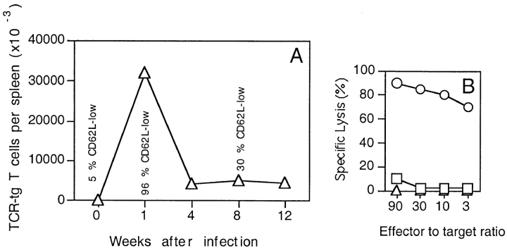
A proper comparison of memory T cells with naive T cells at the single cell level has proven difficult for technical reasons. To be able to compare naive T cells with effector and memory T cells expressing a single defined TCR, we used an adoptive transfer system (19, 27). Spleen cells (106 cells) derived from transgenic mice expressing a class I–restricted TCR (Vα2Vβ8) specific for the LCMV-derived peptide p33 were adoptively transferred into normal C57BL/6 mice, which were infected 2 h later with live virus. The adoptively transferred naive T cells expanded dramatically within 1 wk, differentiated to effector cells, and exhibited high ex vivo cytolytic activity (Fig. 1, A and B). The number of specific CD8+ T cells subsequently declined, but memory T cells remained at a high frequency in the host for at least 90 d (Fig. 1 A). Compared with the effector cells isolated on day 8 after infection, the memory T cells exhibited at least 100-fold reduced ex vivo cytolytic activity (Fig. 1 B). Moreover, although 95% of the effector cells expressed low levels of CD62L, 70% of the memory T cells had reverted back to a CD62Lhigh status (94% of the naive T cells were CD62Lhigh; Fig. 1 A). Similar results were obtained when CD44 expression was analyzed (not shown). Thus, the transferred naive T cells were rapidly activated in vivo after infection, then proliferated and differentiated to lytic effector cells. After elimination of the virus at day 6 to day 8 after immunization (not shown), a proportion of the cells survived as memory cells which exhibited a more quiescent status than the effector cells.
Figure 1.
Generation of effector and memory T cells. (A) Spleen cells (106 cells) from naive TCR transgenic mice were adoptively transferred into C57BL/6 recipient mice which were subsequently immunized with LCMV (200 PFU). The number of TCR transgenic T cells present in the spleen at various time points after infection was determined by flow cytometry by staining for CD8+ T cells expressing Vα2. The percentages of cells expressing low levels of CD62L are indicated. (B) Spleen cells were isolated at various time points after infection and tested in a 51Cr-release assay on p33-pulsed target cells. Similar results were obtained using peptide KAVVNIATM, which is specifically recognized by the transgenic TCR. One representative experiment of three is shown. Symbols denote naive (▵), effector (○), and memory (□) T cells.
The requirements for the activation of ex vivo–isolated naive, effector, and memory cells were analyzed in vitro. We found that the three populations required the same density of the virus-derived peptide p33 to proliferate, when B7+ thioglycollate-elicited macrophages were used as APCs, confirming earlier results (19; not shown). However, when B7-mediated costimulation was inhibited by a CTLA-4–Ig fusion molecule, proliferation of naive cells was strongly reduced, whereas almost no effect could be observed for effector and memory cells (Fig. 2 A). Similar results were obtained with different peptide concentrations (not shown). As shown previously for naive T cells (30), proliferation of effector and memory T cells was affected more severely by blocking CD28 during stimulation with the weak agonist A4Y (Fig. 2 B). These results indicate that, when stimulated by professional APCs, naive T cells do not require higher doses of antigen or higher levels of TCR engagement than effector and memory cells. However, in contrast to effector and memory T cells, naive T cells require the triggering of CD28 in order to be maximally activated.
Figure 2.
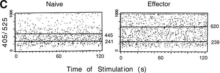
Reduced requirement for CD28 along with enhanced TCR signaling in effector and memory T cells. (A, B) Naive, effector, and memory T cells were stimulated with (A) p33-pulsed (10−7 M) or (B) A4Y-pulsed (10−6 M) thioglycollate-elicited macrophages in the absence (hatched bars) or presence (white bars) of CTLA-4–Ig fusion molecule (10 μg/ml). (C) Naive (left) and effector (right) T cells were stimulated with p33-pulsed (10−7 M) thioglycollate-elicited macrophages. Induction of increased intracellular free Ca2+ concentrations was assessed for CD4− B220− cells forming conjugates with APCs. The top straight line indicates average intracellular free Ca2+ for T cell–APC conjugates formed in the presence of peptide p33. The bottom straight line indicates baseline intracellular free Ca2+ for T cell–APC conjugates formed in the absence of peptide p33. Numerical values for the average OD 405/525 are shown.
CD28 reduces the threshold for T cell activation by amplifying the signals delivered by the TCR, thus inducing a stronger phosphorylation of many proteins involved in T cell activation (29, 31, 32). To investigate whether effector T cells exhibit enhanced TCR-mediated signaling, induction of Ca2+ flux was assessed in naive and effector cells after stimulation with p33-pulsed APCs. Elevation of free intracellular Ca2+ was significantly enhanced in effector cells compared with naive T cells, indicating that the TCR in effector T cells is more efficiently coupled to the signaling machinery than in naive T cells (Fig. 2 C). Similar results were obtained using various peptide concentrations (10−6 to 10−9 M; not shown), whereas intracellular free Ca2+ was low in both naive and effector cells in the absence of specific peptide (Fig. 2 C). Together with the observation that effector and memory T cells have a reduced requirement for the signal amplification by CD28, these results suggest that another mechanism might be able to substitute for CD28 in effector and memory cells. Such a mechanism may be of physiological relevance, since it would allow for the activation of memory T cells in peripheral tissues, where B7 expression is scarce.
Comodulation of CD8 and Triggered TCRs in Effector and Memory but not Naive T Cells.
Interestingly, we observed that, in contrast to effector and memory cells, proliferation of naive T cells could be inhibited by anti-CD8 antibodies (Fig. 3 A). This effect cannot be accounted for by a decreased stability of the TCR–peptide/MHC interaction, since (a) the affinity of the complex was the same in all the cells; (b) proliferation of naive, effector, and memory cells was inhibited by anti-CD8 antibodies when the APCs were pulsed with a weak agonist (Fig. 3 B); and (c) anti-CD8 antibodies did not inhibit T cell–APC conjugate formation (not shown).
Figure 3.
Anti-CD8 blocks proliferation of naive but not effector and memory T cells. Naive, effector, and memory T cells were stimulated with (A) p33-pulsed (10−7 M) or (B) A4Y-pulsed (10−6 M) thioglycollate-elicited macrophages in the absence (hatched bars) or presence (white bars) of anti-CD8 antibodies (3 μg/ml). Proliferation was assessed 30 h later by pulsing cultures with [3H]thymidine for 6 h. One representative experiment of three is shown.
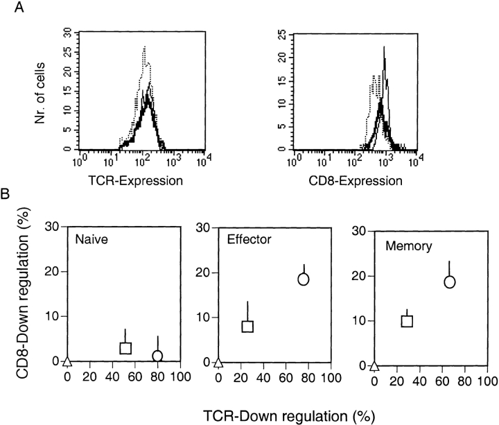
The contribution of coreceptors to T cell activation is not only due to their capacity to bind to the same MHC molecule engaged by the TCR and thus stabilize the TCR–MHC/peptide interaction (33–36), but also to their association with the tyrosine kinase Lck (37), which is involved in many early signaling events (38). It has been shown that in specific human T cell–APC conjugates, the coreceptors are recruited to triggered TCRs and are downregulated and degraded with identical kinetics and with fixed stoichiometry (39). It was possible that the distinct susceptibilities of naive versus effector and memory T cells to anti-CD8 blocking might be due to the distinct association of CD8 with the triggered TCR. Therefore, the association and the comodulation of TCR and CD8 in naive, effector, and memory cells were assessed in the next experiment. Fig. 4 A shows the expression levels of TCR and CD8 on naive, effector, and memory T cells before stimulation. Similar levels of TCR were expressed on the surface of all cell types, whereas CD8 expression was reduced by ∼50% on ex vivo–isolated effector cells. Specifically triggered TCRs have been shown to be internalized rapidly (40). In addition, CD8 has been shown to be associated and co-downmodulated with the TCR in T cell clones (39). To test whether the distinct susceptibility to anti-CD8 antibodies of naive versus effector and memory T cells might reflect a distinct association and co-downmodulation of CD8 with the TCR, the various cell types were stimulated with specific peptide, and downregulation of the TCR and CD8 was monitored at different time points. The TCR was specifically internalized in all three different cell types. In contrast, CD8 expression was not affected in naive T cells, whereas a proportion of CD8 was internalized in effector and memory T cells (Fig. 4 B).
Figure 4.
Co-downregulation of triggered TCRs and CD8 molecules in effector and memory but not in naive cells. (A) Expression of Vα2 (TCR; left) and CD8 (right) was analyzed before stimulation for the different cell types by flow cytometry. Results are shown for CD8+Vα2+ naive (solid lines), effector (dotted lines), and memory (bold lines) T cells. Nr., number. (B) Spleen cells were mixed with peptide p33-pulsed (10−8 M) macrophages, centrifuged, and analyzed immediately (▵) or incubated at 37°C for 1 h (□) or 3 h (○). Cells were stained for the expression of CD8 (APC), Vα2 (FITC), and Vβ8 (PE) and analyzed by flow cytometry. Percent downregulation of Vα2 and CD8 is shown for CD8+Vβ8+Vα2+ cells, and the SEM of triplicate cultures are indicated.
Priming-induced CD8–Lck Association in T Cells.
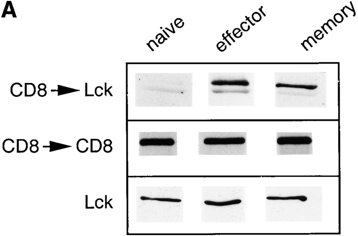
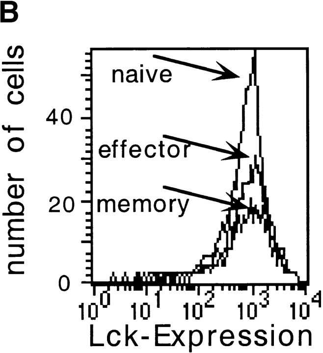
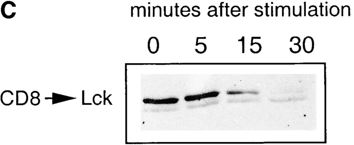
The downmodulation of coreceptor induced by TCR triggering is the consequence of binding of the coreceptor-associated Lck to the CD3 complex (39). Indeed, it has been shown that truncated CD4 molecules that fail to associate with Lck are not downregulated with triggered TCRs (39). This prompted us to analyze whether the lack of CD8 downregulation in naive T cells after triggering is due to a less extensive association of their coreceptors with Lck. Immunoprecipitation of CD8 and probing for the presence of Lck by immunoblotting showed that their association was much higher in both effector and memory T cells compared with naive T cells (Fig. 5 A). Flow cytometry analysis showed that surface CD8 expression was comparable in naive and memory cells and was only twofold reduced in effector cells (Fig. 4 A). Further, total cellular amounts of Lck were not different in the various cell types (Fig. 5 B). These data rule out the possibility that CD8–Lck association is driven by changes in expression levels. Interestingly, after triggering of effector T cells with peptide-pulsed APCs, the amount of CD8-associated Lck decreases in a time-dependent way correlating with TCR internalization, suggesting that the CD8-associated Lck is preferentially used by the triggered TCRs (Fig. 5 C).
Figure 5.
Priming of naive T cells induces targeting of Lck to the coreceptor. (A) Weak association of Lck with CD8 in naive cells. Immunoprecipitation with anti-CD8 antibodies and immunoblotting with anti-Lck antibody. As control, the amount of CD8 immunoprecipitated was detected. In control experiments, the total amount of Lck was determined in a purified population of cells. (B) Expression of Lck was analyzed by flow cytometry in permeabilized CD8+Vα2+ T cells. The average fluorescence of the negative controls for the different cell types (second stage only) were all <50. (C) CD8-associated Lck is preferentially used by TCRs during antigenic stimulation. Effector T cells were stimulated with p33-pulsed APCs and lysed at different times. The samples were then treated as in A.
Taken together, these results demonstrate that in naive cells only a few CD8 molecules are associated with Lck, and that specific priming results in redistribution of the kinase, which becomes preferentially associated with the coreceptors. These results suggest that the different localization of Lck might be responsible for the functional difference between naive and effector/memory T cells. In other words, the less stringent requirements in terms of costimulation for activation of memory and effector T cells may be explained by an Lck-mediated amplification of TCR signaling in these cells.
Constitutive Recycling of CD8 in Naive T Cells.
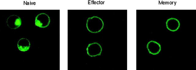
It has been shown that the association of Lck to the coreceptor prevents its targeting to the endocytic pathway and its recycling (41, 42). Therefore, we analyzed the CD8 endocytosis in naive, effector, and memory cells. The cells were stained at 4°C with an FITC-conjugated anti-CD8 antibody and than warmed to 37°C for 1 h. In effector and memory cells, the CD8 staining was distributed on the membrane and was completely absent in the cytoplasm of the cells. In contrast, in naive cells a clear process of endocytosis occurred, as demonstrated by the intracellular distribution of the fluorescence (Fig. 6). Thus, while in effector and memory cells the coreceptor is stably expressed on the membrane, in naive cells CD8 is constitutively internalized (Fig. 6), correlating with the lack of CD8–Lck association (Fig. 5).
Figure 6.
Rapid turnover of CD8 on naive but not effector and memory cells. Purified CD8+ T cells were stained for CD8 (FITC), incubated for 1 h at 37°C, and analyzed by confocal microscopy for CD8 distribution.
Cytosolic Distribution of Lck in Naive T Cells.
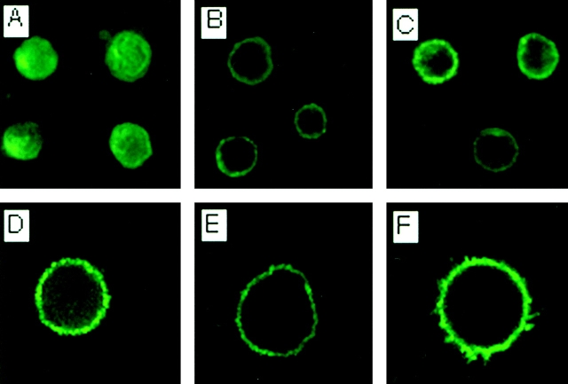
The differential association of Lck with CD8 in naive versus effector and memory T cells might reflect a different distribution of the kinase inside the cell. To investigate this question, we compared the cellular distribution of Lck in naive, effector, and memory T cells. We found that in effector and memory cells, Lck was localized at the plasma membrane, whereas in naive cells Lck had a more homogeneous cytosolic distribution (Fig. 7, A–C). In contrast, CD8 was present only in the cell membrane (Fig. 7, D–F). Thus, the different association of Lck with the CD8 coreceptor in naive versus effector and memory cells is due to a distinctive targeting of the kinase inside the cells.
Figure 7.
Differential distribution of Lck in naive versus effector and memory T cells. Purified CD8+ naive (A and D), effector (B and E), and memory (C and F) T cells were stained for the surface expression of Vα2, fixed and permeabilized, and subsequently stained for the expression of Lck (A–C) or CD8 (D–F). All cells shown had detectable expression of the transgene-encoded TCR (not shown).
Distinct Distribution of Lck in Virus-specific Bona Fide Memory T Cells.
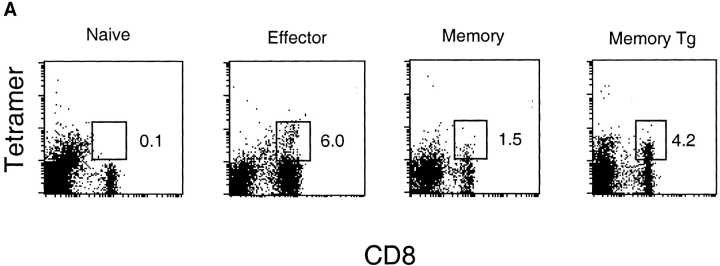
To reveal whether the distinct distribution of Lck was also observed in memory T cells isolated from normal, non-TCR transgenic animals, the recently developed tetramer technology was used for the isolation of virus-specific effector and memory T cells. Mice were infected with LCMV, and peptide p33–specific effector and memory T cells were stained using H-2Db tetramers pulsed with peptide p33 (Fig. 8 A). As a control, TCR transgenic memory T cells generated as described above were also stained. Specific cells were isolated by cell sorting, and the distribution of Lck was analyzed by confocal microscopy. To facilitate analysis of the intracellular distribution of Lck, the nuclei were separately stained using PI (Fig. 8 B). As a source of naive T cells, CD8+, tetramer-negative T cells were isolated from uninfected animals. The intracellular localization of Lck was clearly visible in naive T cells, whereas it was predominantly in the cell membrane in effector and memory T cells.
Figure 8.
Lck is targeted to the membrane in antigen-specific memory T cells isolated from normal mice. C57BL/6 mice were immunized with LCMV, and spleen cells were harvested on day 0 (naive), day 8 (effector), or day 45 (memory). As a positive control, memory T cells were also generated using the adoptive transfer method described in the legend to Fig. 1 (Tg memory). (A) Specific CD8+ T cells were stained with PE- labeled p33-pulsed H-2Db tetramers and analyzed by flow cytometry. (B) Tetramer-positive T cells were sorted, fixed and permeabilized, and stained for the expression of Lck (green). The nuclei were stained using PI (red).
Discussion
Using an adoptive transfer system, we compared early and late events after TCR triggering in naive, effector, and memory cells. In naive T cells, TCR triggering induced a weak calcium flux, and optimal proliferation required costimulatory signals via CD28. In contrast, CD28 costimulation did not affect effector and memory T cell responses. These results strongly support the concept that T cell priming requires the presentation of antigenic peptides by professional APCs, which express high levels of costimulatory molecules, whereas activation of effector and memory cells can also be achieved by nonprofessional APCs.
Since naive, effector, and memory T cells expressed the same TCR, our results indicate that the reduced requirement for costimulation and the enhanced TCR signaling are not due to increased TCR affinities but rather reflect an improved signaling cascade. Moreover, many recent findings indicate that CD28 engagement, in addition to delivering accessory signals, facilitates T cell activation by enhancing the signals delivered by the triggered TCRs (29, 32, 39). We therefore suggest that effector and memory T cells do not require the CD28-mediated costimulation because they acquire an improved signal transduction machinery amplifying TCR signaling by a different mechanism.
In effector and memory cells, Lck, a kinase critically involved in TCR signaling (38), is targeted to the membrane and associated with the coreceptor. Surprisingly, however, in naive cells only a few CD8 molecules are associated with Lck and the kinase is homogeneously distributed inside the cell. Thus, in effector and memory T cells, Lck is located in a strategically important position at the cell surface and therefore is able to rapidly react to TCR triggering. In support of this notion, it has been recently shown that Lck must be localized to the plasma membrane to function properly in TCR signaling (43).
The mechanism of this developmentally regulated targeting of Lck to the plasma membrane in effector and memory T cells is presently not known. It is possible that membranes of effector and memory T cells may be enriched in glycolipid microdomains, which preferentially bind src kinases (44, 45). However, preliminary evidence suggests that effector but not memory T cells are enriched in such glycolipid microdomains (not shown), indicating that a different mechanism is responsible for the targeting of Lck to the cell membrane in memory T cells. Interestingly, it has recently been shown that targeting of Lck to the plasma membrane requires the attachment of S-acyl groups to Cys3 and Cys5 of the molecule (43). Therefore, it is possible that in T cells the posttranslational S-acylation of Lck is the mechanism that regulates the intracellular localization of Lck. This notion is supported by the observation that palmitylation of other src family members can occur reversibly and therefore may represent an important mechanism for the regulation of their activity (46, 47). Moreover, it has recently been shown that various palmitylated signaling molecules are recruited to glycolipid microdomains in the plasma membrane upon activation of T cells (48, 49), further indicating that T cell activation may alter the intracellular distribution of signaling molecules.
During the interaction of T cell clones with APCs, the coreceptors are recruited to triggered TCRs and are downregulated with identical kinetics. This process is the consequence of binding of coreceptor-associated Lck to ZAP-70/ζ and takes place whenever the TCR is triggered (39). We found that in naive mouse T cells, the CD8 coreceptor molecules are not downregulated with the triggered TCRs. Nevertheless, the results do not exclude that few CD8 molecules are associated with Lck and are recruited to the TCR also in naive cells. However, the limited sensitivity of the method does not allow the measurement of their downregulation or degradation. This is also suggested by the fact that proliferation of naive T cells induced by the wild-type agonist can be selectively inhibited by anti-CD8 antibodies. The fact that the responses of effector and memory cells to the strong agonist are not affected by anti-CD8 antibodies is in line with previous findings using T cell clones (39). Indeed, the intracellular interaction between Lck and the CD3 components does not require the extracellular binding of CD8 to the cognate MHC molecule, probably because in mouse (50) as well as in human (39) T cells, a fraction of TCRs are constitutively associated with the coreceptor via Lck. The fact that proliferation of naive T cells can be inhibited by anti-CD8 antibodies might therefore be explained by the lack of constitutive association between CD8 and TCR/CD3 in these cells. Therefore, few Lck-associated CD8 molecules are not easily available and likely need to bind the MHC molecules to be recruited to the T cell–APC contact region.
It has been shown that effector T cells (21) and also memory T cells (19) are more rapidly committed for proliferation than naive T cells. According to our findings, effector and memory T cells might reach thresholds for proliferation more rapidly than naive T cells, due to the Lck-mediated amplification of the signals delivered by the TCRs. In addition, in contrast to naive T cells, memory T cells have been reported to receive a survival signal from noncognate MHC class I molecules (14), a finding which is possibly also related to the enhanced TCR signaling machinery present in memory T cells.
In B cells, the high rate of somatic mutation in the Ig variable regions may lead to the generation of memory cells with high-affinity receptors. This study indicates that in T cells, the “memory bonus” is not offered by the receptor itself but by its enhanced capacity to transduce activation signals. To our knowledge, this finding represents the first evidence of a biochemically controlled process that functionally differentiates memory from naive T cells.
Acknowledgments
We thank Klaus Karjalainen and Jeff Bluestone for critical reading of the manuscript, and Barbara Ecabert for technical assistance.
Abbreviations used in this paper
- CTLA-4
cytotoxic T lymphocyte–associated antigen 4
- LCMV
lymphocytic choriomeningitis virus
- PI
propidium iodide
Footnotes
The Basel Institute for Immunology was founded and is supported by F. Hoffmann-La Roche, Basel, Switzerland.
References
- 1.Doherty PC, Hou S, Tripp RA. CD8+ T-cell memory to viruses. Curr Opin Immunol. 1994;6:545–552. doi: 10.1016/0952-7915(94)90139-2. [DOI] [PubMed] [Google Scholar]
- 2.Sprent J, Tough DF. Lymphocyte life-span and memory. Science. 1994;265:1395–1400. doi: 10.1126/science.8073282. [DOI] [PubMed] [Google Scholar]
- 3.Ahmed R, Gray D. Immunological memory and protective immunity: understanding their relation. Science. 1996;272:54–60. doi: 10.1126/science.272.5258.54. [DOI] [PubMed] [Google Scholar]
- 4.Zinkernagel RM, Bachmann MF, Kündig TM, Oehen S, Hengartner H. On immunological memory. Annu Rev Immunol. 1996;14:333–367. doi: 10.1146/annurev.immunol.14.1.333. [DOI] [PubMed] [Google Scholar]
- 5.Dutton RW, Bradley LM, Swain SL. T cell memory. Annu Rev Immunol. 1998;16:201–223. doi: 10.1146/annurev.immunol.16.1.201. [DOI] [PubMed] [Google Scholar]
- 6.Parker DC. T cell-dependent B cell activation. Annu Rev Immunol. 1993;11:331–360. doi: 10.1146/annurev.iy.11.040193.001555. [DOI] [PubMed] [Google Scholar]
- 7.Foy TM, Aruffo A, Bajorah J, Buhlmann JE, Noelle JN. Immune regulation by CD40 and its ligand. Annu Rev Immunol. 1996;14:591–617. doi: 10.1146/annurev.immunol.14.1.591. [DOI] [PubMed] [Google Scholar]
- 8.Mullbacher A, Flynn K. Aspects of cytotoxic T cell memory. Immunol Rev. 1996;150:113–127. doi: 10.1111/j.1600-065x.1996.tb00698.x. [DOI] [PubMed] [Google Scholar]
- 9.Hou S, Hyland L, Ryan KW, Portner A, Doherty PC. Virus-specific CD8+ T-cell memory determined by clonal burst size. Nature. 1994;369:652–654. doi: 10.1038/369652a0. [DOI] [PubMed] [Google Scholar]
- 10.Lau LL, Jamieson BD, Somasundaram T, Ahmed R. Cytotoxic T-cell memory without antigen. Nature. 1994;369:648–652. [PubMed] [Google Scholar]
- 11.Mullbacher A. The long-term maintenance of cytotoxic T cell memory does not require the persistence of antigen. J Exp Med. 1994;179:317–321. doi: 10.1084/jem.179.1.317. [DOI] [PMC free article] [PubMed] [Google Scholar]
- 12.Bruno L, Kirberg J, von Boehmer H. On the cellular basis of immunological T cell memory. Immunity. 1995;2:37–43. doi: 10.1016/1074-7613(95)90077-2. [DOI] [PubMed] [Google Scholar]
- 13.Bachmann MF, Kündig TM, Hengartner H, Zinkernagel RM. Protection from immunopathological consequences of a viral infection by activated but not resting cytotoxic T cells: T cell memory without memory T cells? . Proc Natl Acad Sci USA. 1997;94:640–645. doi: 10.1073/pnas.94.2.640. [DOI] [PMC free article] [PubMed] [Google Scholar]
- 14.Tanchot C, Lemonnier FA, Perarnau B, Freitas AA, Rocha B. Differential requirements for survival and proliferation of CD8 naive or memory T cells. Science. 1997;276:2057–2062. doi: 10.1126/science.276.5321.2057. [DOI] [PubMed] [Google Scholar]
- 15.Selin LK, Nahill SR, Welsh RM. Cross-reactivities in memory cytotoxic T lymphocyte recognition of heterologous viruses. J Exp Med. 1994;179:1933–1943. doi: 10.1084/jem.179.6.1933. [DOI] [PMC free article] [PubMed] [Google Scholar]
- 16.Tough DF, Borrow P, Sprent J. Induction of bystander T cell proliferation by viruses and type I interferon in vivo. Science. 1996;272:1947–1950. doi: 10.1126/science.272.5270.1947. [DOI] [PubMed] [Google Scholar]
- 17.Ehl S, Hombach J, Aichele P, Hengartner H, Zinkernagel RM. Bystander activation of cytotoxic T cells: studies on the mechanism and evaluation of in vivo significance in a transgenic mouse model. J Exp Med. 1997;185:1241–1251. doi: 10.1084/jem.185.7.1241. [DOI] [PMC free article] [PubMed] [Google Scholar]
- 18.Zhang X, Sun S, Tough DF, Sprent J. Potent and selective stimulation of memory-phenotype CD8+ T cells in vivo by IL-15. Immunity. 1998;8:591–599. doi: 10.1016/s1074-7613(00)80564-6. [DOI] [PubMed] [Google Scholar]
- 19.Bachmann MF, Barner M, Viola A, Kopf MF. Distinct kinetics of cytokine production and cytolysis in effector and memory T cells after viral infection. Eur J Immunol. 1999;29:291–299. doi: 10.1002/(SICI)1521-4141(199901)29:01<291::AID-IMMU291>3.0.CO;2-K. [DOI] [PubMed] [Google Scholar]
- 20.Croft M, Bradley LM, Swain S. Naive versus memory CD4+ T cell responses to antigen: memory cells are less dependent on accessory cell costimulation and respond to many antigen-presenting cell types including resting B cells. J Immunol. 1994;152:2675–2685. [PubMed] [Google Scholar]
- 21.Iezzi G, Karjalainen K, Lanzavecchia A. The duration of antigenic stimulation determines the fate of naive and effector T cells. Immunity. 1998;8:89–95. doi: 10.1016/s1074-7613(00)80461-6. [DOI] [PubMed] [Google Scholar]
- 22.McHeyzer-Williams MG, Davis MM. Antigen-specific development of primary and memory T cells in vivo. Science. 1995;268:106–111. doi: 10.1126/science.7535476. [DOI] [PubMed] [Google Scholar]
- 23.Bachmann MF, Speiser DE, Ohashi PS. Functional maturation of an anti-viral cytotoxic T cell response. J Virol. 1997;71:5764–5768. doi: 10.1128/jvi.71.8.5764-5768.1997. [DOI] [PMC free article] [PubMed] [Google Scholar]
- 24.Murali-Krishna K, Altman JD, Suresh M, Sourdive DJ, Zajac AJ, Miller JD, Slansky J, Ahmed R. Counting antigen-specific CD8 T cells: a reevaluation of bystander activation during viral infection. Immunity. 1998;8:177–187. doi: 10.1016/s1074-7613(00)80470-7. [DOI] [PubMed] [Google Scholar]
- 25.Altman JD, Moss PAH, Goulder PJR, Barouch DH, McHeyzer-Williams MG, Bell JI, McMichael AJ, Davis MM. Phenotypic analysis of antigen-specific T lymphocytes. Science. 1996;274:94–96. [PubMed] [Google Scholar]
- 26.Pircher HP, Bürki K, Lang R, Hengartner H, Zinkernagel RM. Tolerance induction in double specific T-cell receptor transgenic mice varies with antigen. Nature. 1989;342:559–561. doi: 10.1038/342559a0. [DOI] [PubMed] [Google Scholar]
- 27.Zimmermann C, Brduscha-Riem K, Blaser C, Zinkernagel RM, Pircher H. Visualization, characterization, and turnover of CD8+memory T cells in virus- infected hosts. J Exp Med. 1996;183:1367–1375. doi: 10.1084/jem.183.4.1367. [DOI] [PMC free article] [PubMed] [Google Scholar]
- 28.Ledbetter JA, Herzenberg LA. Xenogeneic monoclonal antibodies to mouse lymphoid differentiation antigens. Immunol Rev. 1979;47:63–90. doi: 10.1111/j.1600-065x.1979.tb00289.x. [DOI] [PubMed] [Google Scholar]
- 29.Bachmann MF, McKall-Faienza K, Schmits R, Bouchard D, Beach J, Speiser DE, Mak TW, Ohashi PS. Distinct roles for LFA-1 and CD28 during activation of naive T cells: adhesion versus costimulation. Immunity. 1997;7:549–557. doi: 10.1016/s1074-7613(00)80376-3. [DOI] [PubMed] [Google Scholar]
- 30.Bachmann MF, Sebzda E, Kündig TM, Shahinian A, Speiser D, Mak TW, Ohashi PS. T cell responses are governed by avidity and costimulatory thresholds. Eur J Immunol. 1996;26:2017–2022. doi: 10.1002/eji.1830260908. [DOI] [PubMed] [Google Scholar]
- 31.Viola A, Lanzavecchia A. T cell activation determined by T cell receptor number and tunable thresholds. Science. 1996;273:104–106. doi: 10.1126/science.273.5271.104. [DOI] [PubMed] [Google Scholar]
- 32.Tuosto L, Acuto O. CD28 affects the earliest signaling events generated by TCR engagement. Eur J Immunol. 1998;28:2131–2142. doi: 10.1002/(SICI)1521-4141(199807)28:07<2131::AID-IMMU2131>3.0.CO;2-Q. [DOI] [PubMed] [Google Scholar]
- 33.Doyle C, Strominger JL. Interaction between CD4 and class II MHC molecules mediates cell adhesion. Nature. 1987;330:256–259. doi: 10.1038/330256a0. [DOI] [PubMed] [Google Scholar]
- 34.Norment AM, Salter RD, Parham P, Engelhard VH, Littman DR. Cell-cell adhesion mediated by CD8 and MHC class I molecules. Nature. 1988;336:79–81. doi: 10.1038/336079a0. [DOI] [PubMed] [Google Scholar]
- 35.Luescher IF, Vivier E, Layer A, Mahiou J, Godeau F, Malissen B, Romero P. CD8 modulation of T cell antigen receptor-ligand interactions on living cytotoxic T lymphocytes. Nature. 1995;373:353–356. doi: 10.1038/373353a0. [DOI] [PubMed] [Google Scholar]
- 36.Garcia KC, Scott CA, Brunmark A, Carbone FR, Peterson PA, Wilson IA, Teyton L. CD8 enhances formation of stable T-cell receptor/MHC class I molecule complexes. Nature. 1996;384:577–581. doi: 10.1038/384577a0. [DOI] [PubMed] [Google Scholar]
- 37.Veillette A, Bookman MA, Horak EM, Bolen JB. The CD4 and CD8 T cell surface antigens are associated with the internal membrane tyrosine-protein kinase p56lck. Cell. 1988;55:301–308. doi: 10.1016/0092-8674(88)90053-0. [DOI] [PubMed] [Google Scholar]
- 38.Weiss A, Littman DR. Signal transduction by lymphocyte antigen receptors. Cell. 1994;76:263–274. doi: 10.1016/0092-8674(94)90334-4. [DOI] [PubMed] [Google Scholar]
- 39.Viola A, Salio M, Tuosto L, Linkert S, Lanzavecchia A. Quantitative contribution of CD4 and CD8 to T cell antigen receptor serial triggering. J Exp Med. 1997;186:1775–1779. doi: 10.1084/jem.186.10.1775. [DOI] [PMC free article] [PubMed] [Google Scholar]
- 40.Valitutti S, Müller S, Cella M, Padovan E, Lanzavecchia A. Serial triggering of many T cell receptors by a few peptide-MHC complexes. Nature. 1995;375:148–151. doi: 10.1038/375148a0. [DOI] [PubMed] [Google Scholar]
- 41.Pelchen-Matthews A, Armes JE, Marsh M. Internalisation and recycling of CD4 transfected into HeLa and NIH-3T3 cells. EMBO (Eur Mol Biol Organ) J. 1989;8:3641–3649. doi: 10.1002/j.1460-2075.1989.tb08538.x. [DOI] [PMC free article] [PubMed] [Google Scholar]
- 42.Pelchen-Matthews A, da Silva RP, Bijlmakers MJ, Signoret N, Gordon S, Marsh M. Lack of p56lck expression correlates with CD4 endocytosis in primary lymphoid and myeloid cells. Eur J Immunol. 1998;28:3639–3647. doi: 10.1002/(SICI)1521-4141(199811)28:11<3639::AID-IMMU3639>3.0.CO;2-Q. [DOI] [PubMed] [Google Scholar]
- 43.Kabouridis PS, Magee AI, Ley SC. S-acylation of Lck protein tyrosine kinase is essential for its signalling function in T lymphocytes. EMBO (Eur Mol Biol Organ) J. 1997;16:4983–4998. doi: 10.1093/emboj/16.16.4983. [DOI] [PMC free article] [PubMed] [Google Scholar]
- 44.Shenoy-Scaria AM, Gauen LKT, Kwong J, Shaw AS, Lublin DM. Palmitylation of an amino-terminal cysteine motif of protein tyrosine kinase p56lck and p59fyn mediates interaction with glycosyl-phosphatidylinositol- anchored proteins. Mol Cell Biol. 1993;13:6385–6392. doi: 10.1128/mcb.13.10.6385. [DOI] [PMC free article] [PubMed] [Google Scholar]
- 45.Rodgers W, Crise B, Rose JK. Signals determining protein tyrosine kinase and glycosyl-phosphatidyl- anchored protein targeting to a glycolipid-enriched membrane fraction. Mol Cell Biol. 1994;14:5384–5391. doi: 10.1128/mcb.14.8.5384. [DOI] [PMC free article] [PubMed] [Google Scholar]
- 46.Mumby SM. Reversible palmitylation of signaling proteins. Curr Opin Immunol. 1997;9:148–154. doi: 10.1016/s0955-0674(97)80056-7. [DOI] [PubMed] [Google Scholar]
- 47.Wolven A, Okamura H, Rosenblatt Y, Resh MD. Palmitylation of p59fyn is reversible and sufficient for plasma membrane association. Mol Biol Cell. 1997;8:1159–1173. doi: 10.1091/mbc.8.6.1159. [DOI] [PMC free article] [PubMed] [Google Scholar]
- 48.Zhang W, Trible RP, Samelson LE. LAT palmitylation: its essential role in membrane microdomain targeting and tyrosine phosphorylation during T cell activation. Immunity. 1998;9:239–246. doi: 10.1016/s1074-7613(00)80606-8. [DOI] [PubMed] [Google Scholar]
- 49.Xavier R, Brennan T, Li Q, McCormack C, Seed B. Membrane compartmentation is required for efficient T cell activation. Immunity. 1998;8:723–732. doi: 10.1016/s1074-7613(00)80577-4. [DOI] [PubMed] [Google Scholar]
- 50.Diez-Orejas R, Ballester S, Feito MJ, Ojeda G, Criado G, Ronda M, Portoles P, Rojo JM. Genetic and immunological evidence for CD4-dependent association of p56lck with the alpha beta TCR: regulation of TCR-induced activation. EMBO (Eur Mol Biol Organ) J. 1994;13:90–99. doi: 10.1002/j.1460-2075.1994.tb06238.x. [DOI] [PMC free article] [PubMed] [Google Scholar]


