Abstract
The chemokine thymus and activation-regulated chemokine (TARC; CCL17) is displayed by cutaneous (but not intestinal) venules, and is thought to trigger vascular arrest of circulating skin homing memory T cells, which uniformly express the TARC receptor CC chemokine receptor (CCR)4. Cutaneous T cell–attracting chemokine (CTACK; CCL27), expressed by skin keratinocytes, also attracts cutaneous memory T cells, and is hypothesized to assist in lymphocyte recruitment to skin as well. Here we show that chronic cutaneous inflammation induces CD4 T cells expressing E-selectin binding activity (a marker of skin homing memory cells) in draining lymph node, and that these E-selectin ligand+ T cells migrate efficiently to TARC and to CTACK. In 24 h in vivo homing assays, stimulated lymph node T cells from wild-type mice or, surprisingly, from CCR4-deficient donors migrate efficiently to inflamed skin; and an inhibitory anti-CTACK antibody has no effect on wild-type lymphocyte recruitment. However, inhibition with anti-CTACK monoclonal antibody abrogates skin recruitment of CCR4-deficient T cells. We conclude that CTACK and CCR4 can both support homing of T cells to skin, and that either one or the other is required for lymphocyte recruitment in cutaneous delayed type hypersensitivity.
Keywords: lymphocyte homing, chemokines, inflammation, delayed type hypersensitivity, selectins
Introduction
There is growing evidence that chemotactic cytokines (chemokines) contribute to tissue-specific lymphocyte homing in conjunction with adhesion receptors (1–6). For example, CC chemokine receptor (CCR)4 on T lymphocytes and its ligand thymus and activation-regulated chemokine (TARC) are implicated in lymphocyte–endothelial interactions during lymphocyte recruitment to normal and inflamed cutaneous sites (5). Immunohistochemistry suggests that TARC is constitutively expressed and hyperinducible on cutaneous venules and some other systemic venules, but is absent from vessels at sites of lymphocyte recruitment into the intestines. Moreover, its receptor CCR4 is highly expressed by cutaneous lymphocyte antigen positive (CLA+, E-selectin binding) circulating skin memory lymphocytes, but not by intestinal memory cells, consistent with a role in tissue selective trafficking. Recently, another skin associated chemokine, cutaneous T cell–attracting chemokine (CTACK), has been shown to selectively attract CLA+ cutaneous memory T cells as well. CTACK is constitutively expressed by epidermal skin keratinocytes and has been postulated to be involved in basal T cell trafficking during immunosurveillance (6). Inflammatory chemokines that are upregulated in diverse tissue sites during inflammation, such as ligands for CXC chemokine receptor (CXCR)3, CCR5, or CCR6, also have the potential to contribute to skin homing. However, the significance of these various chemokine/receptor systems to lymphocyte recruitment to skin has not been assessed in physiologic models.
This study was designed to evaluate the importance of the CCR4/TARC and CCR10/CTACK in T cell recruitment to inflamed skin in a model of delayed type hypersensitivity (DTH) in the mouse. Involvement of CCR4 was assessed using CCR4-deficient lymphocytes (7), and that of CTACK using a blocking anti-CTACK mAb, in short term assays of T cell homing. Our results reveal that at least one of these pathways (CCR4 or CCR10/CTACK) must be functional for effective T cell recruitment to skin; but that, in the setting of DTH, their involvement is overlapping so that either is permissive for T cell accumulation. We conclude that in this model of cutaneous inflammation, these tissue-selective chemokines play an essential role that cannot be efficiently replaced by inflammatory chemokines.
Materials and Methods
Antibodies.
Rat anti–mouse antibodies to CTACK (rat IgG2b), CD44-FITC, CD45RB-PE, CD4-allophycocyanin (APC), L-selectin-APC, and streptavidin-Cychrome were purchased from BD PharMingen. E-selectin/human IgM chimera was kindly provided by Dr. Lowe, University of Michigan, Ann Arbor, MI. Biotinylated goat anti–human IgM was from Caltag. Monoclonal rat IgG2a anti–human CD44 antibody Hermes-1 (American Type Culture Collection) and 30H12 (anti–mouse Thy1.2; gift from Dr. Herzenberg, Stanford University, Stanford, CA) were produced from hybridomas in our laboratories.
Animals.
C57Bl/6 and CCR4−/− (7) mice were housed and bred at the animal facility of Palo Alto Veterans Medical Center. B6.PL-Thy1.1 mice were obtained from The Jackson Laboratory. Mice of both sexes were used at age 8–14 wk.
Lymphocyte Isolation.
Lymphocytes from skin draining LN (cervical, and axillary LN) and red cell–depleted splenocytes were isolated as described (1). Mouse peripheral blood was collected and lymphocytes were obtained after sedimentation of erythrocytes in PBS/5 mM EDTA/2% Dextran for 30 min at 37°C and further depleted of red blood cells by lysis. Skin-infiltrating T cells were released from the connective tissue after separating the epidermal sheets as described (4).
For FACS® analysis skin infiltrating T lymphocytes were purified using CD3 selection columns as directed by the manufacturer (R&D Systems). For chemotaxis assays lymphocytes from skin draining LN T cells were incubated at 37°C for 1 h before the assay.
Chemotaxis Assay.
Standard transmigration assays using 5-μm pore transwells inserts (Costar) were performed as described (5, 8). Mouse chemokines TARC (CCL17; R&D Systems), CTACK (CCL27; R&D Systems), macrophage-derived chemokine (MDC; CCL22; PeproTech), and secondary lymphoid tissue chemoattractant (SLC; CCL21; PeproTech) were used at 100 nM. Input and migrated T cell subsets were identified by their homing-receptor and activation marker expression using the E-selectin chimera as well as commercially available monoclonal antibodies against CD4, CD44, and CD45RB; and percent migration of antigenically defined subsets was calculated.
Flow Cytometry.
Identification of E-selectin (CD62E-binding) ligand (E-lig+) has been assessed by binding with murine E-selectin Ig chimera, as described (9). In brief, cells were incubated with E-selectin Ig chimera on ice for 30 min. Nonspecific staining was determined by addition of 5 mM EDTA. Cells were washed and biotinylated goat anti–human IgM was administered. After blocking with 10% rat IgG, cells were stained with streptavidin-Cychrome, CD44-FITC, CD45RB-PE, and CD4-APC. For homing studies, Thy1.2-FITC and CD4-APC were used. Cells were analyzed using CELLQuest™ software (Becton Dickinson).
Immunohistochemistry.
Acetone fixed frozen sections of mouse ears (6 μm) were blocked with 50% rat IgG before incubation with 20 μg/ml rat anti–mouse CD4-PE (R&D Systems) and 20 μg/ml rat anti–mouse Thy1.2-FITC and visualized by confocal microscopy. Stained sections from Thy1.1 recipients of Thy1.2+ donor cells of wild-type or CCR4−/− origin were analyzed by counting the total number of CD4 T cells, and the number of Thy1.2+ CD4 T cells in ∼20 optical fields (40× objective); at least 150 Thy1.2+ CD4 T cells were counted for each recipient type. Mice that received no donor cells served as staining controls. Data represent mean ± SEM of CD4 or Thy1.2+ CD4 T cells per optical field of three independent experiments.
DTH.
DTH reaction was induced by repeated epicutaneous application of 2,4 dinitrofluorobenzene (DNFB; Sigma-Aldrich) (10). Primary immunization was achieved by painting bare abdominal skin on day 1 with 0.5% DNFB (20 μl, in acetone:olive oil, 4:1). On day 7, mice were challenged by application of 0.5% DNFB (20 μl) on the ears to create a secondary DTH response and lymphocytes draining the inflamed skin (from cervical, and axillary LN) were collected 24 h later and used as donor cells for adoptive transfer studies. Congenic recipients did not receive primary immunization and were challenged only (20 μl of 0.5% DNFB on both ears) just after transfer of donor cells. Ear thickness (measured with Ames calipers) was measured at the time of challenge (0 h, baseline) and at 24 h after donor cell (or no donor control) administration and ear painting, and the difference (swelling) was calculated. Donor cell–dependent swelling was the difference between swelling of ears of donor cell recipients, and of “no donor control” mice.
Adoptive Transfer/Homing Assay.
The in vivo homing of wild-type and CCR4−/− lymphocytes to inflamed skin was compared using the Thy1 congenic system. B6.Thy1.1 mice were used as recipients, whereas CCR4−/− and C57Bl/6 T cells (Thy1.2 allotype) served as donor T cells. 5–10 × 107donor T cells, obtained from LN draining the inflamed skin 24 h after the DNFB treatment on day 7 of initially primed animals (described above), were retro-orbitally injected into unprimed congenic recipients in 100–200 μl saline (after labeling with 500 nM CFSE for proliferation studies). Immediately after the adoptive transfer both ears of the unprimed recipients were challenged with the immunogen (20 μl of 0.5% DNFB) to create a “secondary” immune response mediated by the transferred donor cells. Homing of adoptively transferred lymphocytes to skin and other organs was evaluated after 24 h (control animals received no donor cells). The readout for DTH was ear swelling (see above). Additionally, FACS® analysis was performed to identify donor-derived CD4 T cells as described. To assess the role of CTACK, 100 μg (2 mg/ml) monoclonal rat anti–mouse CTACK antibody was injected twice intravenously (6 and 1 h in advance of donor cell injection). Control animals were treated with isotype control antibody, Hermes 1 (American Type Culture Collection). Data are expressed as the relative number of donor T cells (percent Thy1.2+ in CD4 population) in each tissue normalized to that of donor T cells in draining LN (to facilitate comparisons between experiments). The data presented are the mean ± SEM of results from seven experiments.
Statistical Analysis.
For statistical comparison of two samples, a two-tailed Student's t test was used when applicable.
Results and Discussion
E-lig+ T Cells Induced during Cutaneous Inflammation Migrate to TARC, MDC, and CTACK.
T cells are essential participants in pathologic cutaneous inflammatory reactions including contact hypersensitivity, and inflammatory disorders such as psoriasis and atopic dermatitis. T cells recruited in these diverse disorders share a number of common features, associated with important mechanisms for tissue selective homing to skin. These include expression of high levels of the E-selectin binding cutaneous lymphocyte antigen (10–12) and, based on recent studies, expression of the chemokine receptors CCR4 and probably CCR10 (5, 6). Although binding to vascular E-selectin has been shown to be important for T cell recruitment and T cell–mediated inflammation in skin in physiologic models (10, 13), involvement of CCR4 and its vascular ligand TARC, and of CTACK and its lymphocyte receptor CCR10 in skin lymphocyte homing have been proposed based on indirect if compelling studies of their patterns of expression in vivo and their proadhesive/or chemotactic activities in vitro. The present study was designed to assess directly the involvement of these chemokine/receptor pairs in physiologic assays of T cell homing to inflamed skin.
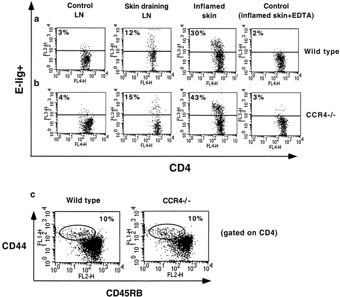
We initially asked whether skin homing T cells in the mouse (as in the human), migrate efficiently to TARC and CTACK. As determined by FACS® analyses, only ∼3% of T cells in normal LN bind E-selectin; but E-lig+ memory T cells (CD4+CD44highCD45RBlow) are abundant in skin inflamed by application of DNFB (36 ± 6% of skin CD4 memory T cells; n = 6) and in LN draining inflamed skin (12 ± 3%, n = 6; Fig. 1 a). Importantly, induction of E-lig+ T cells in LN, and the accumulation of T cells and the frequency of E-lig+ T cells in inflamed ears, were not substantially altered in CCR4-deficient mice (13 ± 3% in draining LN, and 50 ± 5% in inflamed skin, n = 6; Fig. 1 b). The fraction of CD4 T cells of memory phenotype (CD44highCD45RBlow) in skin-draining LN was also similar (Fig. 1 c), representing 10 ± 1% and 12 ± 1% (mean ± SEM, n = 10) of the CD4 pool in both wild-type and CCR4−/− mice. In conclusion, our experiments show that CCR4 is not required for induction of memory T cells with skin homing (E-lig+) phenotype, nor for the accumulation of T cells in inflamed skin.
Figure 1.
Generation of E-lig+ CD4 memory T cells after cutaneous immunization with DNFB. T lymphocytes from draining LN and inflamed skin of wild-type (a) and CCR4−/− mice (b) treated with immunogen (DNFB) were isolated and analyzed by flow cytometry for E-lig+ expression in comparison to LN cells from untreated mice. Induction of E-lig+ T cells in LN, and the localization of E-lig+ T cells in inflamed ears, were not significantly altered in CCR4-deficient mice (b). The fraction of CD4 T cells of memory phenotype in skin-draining LNs was also similar (c). T cells are gated on memory T cells (CD4+ CD44highCD45RBlow). Additional controls (E-selectin Ig chimera + EDTA on LN cells) were also negative (not shown). The data are representative of 6 (a and b) and 10 (c) experiments.
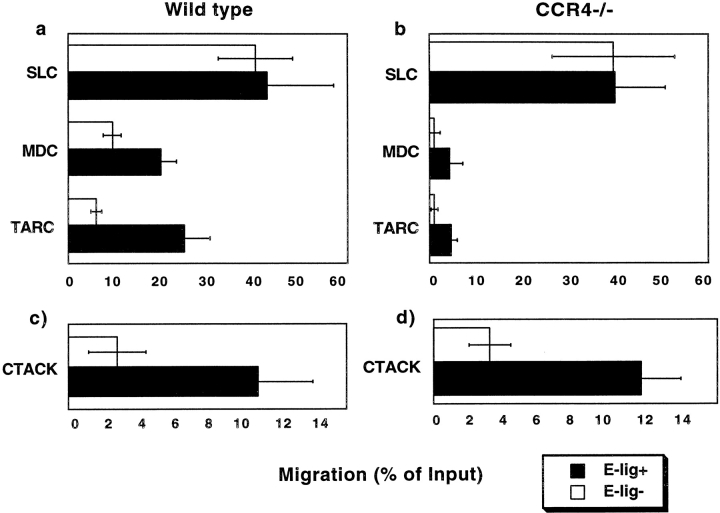
In transwell chemotaxis assays, the E-lig+ subset of T cells derived from draining LN migrates efficiently to CCR4 ligands TARC and MDC. Migration is selective, in that the E-lig-memory phenotype T cells (and naive T cells) respond relatively poorly to the CCR4 ligands (Fig. 2 a), though they migrate well to the CCR7 ligand SLC (Fig. 2 a). Migration to TARC and MDC is CCR4 dependent, as T cells from CCR4-deficient mice do not respond to these CCR4 ligands, but retain their responsiveness to SLC (Fig. 2 b). In addition, both wild-type and CCR4-deficient E-lig+ but not E-lig- CD4 memory T cells migrate to CTACK (Fig. 2, c and d). Thus CTACK, as well as TARC and MDC, attract E-lig+ memory T cells in mouse, as in man.
Figure 2.
Migration of E-lig+ CD4 memory T cells to TARC, MDC, and CTACK. T lymphocytes from LN draining a cutaneous DTH site were isolated and migrated to TARC, MDC, CTACK, or SLC in a transwell chemotaxis assay (background migration to medium alone is subtracted). Both, E-lig+ and E-lig− wild-type (a) and CCR4−/− (b) CD4 memory T cells migrate equally well to SLC. Chemotaxis to MDC and TARC correlates with E-lig+ expression and is CCR4 dependent as CCR4−/− cells do not respond (a and b). Similarly, E-lig+ but not E-lig− wild-type and CCR4−/− CD4 memory T cells are attracted by CTACK (c and d). The data are mean ± SEM of three to seven experiments.
CCR4-deficient Lymphocytes Home Efficiently to Inflamed Skin.
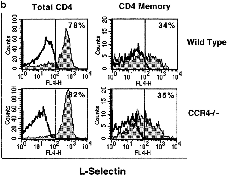
We next wanted to assess the physiologic importance of CCR4 for lymphocyte homing to cutaneous inflammatory sites (Fig. 3). To this end, we adoptively transferred wild-type C57Bl/6 and CCR4−/− donor T cells expressing the Thy1.2 marker into B6.Thy1.1 recipients whose ears were stimulated by painting with DNFB at the time of cell transfer. Donor T cells were obtained from LN draining sites of DTH (24 h after elicitation), to enhance the numbers of E-lig+, skin homing T cells transferred. 24 h after transfer, recipient LN, spleen, blood, and skin lymphocytes were isolated, and the percentage of CD4 T cells expressing the donor Thy1.2 allotype was determined by FACS®. To facilitate comparisons between experiments, homing to different organs was normalized to the homing of donor T cells to LN (primarily naive cells; ∼80% L-selectin+ in both wild-type [81 ± 1%, mean ± SEM, n = 3] and CCR4−/− donors [81 ± 2%, mean ± SEM, n = 3]; Fig. 3 b).
Figure 3.
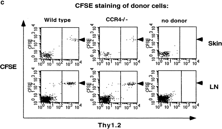
CCR4 is not essential for T cell homing to inflamed skin in DTH. Adoptively transferred wild-type and CCR4−/− donor T cells (Thy1.2+) home with the same efficiency to the inflamed skin of Thy1.1 recipients (a). Background (false Thy1.2+ cells in recipients with no donor cells, <0.2%) has been subtracted. Data are percentage of donor (Thy1.2+) T cells among CD4 T cells, normalized to LN values (mean ± SEM of seven experiments). Donor Thy1.2+ CD4 cells ranged from 2 to 10% of LN CD4 cells in the different experiments presented here. SPL, spleen. (b) L-selectin staining of wild-type and CCR4−/− donor LN T cells (primarily naive cells; representative results, n = 3). (c) CFSE labeling of donor cells revealed that few LN- or skin-homing T cells had undergone division in the course of our experiments.
As shown in Fig. 3 a (left panel), donor preimmunized LN T cells localized more efficiently than endogenous (non-presensitized) host T cells to inflamed skin: although donor T cells comprised on average only ∼5 ± 1% of total LN CD4 cells in recipients' LN (with no significant difference between wild-type and CCR4−/− donors) 24 h after adoptive transfer, they represented up to 50% (∼15–50%) of T cells in DNFB painted ears. Surprisingly, preimmunized CCR4−/− T cells accumulated in skin at least as well (Fig. 3 a, right panel) as wild-type immunized T cells. At this 24 h time point after transfer, accumulation of donor cells is likely dominated by recruitment from the blood, although transit time and recirculation into draining lymph may also contribute. Proliferation of homed cells can contribute to local cell numbers in inflammation, as well, but CFSE labeling of donor cells revealed that few LN- or skin-homing T cells had undergone division in the course of our experiments (Fig. 3 c).
The ability of CCR4−/− T cells to home efficiently to skin was unexpected, and suggested either that CCR4 is not involved in lymphocyte recruitment to inflamed skin, or that its role(s) can be fulfilled by other chemokines acting in parallel in a redundant fashion.
Effect of Anti-CTACK Antibody on T Cell Recruitment to Skin.
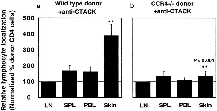
To determine if CTACK mediates T cell recruitment in the DTH model, we administered an inhibitory anti-CTACK mAb before the transfer of Thy1.2+ donor cells. As shown in Fig. 4 a, the anti-CTACK antibody had no effect on the fraction of ear localized CD4 cells of wild-type donor origin. Surprisingly, however, administration of anti-CTACK mAb dramatically reduced the skin localization of CCR4-deficient T cells (Fig. 4 b). Administration of the isotype control mAb had no effect on wild-type or CCR4-deficient T cell recruitment to the skin (data pooled from experiments with control mAb (n = 3) and no pretreatment of recipients (n = 4; Fig. 3 a).
Figure 4.
Anti-CTACK antibody inhibits donor CCR4−/− but not wild-type T cell recruitment to inflamed skin. Administration of anti-CTACK mAb (a) or control irrelevant antibody (not shown) had no effect on the percentage of adoptively transferred wild-type donor T cells (Thy1.2+) among skin CD4 cells (compared with panel a). In contrast, anti-CTACK dramatically inhibits skin homing of CCR4-deficient lymphocytes (b). Data are percentage of donor (Thy1.2+) T cells among CD4 T cells in each tissue, normalized to LN values (mean ± SEM of seven experiments). The proportion of donor Thy1.2+ CD4 cells among LN CD4 cells (which defines a relative localization of 100% in each experiment) ranged from 1 to 13%.
In addition, we used immunohistology as an independent means to assess the relative and absolute localization of donor cells in recipients' ears. Immunofluorescence staining revealed no significant difference in the number of Thy1.2+ CD4 wild-type donor cells localizing in inflamed ears after treatment with anti-CTACK antibody compared with the control mAb (6 ± 0.4 versus 5 ± 0.4 Thy1.2+ CD4 T cells per 40× microscope field; mean ± SEM, n = 3). Importantly, the number of Thy1.2+ CD4 cells of CCR4−/− origin was greatly reduced in anti-CTACK treated recipients (by 77%: 2 ± 0.5 CCR4−/− Thy1.2+ CD4 T cells per 40× field in anti-CTACK treated recipients, versus 7 ± 0.7 cells per field in control mAb treated animals; mean ± SEM, n = 3). In addition, we observed no significant differences in the numbers of total (donor plus host) CD4 T cells in the inflamed recipient skin after administration of anti-CTACK (in recipients of wild-type cells, there were 61 ± 7 total CD4 T cells per field after anti-CTACK treatment, compared with 68 ± 2 CD4 T cells after control mAb treatment; in recipients of CCR4−/− donor cells 64 ± 5 and 61 ± 10, respectively; mean ± SEM, n = 3). Thus, anti-CTACK treatment had little or no effect on endogenous CD4 T cell numbers in skin. We conclude that the effect of anti-CTACK antibody on lymphocyte homing is specific in that the antibody had little or no effect on wild-type lymphocyte homing to skin, or on CCR4−/− or wild-type lymphocyte homing to other organs; and it is potent, in that it dramatically reduces CD4 T cell homing to skin in the absence of CCR4.
Consequently, as for CCR4, CTACK is not essential for lymphocyte homing to skin: only simultaneous inhibition of CCR4 and CTACK (CCR10) responses effectively blocks lymphocyte recruitment. We conclude that memory T cell recruitment to inflamed skin requires either CCR4 or CTACK participation, but that either pathway (CCR4/TARC or CCR10/CTACK) alone can mediate efficient T cell accumulation over the 24 h recruitment period.
Effect of Anti-CTACK on T Cell–dependent Ear Swelling Induced by Adoptive Transfer of Wild-Type or CCR4-deficient Lymphocytes.
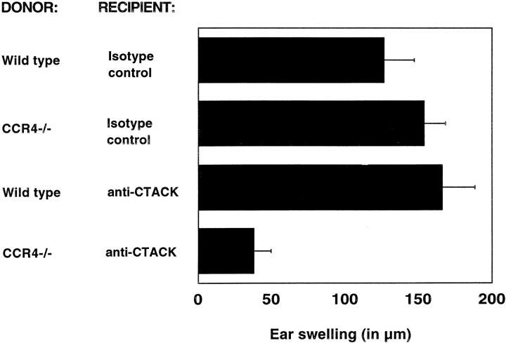
Adoptive transfer of immunized LN cells confers T cell-dependent enhanced ear swelling, the hallmark of DTH, in response to DNFB challenge (14). To determine if this T cell–dependent swelling requires CCR4 or CTACK, the ability of CCR4-deficient lymphocytes to mediate swelling, and the effect of anti-CTACK mAb on wild-type and CCR4-deficient donor T cell–mediated responses was assessed (Fig. 5). Paralleling the results for T cell homing, neither CCR4 deficiency of transferred LN cells, nor anti-CTACK mAb in the setting of wild-type cell transfer, substantially inhibited swelling. However, anti-CTACK dramatically and consistently inhibited swelling induced by CCR4-deficient cells.
Figure 5.
CCR4 and CTACK-dependent reduction of ear swelling in a cutaneous DTH reaction. Thy1.1 congenic recipients were treated with anti-CTACK mAb or the isotype control before the adoptive transfer of immunized Thy1.2+ donor LN cells. Recipient or control mouse ears were stimulated by local application of DNFB. Swelling induced by transferred CCR4−/− but not by wild-type immune T cells is blocked with a mAb against CTACK. Background swelling of DNFB treated recipients without donor cells has been subtracted (mean ± SEM of seven experiments).
The simplest interpretation of our studies is that both CCR4 and its ligands, and CTACK and its receptor CCR10, play overlapping and redundant roles in T cell recruitment into the inflamed ear, potentially mediating both arrest of T cells rolling on cutaneous endothelium, and extravasation of arrested T cells in response to an extravascular gradient. Immunohistochemically defined display of TARC by cutaneous venules, in combination with the ability of CCR4 ligands to trigger CLA+ T cell adhesion (5), has led us to propose that CCR4 can trigger the integrin-dependent arrest of lymphocytes rolling on cutaneous venules in vivo. However CCR4 ligands, especially MDC, can be highly expressed by activated macrophages and other cells within tissues, so that existence of a gradient of CCR4 ligands capable of supporting extravasation of endothelial bound T cells has not been excluded. Conversely, CTACK is highly and selectively expressed by keratinocytes, leading to the hypothesis that CTACK might support diapedesis and epidermotropism of skin homing T cells (6, 15). However, chemokines can be transported across endothelium to participate in leukocyte arrest (2, 16, 17); thus, keratinocyte-derived CTACK may also have a role at the vascular lumen. Roles in retaining homed cells in the inflamed ear are possible, as well. Novel techniques permitting in situ analyses of T cell interactions in inflamed ears will be required to determine precisely the level(s) at which CCR4, CTACK, and inflammatory chemokines participate in the process of lymphocyte recruitment. Importantly, our results also indicate that inflammatory chemokines induced in this model of cutaneous inflammation cannot by themselves support efficient recruitment of T cells.
In conclusion, our studies directly demonstrate the involvement of tissue-selective chemokine/receptor pathways - CCR4 and its ligands, and CTACK, presumably acting through its receptor CCR10 - in lymphocyte recruitment to skin and in cutaneous inflammation. Our findings suggest further that, in some models of cutaneous inflammation, these chemokine/receptor pathways can efficiently substitute for each other, but that either one or the other is required. Of course, the relative importance of CCR4 and CCR10 (and the extent of their redundancy) during skin lymphocyte homing may vary under different circumstances. Inhibition of these pathways, singly or in combination, may offer a novel approach to therapeutic modulation of cutaneous inflammatory disorders.
Acknowledgments
We thank J. Twelves and E. Resurreccion for excellent technical assistance, and members of our laboratory for helpful discussion of the ongoing work. We also like to acknowledge the contributions of Tim Wells and Arlene J. Hoogerwerf.
This work was supported by Grant RE1458/1-1 from the German Research Society (Deutsche Forschungsgemeinschaft [DFG]), by grants from the National Institutes of Health to E.C. Butcher, and AI46784 to J.J. Campbell, and by an Award from the Department of Veterans Affairs.
References
- 1.Warnock, R.A., J.J. Campbell, M.E. Dorf, A. Matsuzawa, L.M. McEvoy, and E.C. Butcher. 2000. The role of chemokines in the microenvironmental control of T versus B cell arrest in Peyer's patch high endothelial venules. J. Exp. Med. 191:77–88. [DOI] [PMC free article] [PubMed] [Google Scholar]
- 2.Stein, J.V., A. Rot, Y. Luo, M. Narasimhaswamy, H. Nakano, M.D. Gunn, A. Matsuzawa, E.J. Quackenbush, M.E. Dorf, and U.H. von Andrian. 2000. The CC chemokine thymus-derived chemotactic agent 4 (TCA-4, secondary lymphoid tissue chemokine, 6Ckine, exodus-2) triggers lymphocyte function-associated antigen 1-mediated arrest of rolling T lymphocytes in peripheral lymph node high endothelial venules. J. Exp. Med. 191:61–76. [DOI] [PMC free article] [PubMed] [Google Scholar]
- 3.Pan, J., E.J. Kunkel, U. Gosslar, N. Lazarus, P. Langdon, K. Broadwell, M.A. Vierra, M.C. Genovese, E.C. Butcher, and D. Soler. 2000. A novel chemokine ligand for CCR10 and CCR3 expressed by epithelial cells in mucosal tissues. J. Immunol. 165:2943–2949. [DOI] [PubMed] [Google Scholar]
- 4.Kunkel, E.J., J.J. Campbell, G. Haraldsen, J. Pan, J. Boisvert, A.I. Roberts, E.C. Ebert, M.A. Vierra, S.B. Goodman, M.C. Genovese, et al. 2000. Lymphocyte CC chemokine receptor 9 and epithelial thymus-expressed chemokine (TECK) expression distinguish the small intestinal immune compartment: Epithelial expression of tissue-specific chemokines as an organizing principle in regional immunity. J. Exp. Med. 192:761–768. [DOI] [PMC free article] [PubMed] [Google Scholar]
- 5.Campbell, J.J., G. Haraldsen, J. Pan, J. Rottman, S. Qin, P. Ponath, D.P. Andrew, R. Warnke, N. Ruffing, N. Kassam, L. Wu, and E.C. Butcher. 1999. The chemokine receptor CCR4 in vascular recognition by cutaneous but not intestinal memory T cells. Nature. 400:776–780. [DOI] [PubMed] [Google Scholar]
- 6.Morales, J., B. Homey, A.P. Vicari, S. Hudak, E. Oldham, J. Hedrick, R. Orozco, N.G. Copeland, N.A. Jenkins, L.M. McEvoy, and A. Zlotnik. 1999. CTACK, a skin-associated chemokine that preferentially attracts skin-homing memory T cells. Proc. Natl. Acad. Sci. USA. 96:14470–14475. [DOI] [PMC free article] [PubMed] [Google Scholar]
- 7.Chvatchko, Y., A.J. Hoogewerf, A. Meyer, S. Alouani, P. Juillard, R. Buser, F. Conquet, A.E. Proudfoot, T.N. Wells, and C.A. Power. 2000. A key role for CC chemokine receptor 4 in lipopolysaccharide-induced endotoxic shock. J. Exp. Med. 191:1755–1764. [DOI] [PMC free article] [PubMed] [Google Scholar]
- 8.Campbell, J.J., E.P. Bowman, K. Murphy, K.R. Youngman, M.A. Siani, D.A. Thompson, L. Wu, A. Zlotnik, and E.C. Butcher. 1998. 6-C-kine (SLC), a lymphocyte adhesion-triggering chemokine expressed by high endothelium, is an agonist for the MIP-3beta receptor CCR7. J. Cell Biol. 141:1053–1059. [DOI] [PMC free article] [PubMed] [Google Scholar]
- 9.Knibbs, R.N., R.A. Craig, P. Maly, P.L. Smith, F.M. Wolber, N.E. Faulkner, J.B. Lowe, and L.M. Stoolman. 1998. Alpha(1,3)-fucosyltransferase VII-dependent synthesis of P- and E-selectin ligands on cultured T lymphoblasts. J. Immunol. 161:6305–6315. [PubMed] [Google Scholar]
- 10.Tietz, W., Y. Allemand, E. Borges, D. von Laer, R. Hallmann, D. Vestweber, and A. Hamann. 1998. CD4+ T cells migrate into inflamed skin only if they express ligands for E- and P-selectin. J. Immunol. 161:963–970. [PubMed] [Google Scholar]
- 11.Picker, L.J., S.A. Michie, L.S. Rott, and E.C. Butcher. 1990. A unique phenotype of skin-associated lymphocytes in humans. Preferential expression of the HECA-452 epitope by benign and malignant T cells at cutaneous sites. Am. J. Pathol. 136:1053–1068. [PMC free article] [PubMed] [Google Scholar]
- 12.Butcher, E.C., and L.J. Picker. 1996. Lymphocyte homing and homeostasis. Science. 272:60–66. [DOI] [PubMed] [Google Scholar]
- 13.Austrup, F., D. Vestweber, E. Borges, M. Lohning, R. Brauer, U. Herz, H. Renz, R. Hallmann, A. Scheffold, A. Radbruch, and A. Hamann. 1997. P- and E-selectin mediate recruitment of T-helper-1 but not T-helper-2 cells into inflammed tissues. Nature. 385:81–83. [DOI] [PubMed] [Google Scholar]
- 14.Gocinski, B.L., and R.E. Tigelaar. 1990. Roles of CD4+ and CD8+ T cells in murine contact sensitivity revealed by in vivo monoclonal antibody depletion. J. Immunol. 144:4121–4128. [PubMed] [Google Scholar]
- 15.Campbell, J.J., and E.C. Butcher. 2000. Chemokines in tissue-specific and microenvironment-specific lymphocyte homing. Curr. Opin. Immunol. 12:336–341. [DOI] [PubMed] [Google Scholar]
- 16.Middleton, J., S. Neil, J. Wintle, I. Clark-Lewis, H. Moore, C. Lam, M. Auer, E. Hub, and A. Rot. 1997. Transcytosis and surface presentation of IL-8 by venular endothelial cells. Cell. 91:385–395. [DOI] [PubMed] [Google Scholar]
- 17.Baekkevold, E.S., T. Yamanaka, R.T. Palframan, H.S. Carlsen, F.P. Reinholt, U.H. von Andrian, P. Brandtzaeg, and G. Haraldsen. 2001. The CCR7 ligand ELC (CCL19) is transcytosed in high endothelial venules and mediates t cell recruitment. J. Exp. Med. 193:1105–1112. [DOI] [PMC free article] [PubMed] [Google Scholar]


