Abstract
Complement receptor type 3 (CR3, CD11b/CD18) serves as a receptor for a number of endogenous ligands and infectious organisms, and is involved in adhesion and host defense functions. Here, we report that signaling via CR3 plays an important role in regulating production of interleukin-12 (IL-12), a key mediator of cell-mediated immunity (CMI). We demonstrate with a variety of stimuli a dose-dependent, specific downregulation of IL-12 secretion by human monocytes in vitro after exposure to antibodies to CR3 (anti-CD11b and anti-CD18), as well as to the natural CR3 ligands, iC3b, and Histoplasma capsulatum. CR3 antibodies also suppressed interferon-γ (IFN-γ) production in cultures of human peripheral blood mononuclear cells (PBMC). We determined that one mechanism by which CR3 antibodies may suppress IL-12 production is by the inhibition of IFN-γ–induced tyrosine phosphorylation. Finally, in a murine model of IL-12–dependent septic shock, we provide evidence that administration of CR3 antibodies leads to suppression of IL-12 and IFN-γ in vivo. Our studies thus define a novel role for CR3 in regulating CMI functions via IL-12.
CR3 is a heterodimeric molecule which, like LFA-1 (CD11a/CD18) and CR4 (CD11c/CD18), belongs to the β2-integrin family of cell adhesion molecules (1–5). CR3 is expressed mainly on polymorphonuclear leukocytes, monocytes and macrophages, and natural killer cells (1, 2), and interacts with a variety of ligands including complement fragment iC3b, intercellular adhesion molecule 1 (ICAM1)1, fibrinogen, and β-glucan (2, 4). Moreover, it mediates the binding of opsonized or unopsonized infectious agents such as Histoplasma capsulatum, Leishmania major, group B streptococci, Bordetella pertussis, Candida albicans, and several mycobacteria (2, 5–7).
Previous studies have shown that CR3 is involved in several monocyte and macrophage functions including transmigrational adhesion (2, 4, 8), phagocytosis, nitric oxide production and the generation of a respiratory burst (2, 3, 5, 8, 10). In addition, CR3 signaling may indirectly affect T cell function as the administration of antibodies to CR3 to animals suppresses delayed type hypersensitivity (DTH) reactions (11, 12), and fatally potentiates infections with Listeria monocytogenes or Toxoplasma gondii (13, 14). Since these phenomena are dependent on an intact cell-mediated immunity (CMI) and Th1 responses, we explored the role of CR3 signaling in regulating cytokines that mediate such responses.
Materials and Methods
Cell Isolation, Cell Culture Conditions and Assessment of Cytokine Production.
Human monocytes were obtained from healthy donors (total n = 14) by standard leukapheresis and were highly purified (95–99%) by counterflow centrifugal centrifugation (15). Cell purity was checked by flow cytometry analysis using monoclonal antibodies to CD14 and CD11b (Becton Dickinson, San Jose, CA). Monocytes were cultured at 2 × 10⁶ cells/ml for 24 h in 1 ml of RPMI 1640 medium (Whittaker, Walkersville, MD) supplemented with 10% fetal calf serum (Whittaker), 100 μg/ml penicillin, 100 μg/ml streptomycin and 0.03% glutamine and stimulated at the beginning of the culture with the following substances as indicated: heat-killed, formalin-fixed Staphylococcus aureus, Cowan I strain (SAC; Calbiochem, Cambridge, MA), LPS (Escherichia coli serotype 0127:B8; Sigma Chem. Co., St. Louis, MO) (16, 17), recombinant IFN-γ (Genzyme, Cambridge, MA), CD40L trimer (kindly provided by Immunex Corporation, Seattle, WA), IFN-α (Endogen, Cambridge, MA), or anti–IFN-γ (Endogen). One experiment was typically performed with the monocytes from one donor, and there was individual variability in IL-12 production (e.g., range for SAC plus IFN-γ stimulation 380–3,300 pg/ml IL-12 p70, mean ∼1,750 pg/ml). Monocytes were incubated with heat-killed Histoplasma capsulatum (HC), (strain GS-57, kindly provided by Dr. R. Seder, Lymphokine Regulation Unit, LCI, NIAID, NIH) or iC3b-SRBC (2 × 10⁷/ml) 2 h before stimulation as indicated. iC3b-SRBC were prepared by sequential addition of anti-sheep erythrocyte antibodies (IgM; PharMingen, San Diego, CA) and C5-deficient human serum (Sigma) to SRBC as described (18, 19). The presence of iC3b on the cell surface was verified by flow cytometry showing a >100fold increase in fluorescence intensity with a monoclonal antibody to the iC3b neoantigen (Quidel, San Diego, CA). Human PBMC were isolated by Ficoll-Hypaque density gradient centrifugation from leukocyte concentrates prepared by automated leukapheresis of healthy donors (total n = 5) as described (15). PBMC were cultured similar to monocytes, except that they were cultured at 1 × 10⁶ cells/ml for 48h and stimulated with PHA (Sigma). In some experiments, recombinant human IL-12, or recombinant IL-2 (PharMingen) were added as indicated.
Culture supernatants were assessed in duplicate by ELISA using kits or antibody pairs for IL-12 p70, TNF-α (both from R&D Systems), IL-12 p40, IL-10 (both from PharMingen), IL-6, IL-1β, IFN-α, IFN-γ (all from Endogen, Cambridge, MA), and TGF-β1 (Genzyme). Unstimulated cultures (with or without added integrin antibodies) contained cytokines below the detection limit (IL-12 p70 >5 pg/ml, IL-12 p40 >20 pg/ml, IFN-α >10 pg/ml, IL-10 >100 pg/ml, TGF-β >50 pg/ml, IL-6 >50 pg/ml, IL-1β >25 pg/ml). For TNF-α, the detecting limit was >25 pg/ml and the background was below 200 pg/ml. Integrin antibodies were added at the beginning of the culture at the indicated concentrations and were obtained in a preservative-free preparation or dialyzed overnight before use. The antibodies and monocyte stimulants (e.g., CD40L trimer) contained low endotoxin levels as per information by the manufacturer and as demonstrated in appropriate assays performed in the presence of 1–5 μg/ml of polymyxin B (Sigma) which revealed little or no effect on IL-12 and TNF-α production. Antibodies were bound to plastic culture plates by overnight incubation at 10 μg/ml in 100 μl of carbonate buffer at 4°C followed by washing with PBS, and bound to polystyrene beads (goat anti–mouse IgG, Dynabeads M450, Dynal, Lake Success, NY) according to the manufacturer's instructions. L cells stably transfected with a plasmid expressing the FcγRII receptor (CD32) as previously described (20, 21) were incubated with 10 μg/ml of antibody at 37°C for 1 h, washed, and then used for the assays. Integrin antibodies were obtained from the American Type Culture Collection (ATCC, Rockville, MD; clone LM2/1 [18]; murine IgG1; and clone M1/ 70, rat IgG2b), Monosan (Amsterdam, Netherlands; clone MEM48, murine IgG1), PharMingen (clones G43-25B, murine IgG2b, B-ly6, murine IgG1, 44, murine IgG1, 107.3, murine IgG1, G555-178, murine IgG2a), Becton Dickinson (clone D12, murine IgG2a), and Caltag (San Francisco, CA; clone CLB-LFA-1/1, murine IgG1).
Western Blotting.
Human monocytes (2.5 × 10⁷ in 5 ml per condition) were either untreated, treated with anti-CR3 (clone LM2/1, 10 μg/ml) for 20 min, treated with recombinant IFN-γ (1 μg/ml) for 5 min, or treated with LM2/1 for 20 min followed by IFN-γ for 5 min. Cells were then solubilized in a buffer consisting of 1% Triton X (Pierce Chem. Co., Rockford, IL), 150 mM NaCl, 50 mM Tris, pH 8.0, 50 mM Na-pyrophosphate, 2.5 mM aprotinin, 2.5 mM leupeptin, 1 mM phenylmethylsulfonyl fluoride, and 1 mM NaVO3 (all from Sigma). Protein concentration of the cell lysates was estimated by measuring the optical density at 280 nm based on the assumption that optical density of 1.4 corresponds to 1 mg/ml. Cell lysates were analyzed using Tris-glycine gels (Novex, San Diego, CA) with equivalent amounts of protein loaded in each lane, followed by transfer to nitrocellulose membranes (Schleicher and Schuell, Keene, NH). After overnight blocking in 5% nonfat dry milk, 150 mM NaCl, 50 mM Tris, and 0.05% Tween 20, membranes were incubated with horseradish peroxidase (HRP)-conjugated phosphotyrosine antibodies (Santa Cruz Biotechnology, Santa Cruz, CA) for 2 h. Membranes were then washed and developed with enhanced chemiluminescence (Super-signal™ substrate; Pierce). Cell lysates from epidermal growth factor–stimulated A431 cells (Upstate Biotechnology Incorporation, Lake Placid, NY) served as positive control for phosphotyrosine and STAT1 (signal transducers and activators of transcription) expression. For reprobing, membranes were stripped in 0.2 M glycine buffer (pH 2.8) for 30 min, washed, exposed to a rabbit polyclonal antibody to STAT1 (Santa Cruz), washed again, incubated with a donkey anti–rabbit HRP-conjugated antibody (Amersham Corp., Arlington Heights, IL), and developed with enhanced chemiluminescence.
Treatment of Animals, LPS Challenge, and Detection of Cytokine Levels.
6–10-wk-old BALB/c mice housed under standard conditions were treated intraperitoneally with 0.5 ml of PBS containing either rat IgG (1 mg per mouse; Sigma), antibodies to CR3 (1 mg of clone M1/70 or 0.5 mg of 5C6; Biosource International, Camarillo, CA), or anti–IL-12 (1 mg, clone C17.8, the hybridoma for which was kindly provided by Dr. G. Trinchieri, The Wistar Institute, Philadelphia, PA). This treatment was followed 1 h later by an intravenous (tail vein) injection of LPS (1 μg per mouse in 100 μl of PBS) or PBS alone, and mice were then either killed either 3 h (for determination of IL-12 p40) or 6 h (for IL-12 p70 and IFN-γ) later by cervical dislocation after blood had been obtained by cardiac puncture. Serum was obtained after 30 min clotting of the blood at 37°C and subsequent centrifugation. Serum levels of IL-12 (p40 and p70) and IFN-γ were assessed by ELISA using antibody pairs from PharMingen. Injection of higher doses of anti-CR3 (e.g., 1 or 3 mg M1/70 per mouse at 12 h and/or 1 h before LPS challenge) had no additional suppressive effects on IL-12 or IFN-γ secretion. IL-12 p70 and IFN-γ were not detectable in mice that had received only PBS and were very low or undetectable at three hours after LPS administration or in anti–IL-12–treated mice (data not shown).
Stastitics.
Statistical significance of differences was determined by the Student's t test where indicated.
Results and Discussion
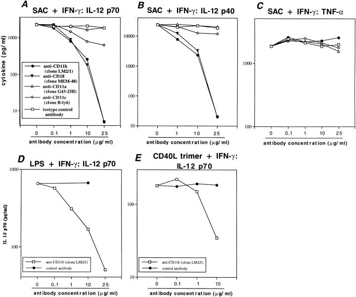
In initial studies we determined whether antibodies to CR3 affect the secretion of IL-12 by highly purified human monocytes stimulated with Staphylococcus aureus cells (SAC) and IFN-γ. We found that exposure of monocytes to monoclonal antibodies that bind to the functionally important I domain of CD11b (clone LM2/1) (18) or to CD18 results in a dose dependent and profound reduction of IL-12 (p70 heterodimer and p40 monomer) secretion, whereas antibodies to CD11a had no effects (Fig. 1, A and B). Similarly, addition of one clone of CD11c antibodies had only modest effects on IL-12 production (Fig. 1, A and B). These cytokine protein levels correlated with downregulated messenger RNA levels for IL-12 p35 and p40 as determined by reverse-transcriptase PCR (data not shown). The blockade of IL-12 production was observed with a variety of monocyte stimulants known to induce IL-12 (e.g., IFN-γ in combination with LPS, SAC or CD40L trimer, Figs. 1, D and E and 2, A–C) and with a panel of monoclonal antibodies to CR3 (Table 1). In contrast, secretion of other monocyte products such as TNF-α, IL-10, TGF-β, IL-6, and IL-1β (Fig. 1 C; Table 1) was not significantly altered by any of the above antibodies (as determined in the same cultures), nor was cell viability (trypan blue exclusion) or cell surface expression of CD14 (flow cytometry; data not shown). The one exception was an anti-CR3–induced reduction of IFN-α secretion (Table 1), a macrophage-derived cytokine previously shown to enhance IL-12–induced Th1 development (22). It is interesting to note that with higher doses of CD11b antibodies (25 μg/ml), we observed a further downregulation of IL-12 (<5 pg/ml) and a twofold increase in IL-6 (5,450 pg/ml), a cytokine dichotomy previously described in individuals with HIV infection (23).
Figure 1.
Effects of various integrin antibodies on secretion of IL-12 and other cytokines by human monocytes. (A–C) Production of (A) heterodimeric IL-12 (p70) and (B) monomeric IL-12 (p40) in cultures of highly purified human monocytes as determined by ELISA is suppressed by antibodies to CD11b and CD18 in a dose dependent manner, but not by anti-CD11a and modestly by anti-CD11c, when stimulated with SAC (0.01%) and IFN-γ (1 μg/ml). Secretion of TNF-α (C) in the same culture supernatants is not altered by such antibodies. Data in A–C represent means of duplicates from one experiment and are representative of five experiments. (D and E) Suppression of IL-12 p70 by anti-CD11b (clone LM2/1) in a dose-dependent fashion after stimulation with LPS (1 μg/ml) plus IFN-γ (1 μg/ml) and CD40L trimer (3 μg/ml) plus IFN-γ (1 μg/ml). One of three experiments is shown.
Figure 2.
Monocytic IL-12 production induced by various stimuli is suppressed by antibodies to CD11b. Secretion of IL-12 as determined by ELISA in cultures of human monocytes after stimulation with (A) LPS and IFN-γ, (B) SAC and IFN-γ, and (C) LPS alone (1 μg/ml), SAC alone (0.01%), SAC plus anti–IFN-γ (10 μg/ml), SAC plus IFN-α (10 ng/ml), and CD40L trimer (3 μg/ml) plus IFN-γ (1 μg/ml). Similar results were obtained with LPS plus increasing doses of IFN-γ, SAC plus increasing doses of IFN-γ, SAC plus TNF-α, TNF-α, and SAC plus anti–IL-10 (data not shown). One of three experiments is shown.
Table 1.
Effects of Integrin Antibodies on the Secretion of a Variety of Cytokines by Highly Purified Human Monocytes
| Cytokine | None | Isotype control | CD11b | CD18 | CD11a | CD11c | ||||||||||||||||
|---|---|---|---|---|---|---|---|---|---|---|---|---|---|---|---|---|---|---|---|---|---|---|
| IgG1 (clone 107.3) | IgG2a (G555-178) | (MEM-48) | (CLB- LFA 1/1) | |||||||||||||||||||
| (LM2/1) | (44) | (M1/70) | (D12) | (G43-25B) | (B-ly6) | |||||||||||||||||
| pg/ml | ||||||||||||||||||||||
| IL-12 (p70) | 2,180 | 2,040 | 2,155 | 260 | 735 | 780 | 670 | 185 | 1,720 | 1,625 | 770 | |||||||||||
| IL-12 (p40) | 23,450 | 21,960 | 19,870 | 2,400 | 18,170 | 13,140 | 19,350 | 3,280 | 21,580 | 22,200 | 12,410 | |||||||||||
| IFN-α | 250 | 265 | ND | 30 | 130 | 85 | 175 | 15 | 250 | 135 | 240 | |||||||||||
| TNF-α | 1,850 | 2,113 | 2,325 | 2,575 | 2,100 | 2,638 | 2,050 | 1,850 | 3,188 | 2,025 | 3,150 | |||||||||||
| IL-10 | 5,200 | 5,750 | 5,700 | 4,550 | 5,500 | 3,400 | 4,300 | 5,500 | 6,700 | 4,850 | 4,950 | |||||||||||
| TGF-β | 2,350 | 2,675 | 2,375 | 3,300 | 2,650 | 2,275 | 2,625 | 3,175 | 2,950 | 2,650 | 2,800 | |||||||||||
| IL-6 | 2,650 | 3,050 | ND | 4,275 | 3,700 | ND | 3,450 | 3,425 | 3,600 | 3,350 | 3,525 | |||||||||||
| IL-1β | 2,025 | 2,138 | ND | 2,050 | 2,025 | ND | 2,363 | 1,875 | 2,338 | 2,200 | 1,875 | |||||||||||
Human monocytes (2 × 10⁶/ml) were incubated with the indicated antibodies and stimulated with SAC (0.01%) plus IFN-γ (1 μg/ml). Cytokine levels from culture supernatants were determined as described in Materials and Methods. Data represent antibody concentration of 10 μg/ml. Data are means of duplicates from one experiment and are representative of three experiments. Percent suppression of IL-12 p70 in all three experiments was comparable, e.g., percent suppression of IL-12 p70 for clone LM2/1 ranged from 76–98%, for clone 44 (53–74%), for M1/70 (41–78%), for D12 (48–85%), for MEM-48 (82–99%), for CLB LFA1/1 (12–22%), for G43 25B (-16–33%), and for B-ly6 (37–79%). The baseline stimulated IL-12 p70 production varied in the three experiments (i.e., with cells from three different donors) from 1,060 to 2,180 pg/ml. ND denotes not determined.
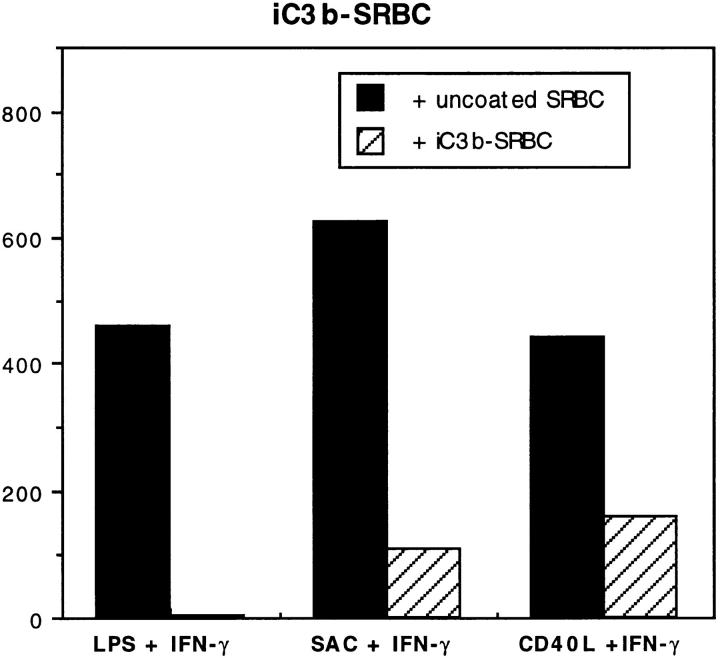
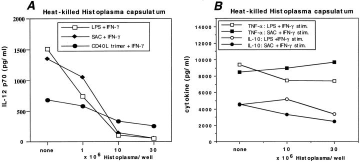
We then studied the ability of natural CR3 ligands to suppress IL-12 production by using iC3b-coated sheep red blood cells (iC3b-SRBC) and heat-killed Histoplasma capsulatum (HC), an organism that binds to CR3 (3, 24). Similar to our findings with anti-CD11b, when human monocytes were incubated with iC3b-SRBC or heat-killed HC and subsequently stimulated with IFN-γ and either SAC, LPS, or CD40L trimer, we observed a downregulation of IL-12 p70 production (Figs. 3 and 4 A), whereas TNF-α and IL-10 levels were largely unaffected (Fig. 4 B). The inhibition of IL-12 production by HC was: (a) not due to direct suppression by IL-10, since the addition of anti–IL-10 antibodies (10 μg/ml) to the cultures only slightly increased the levels of IL-12; and (b) was related to the ability of HC to bind to β2 integrins, since the addition of a CD18 antibody (clone CLB LFA1/1 which did not directly inhibit IL-12 production, see Table 1) to these cultures partially reversed the suppressive effect of HC (threefold increase in IL-12 p70, i.e., to 40% of levels without HC).
Figure 3.
Inhibition of monocyte IL-12 production by a natural ligand to CR3, iC3b-coated SRBC. iC3b-SRBC (2 × 10⁷/ml) were incubated with human monocytes (2 × 10⁶/ml) 2 h before stimulation with LPS (1 μg/ml) plus IFN-γ (1 μg/ml), SAC (0.01%) plus IFN-γ (1 μg/ml), or CD40L trimer (3 μg/ml) plus IFN-γ (1 μg/ml). IL-10 and TNF-α levels in the same culture supernatants were not significantly altered (data not shown). Data are representative of five experiments.
Figure 4.
Secretion of IL-12 p70 by monocytes is suppressed by incubation with heat-killed Histoplasma capsulatum (HC). Heat-killed HC at the indicated concentrations was incubated with human monocytes 2 h before stimulation with LPS (1 μg/ml) plus IFN-γ (1 μg/ml), SAC (0.01%) plus IFN-γ (1 μg/ ml), or CD40L trimer (3 μg/ml) plus IFN-γ (1 μg/ml) (A). (B) IL-10 and TNF-α levels from the same culture supernatants. Data are representative of five experiments, and percent suppression by HC was similar in all instances.
Our findings with iC3b-SRBC expand upon a recent study by Karp et al. (25), who first demonstrated a role for complement regulatory proteins in the regulation of IL-12. It was shown that the in vitro infection of human monocytes with measles virus results in the suppression of IL-12 production and that such suppression was mediated through a measles virus receptor, CD46, which is an important complement regulatory protein that binds C3b (25). Thus, it appears that the binding of C3b to CD46, and the binding of iC3b to CR3, as may occur with the interaction of opsonized organisms with phagocytes, are two mechanisms by which complement components may suppress IL-12 production by human monocytes.
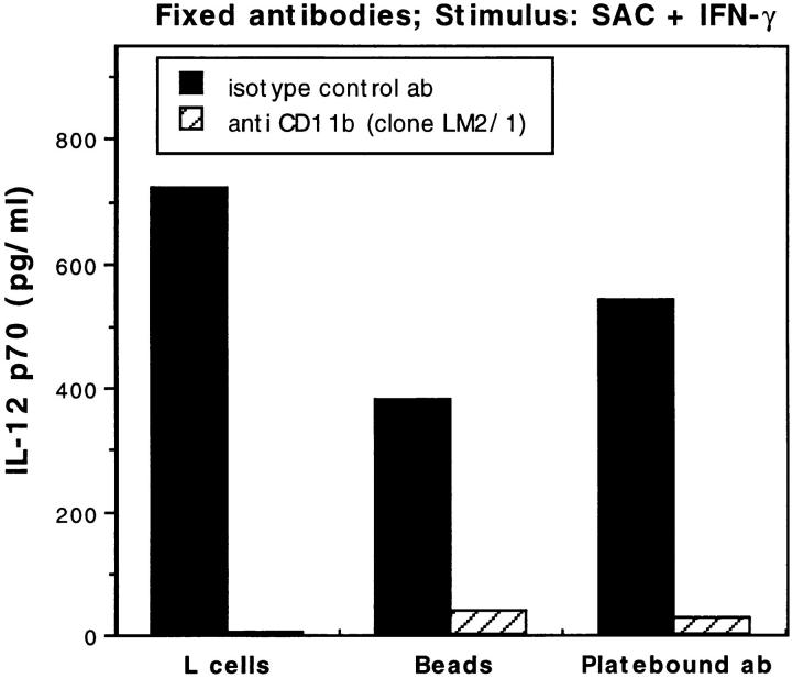
The suppression of IL-12 production by antibodies and ligands to CR3 could be due either to the transmission of a direct inhibitory signal through the CR3 molecule, or to the blocking of a positive signal for IL-12 production, as could be provided through interactions between CR3 and ICAM-1 on neighboring monocytes. To clarify this issue, we initially performed a number of studies which together suggested that important cell–cell interactions, especially via ICAM-1/CR3, are not particularly important for the induction of IL-12, and thus indirectly supported the conclusion that CR3 antibodies act via a direct inhibitory signal. First, we found that the relative production of IL-12 per monocyte after SAC and IFN-γ stimulation was not altered when cells were diluted over large ranges (⩾1:1,000) before culture, i.e., resulting in decreased homotypic interactions (data not shown). Second, the addition of anti–ICAM-1 to monocyte cultures did not suppress IL-12 production (data not shown). And third, antibodies to CD11b (clone LM2/1) immobilized onto plastic culture plates, polystyrene beads coated with anti-mouse IgG (Dynal, Lake Success, NY), or to FcγRII receptor (CD32)-expressing L cells, completely inhibited SAC plus IFN-γ–induced IL-12 production (Fig. 5), suggesting that the membrane-fixed subset of CR3 (i.e., which remains unbound to the solid supports [26]) is not capable of providing a positive signal for IL-12 production through an interaction with ICAM-1 on neighboring monocytes.
Figure 5.
Monocyte IL-12 production is suppressed by antibodies to CD11b (clone LM2/1). Antibodies were bound to culture plates, polystyrene beads, or L cells as described in Materials and Methods. The secretion of IL-12 was determined by ELISA in cultures of human monocytes after stimulation with SAC (0.01%) and IFN-γ (1 μg/ml). One of three experiments is shown.
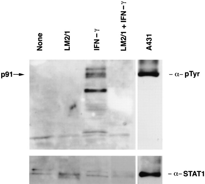
Because these studies provided only indirect evidence that anti-CR3 was acting to transmit a negative signal to monocytes, we next looked more directly for intracellular targets of inhibitory signaling by CR3. Inhibition of tyrosine phosphorylation by CR3 signaling seemed possible since this effect had been previous demonstrated in a system that involves a CR3 ligand. Thus, it has been shown that tyrosine phosphorylation in response to IFN-γ was suppressed in macrophages infected with L. donovani (27), an organism binding to monocyte CR3 by either gp63 or lipophosphoglycan (LPG) (3, 7). Furthermore, a similar phenomenon has been described in human monocytes after the binding of immune complexes (28), and a role for β1integrin engagement in tyrosine dephosphorylation has been recently established (29). Thus, we explored the possibility that reduced production of IL-12 and IFN-α could follow a CR3-induced inhibition of IFN-γ signal transduction. As shown in Fig. 6, we found that incubation of monocytes with IFN-γ alone induced tyrosine phosphorylation of several proteins, one of which was identified as STAT1 (signal transducers and activators of transcription 1) by Western blotting with anti-STAT1 antibodies and by use of a positive tyrosine phosphorylated STAT1 control (Fig. 6). After preincubation with anti-CR3, IFN-γ–induced tyrosine phosphorylation of several proteins, including STAT1 was not seen, suggesting an inhibition of tyrosine phosphorylation, or alternatively, an accelerated dephosphorylation of tyrosine residues by an as yet unidentified protein tyrosine phosphatase.
Figure 6.
Prevention of IFN-γ–induced phosphorylation of STAT1 by anti-CR3. Human monocytes (2.5 × 10⁷ in 5 ml per condition) were either untreated (None), treated with anti-CR3 (LM2/1), treated with recombinant IFN-γ (IFN-γ), or treated with LM2/1 followed by IFN-γ (LM2/1 + IFN-γ). Monocytes were then solubilized and analyzed as described in Materials and Methods. Cell lysates from stimulated A431 cells (A431) served as positive control. The upper panel shows probing of the nitrocellulose membranes with anti-phosphotyrosine antibodies (α-pTyr) and the arrow indicates the expression of p91 (STAT1). The lower panel shows a membrane reprobed with anti-STAT1 (α-STAT1). Data are representative of three similar experiments.
The inhibition of IFN-γ–mediated STAT1 activation (i.e., phosphorylation) presents a potential mechanism by which signaling through CR3 could decrease monocyte IL-12 production, since there are two potential STAT1binding sites (IFN-γ activation sequence [GAS] elements) in the human IL-12 p40 promotor (positions −127 to −119 and −277 to −268 of the published sequence), and since mutations with deletions at at least one of these sites reduced transcription of a reporter gene (30). In addition, it is possible that the JAK-STAT-GAS signal transduction pathway may be important for the regulation of human IL-12 p35 gene transcription that is induced by stimulation with IFN-γ (30), or be involved in the expression of gene products that are indirectly responsible for the upregulation of IL-12 p35 or IL-12 p40 gene transcription or translation. Finally, the possibility that in addition to its effects on IFN-γ signaling, anti-CR3 suppressed IL-12 production through an IFN-γ–independent mechanism as well was supported by the observation that the low level of IL-12 induced by SAC alone, which was unchanged with the addition of anti– IFN-γ, was also inhibited with anti-CR3 (Fig. 2 C). Additional studies are needed to address these possibilities, as well as to address whether signaling via CR3 has effects on transcription factors other than STAT1, such as ets-protein family members, that have been more directly implicated in the regulation of IL-12 p40 in humans (30).
We finally extended our studies on functional effects of CR3 signaling to a murine model of IL-12–dependent septic shock. In this model, the intravenous injection of LPS results in symptoms of septic shock as well as high serum levels of IFN-γ and IL-12, all of which can be inhibited by the systemic administration of anti–IL-12 (31). We chose this in vivo model to study the effects of the antibodies to CR3 because, when compared to others, this model should be much less dependent on cell trafficking. This is because the injected LPS should be rapidly delivered to all lymphoid tissues and the response to LPS is unlikely to depend on significant migration of cells, at least within the first 3 or 6 h, the times at which we measured IL-12 and IFN-γ, respectively.
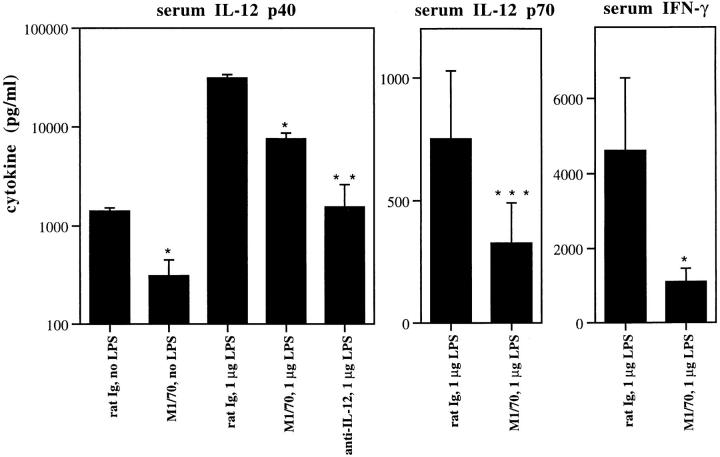
Thus, in our studies, BALB/c mice were given intraperitoneal injections of either CR3 antibodies (1 mg of clone M1/70 or 0.5 mg of clone 5C6 [8, 32]) or control rat IgG 1 h before LPS injection and were killed either 3 or 6 h later, at which time serum was obtained. As shown in Fig. 7, pretreatment with anti-CR3 (clone M1/70) reduced the serum levels of IL-12 (p40 and p70) by more than 4 and 2.5-fold, respectively, and similarly diminished the levels of IFN-γ 4-fold. The effects on IL-12 by treatment with M1/ 70 (or similar effects seen with clone 5C6; data not shown) were, consistent with prior studies of these antibodies (8, 13), not due to elimination of circulating leukocytes since total and differential white blood cell counts were similar in all treatment groups (data not shown). In addition, we found that whole spleen cells from anti-CR3–treated and LPS challenged mice stimulated in vitro with anti-CD3/ anti-CD28 or PHA manifest reduced (two- to fourfold) IFN-γ production when compared to control mice (data not shown). In accordance with these findings, addition of antiCD11b antibodies (10 μl/ml) markedly inhibited IFN-γ production in human monocyte cultures stimulated with either PHA (3,310 pg/ml IFN-γ for isotype control, 1,100 pg/ml for anti-CD11b [LM2/1]), plate-bound anti-CD3 plus anti-CD28 (3,250 pg/ml IFN-γ for isotype control, <10 pg/ml for anti-CD11b) or anti-CD2 plus anti-CD28 (2,450 pg/ml IFN-γ for isotype control, <10 pg/ml for anti-CD11b), but had no significant effect on cell viability or on cell proliferation (as determined by [3H]thymidine incorporation at 72 h). The above effects of anti-CR3 on IFN-γ production were reversed by addition of recombinant human IL-12 (1,100 pg/ml IFN-γ after PHA stimulation with anti-CR3, 3,210 pg/ml after addition of 20 ng/ ml of IL-12) but not by IL-2 indicating the specificity for IL-12.
Figure 7.
Production of IL-12 and IFN-γ in a murine model of septic shock is suppressed by treatment with CR3 antibodies. Pretreatment with 1 mg of antiCR3 (clone M1/70) markedly suppressed the serum levels of IL-12 p40 and p70 as well as serum levels of IFN-γ in BALB/c mice challenged by intravenous LPS (1 μg/mouse). Similar results were obtained with a different anti-CR3 antibody (clone 5C6). Data show the mean ± SD of groups consisting of three separately handled mice. *P <0.001, **P <0.0001, and ***P <0.05 versus the control (i.e., rat Ig treated) group as determined by the Student's t test. Data are representative of five experiments.
These data show that the IL-12 response, crucial for the initiation of most CMI functions (33), may be regulated by CR3 signaling. Thus, they provide insight into mechanisms underlying the ability of CR3 antibodies to abolish DTH reactions (8, 11, 12), to ameliorate Th1 cell–mediated autoimmune diseases including antigen-induced arthritis (34) and experimental allergic encephalomyelitis (35), and to enhance the severity of infections with L. monocytogenes (13) or T. gondii (14). In addition, our studies demonstrating the inhibition of IL-12 by HC and iC3b-SRBC suggest a mechanism for the impaired CMI accompanying infections with CR3 binding microorganisms (e.g., L. major, mycobacteria, HIV) (3, 36). In this regard they may provide a cogent reason for the diminished IL-12 production by mononuclear cells in HIV infected individuals (17) and in leishmaniasis (36). It is possible that CR3-induced suppression of IL-12 responses to the above microorganisms may result in suppressed monocyte nitric oxide production and respiratory burst, thereby explaining their ability to thrive in intracellular compartments.
Finally, our data, together with those recently developed by Karp et al. (25) showing that suppressed IL-12 in measles virus infection is mediated by the CD46 complement regulatory protein, establishes a new locus of control for T cell–mediated immunity via complement components. Thus, in disease states as well as in normal responses to invading microorganisms, signaling through CR3 may play an important role in regulating Th1–Th2 homeostasis. This is important for understanding the pathogenesis of infectious diseases, and may provide a new approach for therapeutic immunointervention targeting IL-12.
Footnotes
We would like to acknowledge Dr. Warren Strober for his helpful discussions and for reviewing the manuscript and thank Drs. Robert Seder, Sharon Wahl, Thomas Wynn, Alan Sher, John O'Shea, and Stephen Straus for helpful comments.
T. Marth is a recipient of a research grant by the Deutsche Forschungsgemeinschaft (Ma 1711/1-1 and 2-1).
1 Abbreviations used in this paper: CMI, cell-mediated immunity; DTH, delayed type hypersensitivity; ECL, enhanced chemiluminescence; HC, Histoplasma capsulatum; HRP, horseradish peroxidase; ICAM-1, intercellular adhesion molecule 1; LPG, lipophosphoglycan; SAC, Staphylococcus aureus cells; STAT1, signal transducers and activators of transcription 1.
References
- 1.Springer TA. Adhesion receptors of the immune system. Nature (Lond) 1990;346:425–434. doi: 10.1038/346425a0. [DOI] [PubMed] [Google Scholar]
- 2.Patarroyo M. Leukocyte adhesion to cells. Molecular basis, physiological relevance, and abnormalities. Scand J Immunol. 1989;30:129–164. doi: 10.1111/j.1365-3083.1989.tb01197.x. [DOI] [PubMed] [Google Scholar]
- 3.Cooper NR. Complement evasion strategies of microorganisms. Immunol Today. 1991;12:327–331. doi: 10.1016/0167-5699(91)90010-Q. [DOI] [PubMed] [Google Scholar]
- 4.Kishimoto TK, Larson RS, Corbi AL, Dustin ML, Staunton DE, Springer TA. The leukocyte integrins. Adv Immunol. 1989;46:149–182. doi: 10.1016/s0065-2776(08)60653-7. [DOI] [PubMed] [Google Scholar]
- 5.Ross GD, Vetvicka V. CR3 (CD11b, CD18): a phagocyte and NK cell membrane receptor with multiple ligand specificities and functions. Clin Exp Immunol. 1993;92:181–184. doi: 10.1111/j.1365-2249.1993.tb03377.x. [DOI] [PMC free article] [PubMed] [Google Scholar]
- 6.Van Strijp JAG, Russell DG, Tuomanen E, Brown EJ, Wright SD. Ligand specificity of purified complement receptor type three (CD11b/CD18, αmβ2, Mac-1): indirect effects of an ARG-GLY-ASP (RGD) sequence. J Immunol. 1993;151:3324–3336. [PubMed] [Google Scholar]
- 7.Antal JM, Cunningham JV, Goodrum KJ. Opsonin-dependent phagocytosis of group B streptococci: role of complement receptor type 3. Infect Immun. 1992;60:1114–1121. doi: 10.1128/iai.60.3.1114-1121.1992. [DOI] [PMC free article] [PubMed] [Google Scholar]
- 8.Rosen, H. 1990. Role of CR3 in induced myelomonocytic recruitment: insights from in vivo monoclonal antibody studies in the mouse. J. Leuk. Biol. 48:465–469. [DOI] [PubMed]
- 9.Goodrum KJ, McCormick LL, Schneider B. Group B streptococcus nitric oxide production in murine macrophages is CR3 (CD11b/CD18) dependent. Infect Immun. 1994;62:3102–3107. doi: 10.1128/iai.62.8.3102-3107.1994. [DOI] [PMC free article] [PubMed] [Google Scholar]
- 10.Anderson DC, Springer TA. Leukocyte adhesion deficiency: an inherited defect in the Mac-1, LFA-1, and p150,95 glycoproteins. Annu Rev Med. 1987;38:175–194. doi: 10.1146/annurev.me.38.020187.001135. [DOI] [PubMed] [Google Scholar]
- 11.Rosen H, Milon G, Gordon S. Antibody to the murine type 3 complement receptor inhibits T lymphocytedependent recruitment of myelomonocyic cells in vivo. J Exp Med. 1989;169:535–549. doi: 10.1084/jem.169.2.535. [DOI] [PMC free article] [PubMed] [Google Scholar]
- 12.Patarroyo M. Adhesion molecules mediating recruitment of monocytes to inflamed tissues. Immunobiol. 1994;191:474–477. doi: 10.1016/S0171-2985(11)80453-5. [DOI] [PubMed] [Google Scholar]
- 13.Rosen H, Gordon S, North RJ. Exacerbation of murine listeriosis by a monoclonal antibody specific for the type 3 complement receptor of myelomonocytic cells. J Exp Med. 1989;170:27–38. doi: 10.1084/jem.170.1.27. [DOI] [PMC free article] [PubMed] [Google Scholar]
- 14.Johnson LL, Gibson GW, Sayles PC. CR3dependent resistance to acute Toxoplasma gondiiinfection in mice. Infect Immun. 1996;64:1998–2003. doi: 10.1128/iai.64.6.1998-2003.1996. [DOI] [PMC free article] [PubMed] [Google Scholar]
- 15.Abrahamsen TG, Carter CS, Read EJ, Rubin M, Goetzman HG, Lizzio EF, Lee YL, Hanson M, Pizzo PA, Hoffman T. Stimulatory effect of counterflow centrifugal elutriation in large-scale separation of peripheral blood monocytes can be reversed by storing cells at 37°C. J Clin Apheresis. 1991;6:48–53. doi: 10.1002/jca.2920060110. [DOI] [PubMed] [Google Scholar]
- 16.Zarewych DM, Kindzelskii AL, Todd RF, III, Petty HR. LPS induces CD14 association with complement receptor type three, which is reversed by neutrophil adhesion. J Immunol. 1996;156:430–433. [PubMed] [Google Scholar]
- 17.Kusunoki T, Hailman E, Juan TS-C, Lichtenstein HS, Wright SD. Molecules from Staphylococcus aureusthat bind CD14 and stimulate innate immune responses. J Exp Med. 1995;182:1673–1682. doi: 10.1084/jem.182.6.1673. [DOI] [PMC free article] [PubMed] [Google Scholar]
- 18.Diamond MS, Garcia-Aguilar J, Bickford JK, Corbi AL, Springer TA. The I domain is a major recognition site on the leukocyte integrin Mac-1 (CD11b/CD18) for four distinct adhesion ligands. J Cell Biol. 1993;120:1031–1043. doi: 10.1083/jcb.120.4.1031. [DOI] [PMC free article] [PubMed] [Google Scholar]
- 19.Ross GD. Analysis of the different types of leukocyte membrane complement receptors and their interaction with the complement system. J Immunol Methods. 1980;37:197–211. doi: 10.1016/0022-1759(80)90307-5. [DOI] [PubMed] [Google Scholar]
- 20.Kelsall BL, Stuber E, Neurath MF, Strober W. Interleukin-12 production by dendritic cells: the role of CD40-CD40L interactions in Th1 T cell responses. Ann NY Acad Sci. 1996;795:116–126. doi: 10.1111/j.1749-6632.1996.tb52660.x. [DOI] [PubMed] [Google Scholar]
- 21.Peltz GA, Trounstein ML, Moore KM. Cloned and expressed human Fc-receptor for OgR mediated antiCD3-dependent lymphoproliferation. J Immunol. 1988;141:1891–1896. [PubMed] [Google Scholar]
- 22.Wenner CA, Güler ML, Macatonia SE, O'Garra A, Murphy KM. Roles of IFN-γ and IFN-α in IL-12-induced T helper cell-1 development . J Immunol. 1996;156:1442–1447. [PubMed] [Google Scholar]
- 23.Chemini J, Starr SE, Frank I, D'Andrea A, Ma X, MacGregor RR, Sennelier J, Trinchieri G. Impaired interleukin-12 production in human immunodeficiency virus-infected patients. J Exp Med. 1994;179:1361–1366. doi: 10.1084/jem.179.4.1361. [DOI] [PMC free article] [PubMed] [Google Scholar]
- 24.Bullock WE, Wright SD. Role of the adherence-promoting receptors, CR3, LFA-1, and p150,95, in binding of Histoplasma capsulatumby human macrophages. J Exp Med. 1987;165:195–210. doi: 10.1084/jem.165.1.195. [DOI] [PMC free article] [PubMed] [Google Scholar]
- 25.Karp CL, Wysocka M, Wahl LM, Ahearn JM, Cuomo PJ, Sherry B, Trinchieri G, Griffin DE. Mechanism of suppression of cell-mediated immunity by measles virus. Science (Wash DC) 1996;273:228–231. doi: 10.1126/science.273.5272.228. [DOI] [PubMed] [Google Scholar]
- 26.Graham IL, Gresham HD, Brown EJ. An immobile subset of plasma membrane CD11b/CD18 (Mac-1) is involved in phagocytosis of targets recognized by multiple receptors. J Immunol. 1989;142:2352–2358. [PubMed] [Google Scholar]
- 27.Nandan D, Reiner NE. Attenuation of gamma interferon-induced tyrosine phosphorylation in mononuclear phagocytes infected with Leishmania donovani:selective inhibition of signaling through Janus kinases and Stat1. Infect Immun. 1995;63:4495–4500. doi: 10.1128/iai.63.11.4495-4500.1995. [DOI] [PMC free article] [PubMed] [Google Scholar]
- 28.Feldman GM, Chuang EJ, Finbloom DS. IgG immune complexes inhibit IFN-γ-induced transcription of the FcγRI gene in human monocytes by preventing the tyrosine phosphorylation of the p91 (Stat1) transcription factor. J Immunol. 1995;154:318–325. [PubMed] [Google Scholar]
- 29.Lu TT, Yan LG, Madri JA. Integrin engagement mediates tyrosine dephosphorylation on platelet-endothelial cell adhesion molecule 1. Proc Natl Acad Sci USA. 1996;93:11808–11813. doi: 10.1073/pnas.93.21.11808. [DOI] [PMC free article] [PubMed] [Google Scholar]
- 30.Ma X, Chow JM, Gri G, Carra G, Gerosa F, Wolf SF, Dzialo R, Trinchieri G. The interleukin-12 p40 gene promoter is primed by interferon-γ in monocytic cells. J Exp Med. 1996;183:147–157. doi: 10.1084/jem.183.1.147. [DOI] [PMC free article] [PubMed] [Google Scholar]
- 31.Wysocka M, Kubin M, Vieira LQ, Ozmen L, Garotta G, Scott P, Trinchieri G. Interleukin-12 is required for interferon-γ production and lethality in lipopolysaccharide-induced shock in mice. Eur J Immunol. 1995;25:672–676. doi: 10.1002/eji.1830250307. [DOI] [PubMed] [Google Scholar]
- 32.Springer T, Galfre G, Secher DS, Milstein C. Monoclonal xenogeneic antibodies to murine cell surface antigens: identification of novel leukocyte differentiation antigens. Eur J Immunol. 1978;8:539–551. doi: 10.1002/eji.1830080802. [DOI] [PubMed] [Google Scholar]
- 33.Trinchieri G. Interleukin-12: A cytokine produced by antigen-presenting cells with immunoregulatory functions in the generation of T-helper cells type 1 and cytotoxic lymphocytes. Blood. 1994;84:4008–4027. [PubMed] [Google Scholar]
- 34.Mazzone A, Ricevuti G. Leukocyte CD11/ CD18 integrins: biological and clinical relevance. Haematologica. 1995;80:161–175. [PubMed] [Google Scholar]
- 35.Gordon EJ, Myers KJ, Dougherty JP, Rosen H, Ron Y. Both anti-CD11a (LFA-1) and anti-CD11b (Mac-1) therapy delay the onset and diminish the severity of experimental autoimmune encephalomyelitis. J Neuroimmunol. 1995;62:153–160. doi: 10.1016/0165-5728(95)00120-2. [DOI] [PubMed] [Google Scholar]
- 36.Carrera L, Gazzinelli RT, Badolato R, Hieny S, Muller W, Kuhn R, Sacks DL. Leishmania promastigotes selectively inhibit interleukin-12 induction in bone marrow-derived macrophages from susceptible and resistant mice. J Exp Med. 1996;183:515–526. doi: 10.1084/jem.183.2.515. [DOI] [PMC free article] [PubMed] [Google Scholar]


