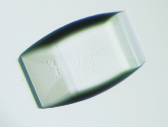
The ligand-binding domain of rat SHPS-1 was purified and crystallized using the vapour-diffusion method with the solution-stirring technique.
Keywords: SHPS-1, immunoglobulin-like domain, CD47, CD47–SHPS-1 system
Abstract
SHPS-1, a receptor-type transmembrane protein, is abundantly expressed in neural and myeloid tissues. The most amino-terminal immunoglobulin-like domain of SHPS-1 plays an important role in a variety of cell functions by binding its ligand CD47. Interaction between SHPS-1 and CD47 is thought to be involved in negative regulation of phagocytosis. The ligand-binding domain of rat SHPS-1 was purified and crystallized using the vapour-diffusion method with the solution-stirring technique. Preliminary X-ray diffraction data were collected from SHPS-1 crystals to 2.8 Å resolution and reduced to primitive hexagonal space group P622. Unit-cell parameters are a = b = 100.5, c = 101.3 Å.
1. Introduction
Phagocytosis is an evolutionarily conserved process that plays a crucial role in diverse biological phenomena including the elimination of pathogens, activation of innate and adaptive immune responses, clearance of apoptotic cells and tissue integrity. Receptor-mediated recognition of target cells is a key step in the early stage of phagocytosis. In mammals, phagocytosis is triggered by the interaction of specific receptors on phagocytic cells, such as macrophages, with their ligands and thus target cells are engulfed. Fc receptors, complement receptors and integrins bind to particles coated by IgG, complement and fibronectin/vitronectin, respectively. On the other hand, SHPS-1 [Src homology 2 domain-containing protein tyrosine phosphatase (SHP) substrate-1; Fujioka et al., 1996 ▶; Yamao et al., 1997 ▶], a receptor-type transmembrane protein, is thought to negatively regulate phagocytosis by binding CD47 (integrin-associated protein, IAP; Oldenborg et al., 2000 ▶; Okazawa et al., 2005 ▶).
SHPS-1, also known as SIRPa (Kharitonenkov et al., 1997 ▶), BIT (Ohnishi et al., 1996 ▶), MFR (Saginario et al., 1998 ▶) and p84 neural adhesion molecule (Comu et al., 1997 ▶), is a transmembrane protein that is especially abundant in macrophages and neurons. The cytoplasmic region of SHPS-1 contains immunoreceptor tyrosine-based inhibition motifs (ITIMs) that upon phosphorylation recruit SHP-1 and SHP-2 (Fujioka et al., 1996 ▶; Kharitonenkov et al., 1997 ▶). The extracellular region of SHPS-1 comprises three Ig-like domains, of which the most amino-terminal domain associates with its ligand CD47. CD47 and SHPS-1 constitute a cell–cell communication system (the CD47–SHPS-1 system) that plays important roles in multiple cellular processes, including cell migration (Liu et al., 2001 ▶; Motegi et al., 2003 ▶), adhesion of B cells (Yoshida et al., 2002 ▶), T-cell activation (Seiffert et al., 2001 ▶; Latour et al., 2001 ▶), macrophage multinucleation (Han et al., 2000 ▶) and dendritic cell maturation (Latour et al., 2001 ▶). In addition, the CD47–SHPS-1 system is implicated in inhibition of IgG, complement or calreticulin-induced phagocytosis. Indeed, CD47-deficient red blood cells are phagocytosed by splenic red pulp macrophages in wild-type mice and rapidly cleared from the bloodstream (Oldenborg et al., 2000 ▶, 2001 ▶). Furthermore, disruption of interaction between CD47 on the target cell and SHPS-1 on the phagocytic cell permitted ingestion of the target cell in a calreticulin/LRP-dependent manner (Gardai et al., 2005 ▶). However, the molecular mechanism by which the CD47–SHPS-1 system inhibits phagocytosis by these cells is unknown.
In the present paper, we describe the crystallization and X-ray diffraction data collection of the ligand-binding domain of SHPS-1, aiming to solve the structure and gain insights into the functional aspects of this protein.
2. Materials and methods
2.1. Protein expression and purification
A cDNA fragment encoding residues Ala31–Leu149 of rat SHPS-1 corresponding to the ligand-binding domain was amplified by the polymerase chain reaction (PCR) from a cDNA clone. The oligonucleotides were designed to introduce BamHI and NotI restriction-endonuclease sites at the 5′- and 3′-end of the PCR products, respectively. PCR products were ligated into the glutathione-S-transferase (GST) gene-fusion vector pGEX-4T-1 (Amersham Biosciences) and sequenced for confirmation. The construct was then transformed into Escherichia coli Origami B cells (Novagen) for IPTG-induced expression. 10 l cultures were grown to an OD600 of 0.5–1.0 at 298 K and IPTG was added to a final concentration of 0.1 mM. Cultures were grown for an additional 40 h at 293 K. Induced bacterial cultures were centrifuged and stored at 243 K. The cell pellet was resuspended in ice-cold lysis buffer containing 20 mM Tris–HCl pH 8.0, 150 mM sodium chloride, 5 mM EDTA, 5% glycerol, 1% NP-40 and the protease inhibitor Complete EDTA-free (Roche Diagnostics) and was then homogenized using an ultrasonic processor. The crude lysate was centrifuged at 48 400g for 30 min at 277 K. Recombinant SHPS-1 was purified using Glutathione Sepharose affinity gels (Amersham Biosciences). After cleavage of the GST portion with thrombin (Amersham Biosciences), SHPS-1 was further purified by HiTrap SP HP column (Amersham Biosciences) cation-exchange chromatography. Clarified concentrate was then separated by FPLC on Superdex 75 column (Amersham Biosciences) and a fraction containing proteins with a molecular weight of ∼13.5 kDa was collected as a finally purified material. The homogeneity of the purified protein was assessed by SDS–PAGE (Laemmli, 1970 ▶). The protein solution was concentrated by ultrafiltration to a final concentration of 10 mg ml−1. The protein concentration was estimated by measuring the absorbance at 280 nm, employing the calculated extinction coefficient of 10 930 M −1 cm−1 (SWISS-PROT; http://www.expasy.ch/). Final recovery of purified SHPS-1 was approximately 1 mg protein per litre of starting culture.
2.2. Crystallization
Crystallization conditions were screened by the conventional hanging-drop vapour-diffusion method (McPherson, 1999 ▶) using Crystal Screen kits (Hampton Research). 2 µl drops consisting of 1 µl protein solution and 1 µl mother liquor were equilibrated against 1.0 ml reservoir solution at 293 K. A few crystals were obtained using solution No. 20 (0.2 M ammonium sulfate, 25% PEG 4000, 0.1 M sodium acetate pH 4.6) of Crystal Screen 1. After optimization of this condition, crystals with dimensions of 0.10 × 0.20 × 0.35 mm were formed in 0.4 M ammonium sulfate, 18–28% PEG 4000, 0.1 M sodium acetate pH 4.6 by the conventional vapour-diffusion method and did not diffract beyond 3.0 Å resolution. Crystallization using the vapour-diffusion method with the solution-stirring technique (Adachi et al., 2004 ▶) under the same reservoir-solution conditions at 293 K improved the crystal quality (Fig. 1 ▶). Although the appearance and size of the crystals obtained using the solution-stirring technique were almost the same, the X-ray resolution was improved.
Figure 1.
An example of crystals of SHPS-1 obtained using the solution-stirring technique. The crystal dimensions are 0.14 × 0.16 × 0.32 mm. The crystal diffracted X-rays to at least 2.8 Å resolution.
2.3. Data collection and processing
Crystals were picked up from the crystallization drop using a nylon-fibre loop and flash-cooled in a nitrogen-gas stream employing a Cryostream 600 (Oxford Cryosystems) and stored in liquid nitrogen until needed for data collection. Diffraction data were collected using synchrotron radiation and a DIP6040 image-plate detector (MAC Science/Bruker AXS) at SPring-8 beamline BL44XU (Hyogo, Japan). Data were collected using an oscillation angle of 1° per frame at a wavelength of 0.90 Å. Each frame was exposed for 10 s. All data were indexed and integrated with MOSFLM (Leslie, 1992 ▶) and reduced with SCALA (Evans, 1993 ▶) from the CCP4 suite (Collaborative Computational Project, Number 4, 1994 ▶).
3. Results and discussion
The crystals obtained using the solution-stirring technique diffracted to better than 2.8 Å resolution. A total of 133 148 measured reflections were merged into 7810 unique reflections with an R merge of 8.8%. They belong to the primitive hexagonal space group P622, with unit-cell parameters a = b = 100.5, c = 101.3 Å. Table 1 ▶ summarizes the data-collection statistics. The Matthews probability calculation suggests the presence of two molecules in the asymmetric unit, with a V M value of 2.7 Å3 Da−1 and a solvent content of 54.7%. We are currently in the process of attempting a molecular-replacement solution using the coordinates of the anti-testosterone Fab fragment (PDB code 1l7t), which shares 35% sequence identity with rat SHPS-1.
Table 1. Data-collection and reduction statistics for SHPS-1 crystals.
Values for the highest resolution shell are in parentheses.
| Wavelength (Å) | 0.90 |
| Unit-cell parameters (Å) | a = b = 100.5, c = 101.3 |
| Space group | P622 |
| Resolution range (Å) | 50.6–2.81 (2.96–2.81) |
| Observed reflections | 133148 |
| Unique reflections | 7810 |
| Completeness (%) | 100.0 (99.9) |
| Rmerge (%) | 8.8 (52.2) |
| 〈I/σ(I)〉 | 6.1 (1.4) |
Acknowledgments
We would like to thank the Crystal Design Project (http://crystal.pwr.eng.osaka-u.ac.jp/sosho.html) for help in high-quality crystallization. We also thank Dr Eiki Yamashita for his support during data collection and processing.
References
- Adachi, H., Matsumura, H., Niino, A., Murakami, S., Takano, K., Kinoshita, T., Warizaya, M., Inoue, T., Mori, Y. & Sasaki, T. (2004). Jpn. J. Appl. Phys.43, L522–L525.
- Collaborative Computational Project, Number 4 (1994). Acta Cryst. D50, 760–763. [Google Scholar]
- Comu, S., Weng, W., Olinsky, S., Ishwad, P., Mi, Z., Hempel, J., Watkins, S., Lagenaur, C. F. & Narayanan, V. (1997). J. Neurosci.17, 8702–8710. [DOI] [PMC free article] [PubMed] [Google Scholar]
- Evans, P. R. (1993). Proceedings of the CCP4 Study Weekend. Data Collection and Processing, edited by L. Sawyer, N. Isaacs & S. Bailey, pp. 114–122. Warrington: Daresbury Laboratory.
- Fujioka, Y., Matozaki, T., Noguchi, T., Iwamatsu, A., Yamao, T., Takahashi, N., Tsuda, M., Takada, T. & Kasuga, M. (1996). Mol. Cell Biol.16, 6887–6899. [DOI] [PMC free article] [PubMed] [Google Scholar]
- Gardai, S. J., McPhillips, K. A., Frasch, S. C., Janssen, W. J., Starefeldt, A., Murphy-Ullrich, J. E., Bratton, D. L., Oldenborg, P. A., Michalak, M. & Henson, P. M. (2005). Cell, 123, 321–334. [DOI] [PubMed] [Google Scholar]
- Han, X., Sterling, H., Chen, Y., Saginario, C., Brown, E. J., Frazier, W. A., Lindberg, F. P. & Vignery, A. (2000). J. Biol. Chem.275, 37984–37992. [DOI] [PubMed] [Google Scholar]
- Kharitonenkov, A., Chen, Z., Sures, I., Wang, H., Schilling, J. & Ullrich, A. (1997). Nature (London), 386, 181–186. [DOI] [PubMed] [Google Scholar]
- Laemmli, U. K. (1970). Nature (London), 227, 680–685. [DOI] [PubMed] [Google Scholar]
- Latour, S., Tanaka, H., Demeure, C., Mateo, V., Rubio, M., Brown, E. J., Maliszewski, C., Lindberg, F. P., Oldenborg, A., Ullrich, A., Delespesse, G. & Sarfati, M. (2001). J. Immunol.167, 2547–2554. [DOI] [PubMed] [Google Scholar]
- Leslie, A. G. W. (1992). Jnt CCP4/ESF–EACBM Newsl. Protein Crystallogr.26
- Liu, Y., Merlin, D., Burst, S. L., Pochet, M., Madara, J. L. & Parkos, C. A. (2001). J. Biol. Chem.276, 40156–40166. [DOI] [PubMed] [Google Scholar]
- McPherson, A. (1999). Crystallization of Biological Macromolecules. Cold Spring Harbor: Cold Spring Harbor Laboratory Press.
- Motegi, S., Okazawa, H., Ohnishi, H., Sato, R., Kaneko, Y., Kobayashi, H., Tomizawa, K., Ito, T., Honma, N., Bühring, H. J., Ishikawa, O. & Matozaki, T. (2003). EMBO J.22, 2634–2644. [DOI] [PMC free article] [PubMed] [Google Scholar]
- Ohnishi, H., Kubota, M., Ohtake, A., Sato, K. & Sano, S. (1996). J. Biol. Chem.271, 25569– 25574. [DOI] [PubMed] [Google Scholar]
- Okazawa, H., Motegi, S., Ohyama, N., Ohnishi, H., Tomizawa, T., Kaneko, Y., Oldenborg, P. A., Ishikawa, O. & Matozaki, T. (2005). J. Immunol.174, 2004–2011. [DOI] [PubMed] [Google Scholar]
- Oldenborg, P. A., Gresham, H. D. & Lindberg, F. P. (2001). J. Exp. Med.193, 855–862. [DOI] [PMC free article] [PubMed] [Google Scholar]
- Oldenborg, P. A., Zheleznyak, A., Fang, Y. F., Lagenaur, C. F., Gresham, H. D. & Lindberg, F. P. (2000). Science, 288, 2051–2054. [DOI] [PubMed] [Google Scholar]
- Saginario, C., Sterling, H., Beckers, C., Kobayashi, R., Solimena, M., Ullu, E. & Vignery, A. (1998). Mol. Cell Biol.18, 6213–6223. [DOI] [PMC free article] [PubMed] [Google Scholar]
- Seiffert, M., Brossart, P., Cant, C., Cella, M., Colonna, M., Brugger, W., Kanz, L., Ullrich, A. & Bühring, H. J. (2001). Blood, 97, 2741–2749. [DOI] [PubMed] [Google Scholar]
- Yamao, T., Matozaki, T., Amano, K., Matsuda, Y., Takahashi, N., Ochi, F., Fujioka, Y. & Kasuga, M. (1997). Biochem. Biophys. Res. Commun.231, 61–67. [DOI] [PubMed] [Google Scholar]
- Yoshida, H., Tomiyama, Y., Oritani, K., Murayama, Y., Ishikawa, J., Kato, H., Miyagawa, J., Honma, N., Nishiura, T. & Matsuzawa, Y. (2002). J. Immunol.168, 3213–3220. [DOI] [PubMed] [Google Scholar]


