Abstract
The sagittal pelvic morphology modulates the individual alignment of the spine. Anatomical angular parameters were described as follows: the “Pelvic Incidence” (PI) and the Jackson’s angle “Pelvic Lordosis” (PR-S1). Significant chains of relationships were expressed connecting these angles with pelvic and spinal positional parameters. This allows an individual assessment of the harmony of the sagittal spinal balance. But in case of spondylolysis with high-grade listhesis, the upper plate of the sacrum shows a dome-shaped deformity. The previous anatomical parameters are therefore imprecise. Indeed, the anterior part of the sacrum being inaccurate, an exact assessment of these angles becomes impossible. Therefore, we propose a new angular parameter named “Femoro-Sacral Posterior Angle” (FSPA): the angle between the posterior wall of the first sacral vertebra, always well definite, and the line connecting the posterior part of the sacral plate to the femoral axis. The validation of this parameter was performed and compared with the classical published parameters. It showed good inter-observer reliability, even with dome-shaped sacral plate. In spite of lower correlation with the positional parameters than those observed with PI or PR-S1, the FSPA appeared to be reliable and precise for an exact evaluation of the sagittal spino-pelvic balance is case of spondylo-listhesis with dome-shaped sacral endplate.
Keywords: Sagittal shape, Pelvis, Spinal balance, Spondylolysis, Sacrum
Introduction
The interdependence between the pelvis and the sagittal spinal curvatures is obvious. The leading part of the pelvic sagittal anatomy in this orientation was well established by Duval-Beaupère thanks to the description of the angle “Pelvic Incidence” (PI) [14]. It is the angle between the line perpendicular to the superior plate of the first sacral vertebra (S1) at its midpoint and the line connecting this point to the middle axis of the femoral heads. It is own for every individual and independent of the position of pelvis. A strict relation was described between this anatomical parameter and the sagittal tilt of the superior plate of the sacrum, and between this sacral tilt and the amount of lumbar lordosis. So the pelvic morphology modulates the sagittal spinal alignment. During and Itoi [1, 7] described also these relations using an angle complementary of the incidence. As well Jackson observed this relation between the pelvic morphology, the sacral inclination and the lumbar curvature. He expressed the pelvic sagittal shape by the “Pelvic Lordosis” (PR-S1) [8–12]. The measurement of these angles is easy in most of the cases. Their reliability and their reproducibility were established. Each author agrees to use as femoral axis the middle point of the line connecting the centers of the two femoral heads.
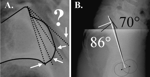
However, the exact assessment of the upper plate of the sacrum (and consequently the values of the anatomical and positional pelvic parameters using it) necessitates a precise visualization of its two extremities. The posterior part of this sacral plate is always very definite. On the other hand, the anterior part of the sacral endplate is imprecise when there is a deformity in the dome, characteristic of the spondylolysis with anterior slip of the fifth lumbar vertebra. The upper sacral plate looks convex and blunted. The exact appraisal of the tilt of the sacral plate and of its anterior extremity is then imprecise (Fig. 1). Such an inaccuracy occurs also in degenerative pathologies. But for these cases precisely, the sagittal alignment of the spine is of first clinical interest for the prognosis of worsening and so for the treatment.
Fig. 1.
Dome-shaped sacrum. The inaccurate visualization of the anterior part of the superior plate of S1 does not allow an exact appraisal of its orientation. The measures of the parameter using this plate are imprecise (“pelvic incidence” varying from 70° to 86°)
Therefore, we suggest here a new angular morphologic pelvic parameter, named “Femoro-Sacral Posterior Angle” (FSPA), using the posterior wall of the first sacral vertebra. This parameter was put in relationship with the already described parameters to demonstrate its reliability for the evaluation of the sagittal spinal balance.
Material and methods
The angular parameters were measured on lateral radiographs (including the femoral heads, the pelvis and the lumbar column) in standing position, with the arms lying on a support.
The data of two population groups were analyzed. The first group comprised a normal population of 145 voluntary subjects free of vertebral disease. There were 73 women and 72 men, with a mean age of 40.7 years (SD 18.7, range 15–76 years). These 145 subjects constituted the control group for measuring the relationship between these parameters in the normal population. They consisted of voluntary medical, nurse or physical therapy students or patients X-rayed for an other pathology than vertebral, mostly for urinary tract exploration. The second group comprised 35 spondylolysis cases, with minor listhesis (Meyerling’s stage I or II) and without any distortion of the superior sacral plate. There were 10 women and 25 men; the mean age was of 24.6 years (SD 9.6, range 15–42 years).
Owing to the repartition of age being different between the two groups, a second normal population was selected from the first cohort according to the range of age (15–42 years). This second normal group was similar in terms of age to the group with spondylolysis.
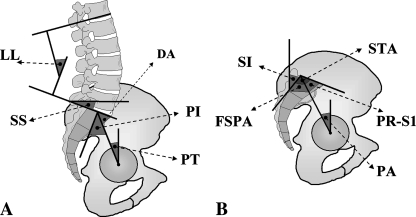
The angular pelvic and spinal parameters were: (Fig. 2).
Fig. 2.
The studied parameters. The parameters described by Duval–Beaupère: pelvic incidence (PI), sacral slope (SS), pelvic tilting (PT), complementary angle of the pelvic incidence, described by During and Itoi (DA), lumbar lordosis (LL). The parameters described by Jackson: pelvic lordosis or Jackson’s angle (PR-S1), pelvic angle (PA), sacral inclination (SI), sacral table angle of S1 (STA), femoro-sacral posterior angle (FSPA)
The PI: value of the angle between the line perpendicular to the superior plate of S1 at its midpoint and the line connecting this point to the axis of the femoral heads.
The During’s angle (DA): it is the complementary angle of the pelvic incidence. This angle was not discussed, being similar to the pelvic incidence.
The sacral slope (SS): value of the angle between the superior plate of S1 and a horizontal line. A vertical sacrum is described by a low value, a horizontal sacrum by a high value.
The pelvic tilting (PT): value of the angle between the vertical and the line connecting the midpoint of the sacral plate to the axis of the femoral heads.
The lumbar lordosis (LL): value of the angle between the upper plate of S1 and the vertebral plate the most tilted backward (in our series always L1 or T12).
The pelvic lordosis or Jackson’s angle (PR-S1): value of the angle between the sacral superior plate and the line connecting the posterior point of sacral plate to the femoral heads axis.
The pelvic angle (PA): value of the angle between the vertical and the line connecting the posterior point of sacral plate to the axis of the femoral heads.
The sacral table angle of S1 (STA): value of the angle between the superior plate of S1 and the posterior side of body of the first sacral vertebra.
The sacral inclination (SI): value of the angle between the posterior side of the body of the first sacral vertebra and the vertical.
Femoro-sacral posterior angle: value of the angle between the line connecting the posterior point of sacral superior plate to the axis of the femoral heads and the posterior side of the body of the first sacral vertebra.
The values of the angles were reported in degree. A positive value was posterior, a negative anterior.
The parameters PI, SS, PT (Fig. 2a) were described by Duval-Beaupère for the analysis of the pelvi-spinal sagittal balance [14]; the parameters PR-S1, PA and SI (Fig. 2b) were the angles expressed by Jackson [8–12]. The STA was firstly described for the assessment of the sacrum of rabbits and recently used to characterize the deformity of the sacrum in spondylolysis cases [6, 18].
The comparisons between parameters were performed using the Student’s test. The relations between parameters were established through the coefficients of correlation.
Furthermore, seven independent observers achieved the measurements of PI, SS, PR-S1 and FSPA on the lateral radiographs of two subjects: one with a normal sacrum and one with a dome-shaped sacrum.
Moreover, the angle FSPA was measured on radiographs from eight cases of spondylolysis with high-grade listhesis, Meyerling’s stage 3 (7 cases) and 4 (1 case). There were four women and four men; the mean age was of 35.2 years (SD=8.7).
Results
The mean values and the standard deviations for the studied parameters are reported in Table 1: column A for the entire normal population, column B for normal subject selected according to the age, column C for the patients with spondylolysis. We observed mean values for PI, SS, PT and lumbar lordosis similar to the values assessed as “normal” firstly established by Duval-Beaupère and corroborated by several authors [3, 14, 19–21]. The value for the pelvic incidence was provided as normal between 43° and 62°. The normal range of value for the sacral slope was from −32° to −49°, from 3° to 18° for the pelvic tilting. The same accordance was observed for the lumbar lordosis (52–75° in numerous works) for all that the limits of the measured curvature were identical.
Table 1.
Mean values and standard deviation of the parameters observed for the entire normal group (column A), the normal subjects selected according to the age (column B), and the spondylolysis group (column C)
| Normal Total pop. n=145 Men 72, Women 73 |
Normal (15–42 years) n=66 Men 33, Women 33 |
Spondylolysis (15–42 years) n=35 Men 25, Women 10 |
Normal subject Total/15–42 years |
Normal subject TT/spondylolysis |
Young normal subject/spondylolysis | |||||||
|---|---|---|---|---|---|---|---|---|---|---|---|---|
| Mean | SD | Mean | SD | Mean | SD | ε Student | Sign | ε Student | Sign | ε Student | Sign | |
| Age (years) | 40.7 | 18.7 | 26.5 | 8.4 | 24.6 | 9.6 | 5.558 | *** | 7.168 | *** | 0.987 | NS |
| Pelvic incidence (PI) | 50.2 | 10.6 | 49.6 | 10.35 | 62. | 11 | 0.389 | NS | 5.731 | *** | 5.546 | *** |
| Sacral slope (SS) | −38.8 | 6.6 | -39 | 6.3 | −47.4 | 8.1 | 0.211 | NS | 5.831 | *** | 5.338 | *** |
| Pelvic tilting (PT) | 11.5 | 5.9 | 10.6 | 5.7 | 14.7 | 3.7 | 1.052 | NS | 4.028 | *** | 4.362 | *** |
| Lumbar lordosis (LL) | 63.7 | 7.9 | 64.4 | 7.8 | 73.1 | 8.8 | 0.682 | NS | 5.782 | *** | 4.914 | *** |
| Jackson’s angle (PR-S1) | 35.2 | 9.6 | 35.8 | 9.2 | 24.3 | 10 | 0.433 | NS | 5.832 | *** | 5.652 | *** |
| Sacral inclination (SI) | 51 | 7.6 | 51.7 | 6.9 | 57.2 | 8.4 | 0.662 | NS | 3.99 | *** | 3.324 | *** |
| Pelvic angle (PA) | 16.1 | 5.1 | 15.2 | 4.9 | 18.3 | 2.8 | 1.221 | NS | 3.464 | *** | 4.043 | *** |
| Sacral table angle (STA) | 102.2 | 5.4 | 102.7 | 5.2 | 99.8 | 5.3 | 0.64 | NS | 2.396 | * | 2.634 | ** |
| Femoro-sacral posterior Angle (FSPA) | 67 | 9.6 | 66.9 | 8.9 | 75.5 | 9.2 | 0.074 | NS | 4.864 | *** | 4.521 | *** |
| A | B | C | D | E | F | |||||||
The values were compared between normal and spondylolysis groups: between the two normal groups (column D), between the spondylolysis group and (column E) the entire normal group, and (column F) the age-selected normal group
NS Not significant
*P<0.05; **P<0.01; ***P<0.001
The mean value reported by Jackson for pelvic lordosis (the Jackson’s angle or PR-S1) was 32° (±9.8). A value for PA was allowed as normal from 0° to 35°. The sacral inclination SI was valued as normal between 40 and 50° [2, 8–12]. The mean values observed for our normal population were so similar to those published.
The comparisons between the two normal groups and the group with spondylolysis were reported in Table 1, columns D, E, F. No significant difference was observed between the values of the entire normal group and the group selected according to the age (between 15 and 42 years) (P>0.1). This reflects the homogeneity of this reference normal population.
On the other hand, significant differences were observed between these two populations and the spondylolysis cases, as for the lordosis angle than for the morphological and positional pelvic parameters. These observations for spondylolysis cases, even with low-grade listhesis, confirm the impact of the sagittal pelvi-spinal balance in this pathology.
The values of the parameters measured by seven different observers on the lateral radiographs of a normal and a dome-shaped sacrum are reported in Table 2. The scattering of the values of PI, SS, PR-S1, described by the standard deviation, was more important when a dome-shaped superior plate of S1 exists. However, the measurement of FSPA was not affected by this distortion. The standard deviations were still 1.2–1.3. It was similar to the scattering of the values of the other parameters measured on the normal radiograph. The reliability of the parameter FSPA was so established identical to the other parameters, even when a distortion of the sacral endplate exists.
Table 2.
Mean values of the parameters measured on two lateral radiographs: one with a normal sacral plate, one with a dome-shaped sacrum
| n=7 | Dome-shaped sacrum | Normal sacral plate | ||
|---|---|---|---|---|
| Mean | SD | Mean | SD | |
| Pelvic incidence (PI) | 78.9 | 5.3 | 50 | 1.3 |
| Sacral slope (SS) | −59.7 | 5 | −32.4 | 1.2 |
| Jackson’s Angle (PR-S1) | 9.6 | 5 | 34.9 | 1 |
| Femoro-sacral posterior Angle (FSPA) | 72.7 | 1.3 | 65.3 | 1.2 |
The SD reflect the scattering of the values when a dome-shaped sacrum and the lack of precision for PI, SS and PR-S1 in these cases exist. For FSPA the precision was observed identical to the others parameters and still reliable with dome-shaped sacrum
The relations between morphological and positional parameter were also analyzed. The significant coefficients of correlation are reported in Table 3. The chain of close interdependence between the morphological (pelvic incidence, Jackson’s angle and FSPA) and the positional pelvic parameters (sacral slope, pelvic tilting, angles SI and PA) was corroborated. The primordial correlation between the pelvic anatomical parameter “pelvic incidence” and the positional parameter “sacral slope” was similar to the reported coefficients (r=0.81–0.86) [14, 20, 21]. A similar relation was observed using the “Jackson’s angle PR-S1”.
Table 3.
Coefficients of correlation (r) between parameters for the entire normal group
| PI | SS | PT | LL | PR-S1 | SI | PA | FSPA | |
|---|---|---|---|---|---|---|---|---|
| Pelvic incidence (PI) | – | 0.8647*** | 0.8287*** | 0.7363*** | 0.9978*** | 0.5668*** | 0.7508*** | 0.8475*** |
| Sacral slope (SS) | 0.8647*** | – | 0.4354*** | 0.8935*** | 0.8613*** | 0.7184*** | 0.3234** | 0.7383*** |
| Pelvic tilting (PT) | 0.8287*** | 0.4354*** | – | 0.3240*** | 0.8383*** | 0.2154* | 0.9851*** | 0.6961*** |
| Lumbar lordosis (LL) | 0.7363*** | 0.8935*** | 0.3240*** | – | 0.7349*** | 0.6305*** | 0.2243* | 0.6162*** |
| Jackson’s angle (PR-S1) | 0.9978*** | 0.8613*** | 0.8283*** | 0.7349*** | – | 0.5545*** | 0.7593*** | 0.8424*** |
| Sacral inclination (SI) | 0.5668*** | 0.7184*** | 0.2154* | 0.6305*** | 0.5545*** | – | 0.127 NS | 0.8472*** |
| Pelvic angle (PA) | 0.7508*** | 0.3234** | 0.9851*** | 0.2243* | 0.7593*** | 0.1127 NS | – | 0.6233*** |
| Femoro-sacral posterior angle (FSPA) | 0.8475*** | 0.7383*** | 0.6961*** | 0.6162*** | 0.8424*** | 0.8472*** | 0.6233*** | – |
NS Not significant
*P<0.05, **P<0.01, ***P<0.001
Similarly, the close relation between the positional pelvic parameters and the lumbar lordosis was significant, although in a lower degree with the pelvic tilting and the angle PA.
We have confirmed the very close relation between the “sacral slope” and the “lordosis”. On the other hand, using the “angle SI” as pelvic positional parameter offers a lower coefficient of correlation with the “lordosis” than using the “sacral slope”.
The direct relation with the “lordosis” was significant both for the “pelvic incidence” and for the “Jackson’s angle PR-S1”. It is in accord with the published coefficients (of r=0.6–0.69).
The “pelvic tilting”, also expressing the pelvic orientation, was highly correlated to the “pelvic incidence”, similarly to the reported coefficients (0.54–0.66). On the other hand, the direct relation was lower (but still significant) between the “pelvic tilting” and the “sacral slope”, and between the “pelvic tilting” and the “lordosis”. Moreover, the relation between the “SI” and the “PA” was not observed significant and poorly significant between the “lordosis” and the “PA”. These observations confer less reliability to the utilization of the “PA” comparatively to the “pelvic tilting” for the evaluation of the sagittal balance.
In spite of the high relation observed between the “sacral slope” and “SI”, the comparison of the relations between these angles (SS and SI) and the “lordosis” showed a better reliability for the “sacral slope”. Moreover, the relations between the “SI” and the “pelvic incidence” and with the “Jackson’s angle PR-S1” were lower than those of the “sacral slope” with the “pelvic incidence” and with the “Jackson’s angle”. So the “sacral slope” seems more reliable to use than “SI”.
We observed that a relationship between the “pelvic incidence” and the “Jackson’s angle” was stronger than between the “FSPA” and the “pelvic incidence” or the “Jackson’s angle”. It is bound to the use of the upper plate of S1 to assess both the pelvic incidence and the Jackson’s angle. Although the coefficients of correlation between FSPA and positional parameters were a bit lower than with PI and PR-S1, they were observed highly significant (P<0.001) with the “sacral slope”, with the “lordosis”, and very more with the “SI”. Consequently, the use of this angle FSPA for the assessment of the relations between the pelvis and the spine may be regarded as reliable, particularly in case of dome-shaped deformity of the upper sacral endplate when this angle is the only one usable. The correlation’s tests were also achieved for the 35 spondylolysis cases. Very significant coefficients were observed between “pelvic incidence” and the Jackson’s angle PR-S1, the “pelvic incidence” and FSPA, the Jackson’s angle and FSPA. The high relationship between the sacral slope and the pelvic incidence or the Jackson’s angle was also observed with FSPA, testifying the reliability of the proposed angle.
The value of FSPA observed in the group of eight cases of spondylolysis with high-stage listhesis was 83.4°, SD 7.5. In spite of the low number of cases, this value was observed significantly different from the value observed for the normal group (t=5.023, P<0.001) and for the group of spondylolysis with low-stage listhesis (t=2.257, P<0.05). This represented the disturbance of the growth of the sacrum in this pathology, as to whether this disturbance was the origin of the consequence.
Discussion
The analysis of the sagittal balance of the spine bases one’s argument not only on the values of parameters, but also more on the assessment of the pelvi-spinal harmony that is specific to each person. Indeed, modifications of the sagittal spinal curvatures have been connected with changes in pelvic orientation and morphology. The significant chain of interdependence was already investigated for normal and pathologic conditions, particularly for the isthmic spondylolysis. The values and the relationship between anatomical and positional parameters observed for our normal population corroborates the already published data [3, 8, 14, 19–21]. The normality of our named “normal” population was thus validated.
Much more, previous research papers have demonstrated the evolution of the pelvic morphology during the childhood. The pelvic incidence of the human fetus was reported comparable to the one observed at the chimp. This angle then quickly increases during the first month after the birth and continues to accent until the age of 10 years. It stays then steady during the adult period. In the same way, the adaptation and the progressive establishment of the sagittal spinal curvatures occur during the childhood [15, 16].
A significant difference in the sagittal pelvic and sacral morphology was already reported between the normal subjects and those suffering from a spondylolysis [4, 6, 12, 17, 22, 23]. This consists of a sagittal widening of the pelvis, expressed by an increase in the values of the anatomical parameters (PI, Jackson’s angle PR-S1, FSPA, STA). From this enlargement emanates an accentuation of the positional pelvic parameters (SS, PT, PA, angle SI) traducing a more tilted sacral plate. Some authors consider an important pelvic incidence as a pejorative factor for the progression of the vertebral listhesis [4, 11, 17]. But others did not observe it as significant [5]. This anatomic alteration permits, however, to suspect a disruption of the growth affecting the first sacral vertebra, leading to a dome-shaped upper sacral plate concomitant to the vertebral listhesis. It looks like epiphyseal injury that produces Blount’s disease or slipped capital femoral epiphysis [3, 23].
A very close correlation was observed between the two anatomical pelvic parameters “pelvic incidence” and “Jackson’s angle”. As well, the relationships between these two anatomical parameters with the positional pelvic and spinal parameters were in the same line of significance. Both describe well identically the same reality and both refer to the upper plate of S1. Only a variation of the “sacral table angle (STA) should be susceptible to induce an alteration in the value of “FSPA” and of the “Jackson’s angle” (in a lesser manner of the “pelvic incidence”) [17]. This STA is indeed the algebraic sum of the “FSPA” and of the “Jackson’s angle”. STA was reported significantly different according to the existence of a spondylolysis with vertebral slip [6, 23].
We have also observed such a significant difference between the normal and the spondylolysis group. As well, the interrelationships were significant between STA and the pelvic anatomical parameters (PI, PR-S1 and FSPA) for the entire normal group. But they were observed less significant for the spondylolysis group.
Otherwise, the accuracy of the measurements using the upper sacral plate is greatly dependent on the radiographic definition of its anterior part. When there is a dome-shaped deformity, it becomes rounded, dropping downward and so difficult to locate precisely. This imprecision will affect the accuracy of the angles as the “sacral slope” or the “Jackson’s angle”, using only the short posterior part of the plate. A similar inaccuracy affects the “pelvic incidence” due to the lack of exact location of the midpoint of the plate. The results of Table 2 expressed this difficulty. The scattering of the values of PI, PR-S1 and SS was important: from 70° to 86° for the PI. On the other hand, the values of the “FSPA” were constant because the posterior wall of S1 was always precisely perceptible, even in this case of dome-shaped plate. The inaccuracy in the values of the PI and PR-S1 in case of spondylolysis contributes to a certain degree to the contradiction observed in the literature; thus, a predictive value for the progression of the listhesis can be assigned to these parameters. Some authors consider a high value of “pelvic incidence” as pejorative, other did not observe it as significant. The observation of a value of FSPA significantly different for the cases of spondylolysis with high-stage listhesis confers on this angle, the only one measurable in these cases, a prognostic value. Its clinical interest is the prediction of a pejorative unfavorable evolution of the slip in case of increased value, and so to specify a surgical indication.
Conclusion
The FSPA was demonstrated to be reliable as anatomic sagittal pelvic parameter even in dome-shaped deformity of the sacral endplate, contrary to PI and PR-S1. Even if the coefficients of correlation between FSPA and the positional parameters were lightly lower than with PI or PR-S1, they were observed highly significant. We suggest using this FSPA angle for the assessment of the sagittal pelvic morphology in case of spondylolysis.
References
- 1.During J, Goudfrooij H, Keesen W, Beeker TW, Crowe A. Toward standards for posture. Postural characteristics of the lower back system in normal and pathologic conditions. Spine. 1985;10:83–87. doi: 10.1097/00007632-198501000-00013. [DOI] [PubMed] [Google Scholar]
- 2.Gardocki RJ, Watkins RG, Williams LA. Measurements of lumbopelvic lordosis using the pelvic radius technique as it correlates with sagittal spinal balance and sacral translation. Spine. 2002;2:421–429. doi: 10.1016/S1529-9430(02)00426-6. [DOI] [PubMed] [Google Scholar]
- 3.Guigui P, Levassor N, Rillardon L, Wodecki P, Cardinne L. Valeur physiologique des paramètres pelviens et rachidiens de l’équilibre sagittal du rachis. Analyse d’une série de 250 volontaires. Rev Chir orthop. 2003;89:496–506. [PubMed] [Google Scholar]
- 4.Hanson DS, Bridwell KH, Rhee JM, Lenke LG. Correlation of pelvic incidence with low- and high-grade isthmic spondylolisthesis. Spine. 2002;27:2026–2029. doi: 10.1097/00007632-200209150-00011. [DOI] [PubMed] [Google Scholar]
- 5.Huang RP, Bohlman HH, Thompson GH, Poe-Kochert C. Predictive value of pelvic incidence in progression of spondylolisthesis. Spine. 2003;28:2381–2385. doi: 10.1097/01.BRS.0000085325.42542.38. [DOI] [PubMed] [Google Scholar]
- 6.Inoue H, Ohomori K, Miyasaka K. Radiographic classification of L5 isthmic spondylolisthesis as adolescent or adult vertebral slip. Spine. 2002;27:831–838. doi: 10.1097/00007632-200204150-00010. [DOI] [PubMed] [Google Scholar]
- 7.Itoi E. Roentgenographic analysis of posture in spinal osteoporotics. Spine. 1991;6:750–756. doi: 10.1097/00007632-199107000-00011. [DOI] [PubMed] [Google Scholar]
- 8.Jackson RP, Hales C. Congruent spinopelvic alignment on standing lateral radiographs of adult volunteers. Spine. 2000;25:2808–2815. doi: 10.1097/00007632-200011010-00014. [DOI] [PubMed] [Google Scholar]
- 9.Jackson RP, Kanemura T, Kawakami N, Hales C. Lumbopelvic lordosis and pelvic balance on repeated standing lateral radiographs of adult volunteers and untreated patients with constant low back pain. Spine. 2000;25:575–586. doi: 10.1097/00007632-200003010-00008. [DOI] [PubMed] [Google Scholar]
- 10.Jackson RP, McManus AC. Radiographic analysis of sagittal plane alignment and balance in standing volunteers and patients with low back pain matched for age, sex, and size. A prospective controlled clinical study. Spine. 1994;19:1611–1618. doi: 10.1097/00007632-199407001-00010. [DOI] [PubMed] [Google Scholar]
- 11.Jackson RP, Peterson MD, McManus AC, Hales C. Compensatory spinopelvic balance over the hip axis and better reliability in measuring lordosis to the pelvic radius on standing lateral radiographs of adult volunteers and patients. Spine. 1998;23:1750–1767. doi: 10.1097/00007632-199808150-00008. [DOI] [PubMed] [Google Scholar]
- 12.Jackson RP, Phipps T, Hales C, Surber J. Pelvic lordosis and alignment in spondylolisthesis. Spine. 2003;28:151–160. doi: 10.1097/00007632-200301150-00011. [DOI] [PubMed] [Google Scholar]
- 13.Labelle H, Roussouly P, Berthonnaud E, Dimnet J, O’Brien M. The importance of spino-pelvic balance in L5-s1 developmental spondylolisthesis: a review of pertinent radiologic measurements. Spine. 2005;30:S27–S34. doi: 10.1097/01.brs.0000155560.92580.90. [DOI] [PubMed] [Google Scholar]
- 14.Legaye J, Duval-Beaupere G, Hecquet J, Marty C. Pelvic incidence: a fundamental pelvic parameter for three-dimensional regulation of spinal sagittal curves. Eur Spine J. 1998;7:99–103. doi: 10.1007/s005860050038. [DOI] [PMC free article] [PubMed] [Google Scholar]
- 15.Mac-Thiong JM, Berthonnaud E, Dimar JR, II, Betz RR, Labelle H. Sagittal alignment of the spine and pelvis during growth. Spine. 2004;29:1642–1647. doi: 10.1097/01.BRS.0000132312.78469.7B. [DOI] [PubMed] [Google Scholar]
- 16.Mangione P, Gomez D, Senegas J. Study of the course of the incidence angle during growth. Eur Spine J. 1997;6:163–167. doi: 10.1007/BF01301430. [DOI] [PMC free article] [PubMed] [Google Scholar]
- 17.Marty C, Boisaubert B, Descamps H, Montigny JP, Hecquet J, Legaye J, Duval-Beaupere G. The sagittal anatomy of the sacrum among young adults, infants, and spondylolisthesis patients. Eur Spine J. 2002;11:119–125. doi: 10.1007/s00586-001-0349-7. [DOI] [PMC free article] [PubMed] [Google Scholar]
- 18.Osterman K, Osterman H. Experimental lumbar spondylolisthesis in growing rabbits. Clin Orthop Relat Res. 1996;332:274–280. doi: 10.1097/00003086-199611000-00035. [DOI] [PubMed] [Google Scholar]
- 19.Roussouly P, Gollogly S, Berthonnaud E, Dimnet J. Classification of the normal variation in the sagittal alignment of the human lumbar spine and pelvis in the standing position. Spine. 2005;30:346–353. doi: 10.1097/01.brs.0000152379.54463.65. [DOI] [PubMed] [Google Scholar]
- 20.Vaz G, Roussouly P, Berthonnaud E, Dimnet J. Sagittal morphology and equilibrium of pelvis and spine. Eur Spine J. 2002;11:80–87. doi: 10.1007/s005860000224. [DOI] [PMC free article] [PubMed] [Google Scholar]
- 21.Vialle R, Levassor N, Rillardon L, Templier A, Skalli W, Guigui P. Radiographic analysis of the sagittal alignment and balance of the spine in asymptomatic subjects. J Bone Joint Surg (Am) 2005;87:260–267. doi: 10.2106/JBJS.D.02043. [DOI] [PubMed] [Google Scholar]
- 22.Whitesides TE, Horton WC, Hutton WC, Hodges L. Spondylolytic spondylolisthesis: a study of pelvic and lumbosacral parameters of possible etiologic effect in two genetically and geographically distinct groups with high occurrence. Spine. 2005;30:S12–S21. doi: 10.1097/01.brs.0000155574.33693.60. [DOI] [PubMed] [Google Scholar]
- 23.Yue WM, Brodner W, Gaines RW. Abnormal spinal anatomy in 27 cases of surgically corrected spondyloptosis: proximal sacral endplate damage as a possible cause of spondyloptosis. Spine. 2005;30:S22–S26. doi: 10.1097/01.brs.0000155572.72287.92. [DOI] [PubMed] [Google Scholar]


