Abstract
Studies describing the movement patterns, relative contributions and kinematic characteristics of the lumbar spine and hip present conflicting results. Differences could be due to sample characteristics, methodological issues and descriptive methods. The purpose of this study was to describe the amount and pattern of lumbar spine and hip movement during flexion and return using a range of kinematic and temporal variables. Our aim is to gain a more complete picture of movement patterns taking place at the hip and lumbar spine in asymptomatic individuals. Ultimately the development of a normative database of movement patterns will help to clarify comparative movement interpretation in patients with lumbar back pain. This study analysed lumbar spine and hip motion in group of young healthy males (n = 20) during the flexion and return movement. A motion analysis system captured continuous movement profiles in the sagittal plane. Each participant performed five trials of flexion and return. The angular and velocity data were averaged and used for statistical and descriptive analysis. The kinematic and temporal variables distinguishing statistically significant differences in the lumbar spine and hip movement patterns are not the same for the flexion and return movement. However, within this group four (20%) demonstrated a pattern angular change between the lumbar spine and hip which was different from the other participants. Even within a healthy group of participants individual differences exist in the lumbar spine and hip movement patterns during flexion and return.
Keywords: Movement patterns, Kinematic, Lumbar spine, Hip, Sagittal
Introduction
Although a traditional research focus has been range of movement (ROM) analysis, low back pain (LBP) is also associated with changes in hip and lumbar spinal movement sequences [2, 20]. Dynamic trunk motion characteristics of angular displacement, velocity and acceleration have been used to document the status of the trunk musculoskeletal control system [1, 5, 11,17, 18] and alterations in these kinematic variables are considered to be important indicators of spinal disorder [8–10, 12, 16–17]. However researchers investigating the relative contribution and patterns of hip and lumbar spine movement report conflicting results between asymptomatic and LBP subjects.
In asymptomatic subjects the lumbar spine dominates angular displacement in the initial stages of flexion with a transition to hip dominance as the movement progresses and with the return from flexion considered to be a reversal of the forward flexion sequence [2–3, 6–7, 13–14, 16, 19, 22]. It is also argued that flexion comprises of the simultaneous contribution of pelvic and lumbar motion with the return movement initiated by the pelvis but completed by the lumbar spine [15]. Trunk flexion has also been described as an initial posterior sway of the pelvis immediately followed by hip flexion, reversal of the lumbar lordosis, and movement completion at the hip [20].
Although it is recognized that chronic LBP subjects have significantly less lumbar movement in all phases of flexion the issue of movement sequences in these individuals is more confusing. Some results indicate that subjects with LBP use less lumbar movement in the middle phase of flexion and initiate lumbar movement earlier in the return from flexion compared with asymptomatic patients [2, 14]. However, other studies are unable to identify any real differences in movement patterns between LPB and asymptomatic subjects [23]. Significant reductions in hip and lumbar peak angular velocity during flexion and return have also been observed in both acute and chronic LBP subjects [4, 9–10, 12, 16, 20].
There is confusion in the literature in the studies reporting hip and lumbar spine movement patterns of low back pain subjects. Some investigators categorise subjects on the basis of whether they have either lumbar or hip dominant movements during flexion [19]. On the other hand, other investigators use the criteria of lumbar and hip dominant patterns based on the ratios of lumbar to hip range of motion through combined flexion and return to the upright position cycle [2, 14]. Furthermore, these categories of movement patterns have not been thoroughly explored throughout continuous flexion and return cycles in asymptomatic subjects and the current study seeks to address this issue by conducting more detailed analysis of movement parameters. Specifically, the study aims to describe and compare the initiation patterns, the relative contribution and peak angular displacement profiles of the lumbar spine and hip region during flexion and return in asymptomatic subjects. The study also aims to examine the velocity and temporal changes during these movements. We hypothesize that characteristic relative movement patterns exist in healthy individuals during flexion at the hip and lumbar spine.
Materials and methods
Sagittal plane analysis of lumbar spine and hip flexion from the upright stance position to full flexion and return was carried out on 20 healthy male participants. None of the participants had any history of previous LBP requiring clinical intervention, previous fracture or surgery to the spine, pelvis, or lower limb. Exclusion criteria also included those individuals with a known history of connective tissue disease and/or neurological disorder.
Participants for the study ranged in age from 19 to 23 years (mean age 20.6 years (±SD, ±1.2 years), with a mean height of 1.8 m (±0.1 m) ranging from 1.65 to 1.89 m, and a mean weight of 77.3 kg (±11.5 kg) ranging from 51.0 to 100.0 kg. The body mass index (BMI) for the group ranged from 17.8 to 31.7 kg/m2 (mean 24.3 ± 3.0 kg/m2). Approval for the study was granted by the Ethics Committee of the University with written informed consent given by the participants prior to testing.
Apparatus
A three-dimensional (3-D) Motion Analysis System1 (MAS) with a set of 12 opto-electric cameras supported by EVaRT™ 4.0 software was used to record the sagittal movement pathways at a sampling rate of 60 Hz. The preliminary calibration estimate of each camera within the MAS to detect a single point in the x-axis, in keeping the agreed acceptable error for this sytstem, was 0.45 mm (±0.14 mm).
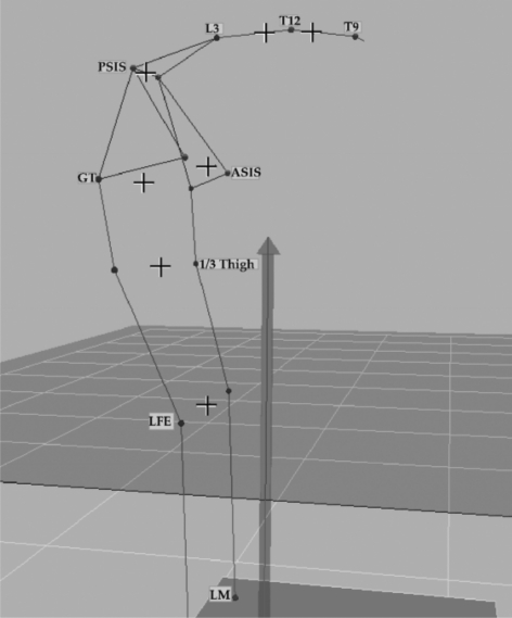
Twelve reflective markers were placed on lower limb, pelvic and spinal landmarks by an experienced musculoskeletal physiotherapist. The landmarks served to define a right-handed local co-ordinate system comprising the T12 and L3 spinous processes, and bilaterally: the anterior and posterior superior iliac spines (ASIS, PSIS), greater trochanters (GT), lateral femoral epicondyles (LFE). Another point was located at one-third of the distance and in line between the GT and LFE (1/3 thigh) (Fig. 1).
Fig. 1.
Captured image from motion analysis system at the end of flexion showing reflective surface skin markers and virtual markers used to define the lumbar and hip angles. Abbreviations: LM lateral malleolus, LFE lateral femoral epicondyle, 1/3 Thigh (point taken 1/3 of the distance between the greater trochanter and the lateral femoral epicondyle), GT greater trochanter, ASIS anterior superior iliac spine, PSIS posterior superior iliac spine, L3 spinous process of 3rd lumbar vertebra, T9 and T12 spinous processes of 9th and 12th thoracic vertebrae. Note + represents virtual markers
The hip angle was constructed by the angular displacement of two lines relative to each other and was used as an indicator of pelvic movement in relation to the femur. A line for the hip angle was created from virtual markers between the two ASISs and PSISs with a second line taken from virtual markers between the midpoint of the two LFEs and the corresponding 1/3 thigh markers (Fig. 1). The lumbar spine angle was taken between a line through the virtual markers T11 and L1 and another line through virtual markers of the two ASISs and PSISs (Fig. 1).
Following collection of the anthropometric measurements, a standardised preparatory session of walking on a treadmill for 5 min at their own pace was undertaken by each participant. Following a demonstration of the required movement tasks, participants undertook the task of flexion (and return) and extension (and return) (performed in a randomised order). They were instructed to complete each of these sequences in their own comfortable pace within a 10 s timeframe. In order to standardise foot placement for the recording session, the 2 ft were placed at a distance of one-tenth the participant’s body height [4]. At the sound of an audible signal the participant was instructed to bend forward as far as possible without bending their knees while holding the maximum achievable flexed position for a count of two before returning to their upright position. The movement sequence was randomised between flexion (and return) and extension (and return) and five trials of each movement was recorded for each participant.
In the post-processing collection of data the individual reflective marker paths of the recorded motion sequences were observed for possible error before smoothing with a 6 Hz Butterworth filter. The kinematic data describing the flexion and return cycles for each of the five trials were normalized to 100 data points with respect to time then transferred to a Microsoft Excel (Version 2005) spreadsheet for statistical analysis.
The data for each participant were averaged and pooled for analysis. The kinematic variables describing the movement in the sagittal plane during flexion and return motion of the hip and lumbar spine were expressed in terms of angular displacement (deg) and angular velocity (deg/s) (±95% confidence intervals). The time taken for the hip and lumbar spine during flexion and return was expressed as a percentage of the total movement task time. The mean angular displacement and velocity patterns were presented graphically and descriptively. Paired t tests were used to determine significant differences (P < 0.05) between the lumbar and hip variables during flexion and return with respect to movement initiation, time to reach peak velocities and peak angular displacement
Results
The results of all the kinematic displacements and temporal variables are given in Table 1. The absolute mean maximum angular displacement of the hip (69.2 ± 10.3°) during flexion and return cycle was significantly greater (P < 0.001, mean difference = 13.3°) than that of the lumbar spine (55.9 ± 5.8°) (Table 1).
Table 1.
Summary of the kinematic and temporal variables during flexion and return (n = 20) derived from absolute mean peak values
| Lumbar | SD | Hip | SD | Mean difference | 95% CI | P value | |
|---|---|---|---|---|---|---|---|
| Kinematic variables | |||||||
| Peak angular displacement (deg) | 55.9 | 5.8 | 69.2 | 10.3 | 13.3 | 6.1 to 20.5 | 0.001** |
| Peak flexion velocity (deg/s) | 43.5 | 7.2 | 49.0 | 11.7 | 5.5 | −0.5 to 11.7 | 0.071 |
| Peak return velocity (deg/s) | −36.7 | 8.3 | −43.4 | 12.7 | 6.7 | 2.7 to 12.8 | 0.002* |
| Temporal variables | |||||||
| Time to reach flexion initiation (%) | 16.0 | 3.5 | 25.9 | 4.3 | 9.9 | 7.7 to 12.1 | 0.001** |
| Time to reach flexion peak velocity (%) | 43.0 | 7.4 | 56.3 | 4.8 | 13.3 | 8.9 to 17.7 | 0.001** |
| Time to reach return initiation (%) | 12.1 | 6.1 | 7.4 | 2.2 | 4.7 | 1.4 to 8.0 | 0.009* |
| Time to reach return peak velocity (%) | 41.9 | 6.3 | 36.7 | 7.6 | 5.2 | 1.6 to 8.8 | 0.007* |
SD standard deviation, CI confidence interval
*P < 0.05, **P < 0.001
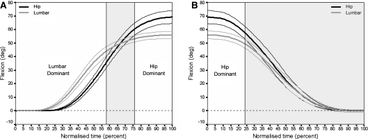
Analysis of the group mean sagittal angular displacement values and its respective CI of the lumbar spine and hip during forward flexion cycle (Fig. 2a) showed that the lumbar spine dominated the movement between 22 and 58% of the movement with both the lumbar spine and hip contributing equally between 59 and 76% of the cycle. The hip was found to dominate the final 24% of the forward flexion cycle.
Fig. 2.
Group mean sagittal angular displacement profiles (deg ±95% confidence intervals) about the lumbar spine and hip during a flexion from and b return to normal stance (n = 20)
The CI of mean sagittal angular displacement values revealed that the hip dominated the initial 22% of the return movement with both the hip and lumbar spine contributing equally to the remaining part of the cycle. (Fig. 2b).
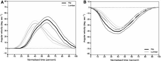
The group mean sagittal angular velocity profiles about the lumbar spine and hip for flexion and return are given in Fig. 3a, b.
Fig. 3.
Group mean sagittal angular velocity profiles (deg/s ±95% confidence intervals) about the lumbar spine and hip during a flexion from and b return to normal stance (n = 20)
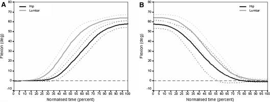
Further analysis of each subject data revealed a sub-group of four subjects did not exhibit the group mean movement pattern described above (Fig. 4). These four subjects exhibited a pattern of lumbar dominance during flexion and return with the absolute mean maximum angular displacement value being greater for the lumbar spine (64.0 ± 3.4°) than the hip (57.8 ± 3.9°).
Fig. 4.
Sub-group mean sagittal angular displacement (deg. ±95% confidence intervals) about the lumbar spine and hip during a flexion from and b return to normal stance (n = 4)
Forward cycle
The point of initiation was determined by a 0.5° change in the angular displacement values of the hip and lumbar spine during the flexion and return movement sequences. Absolute mean angular velocity values were examined with respect to the percentage of task time and it was found that flexion was initiated by the lumbar spine (16.0 ± 3.5%) significantly earlier (P < 0.001) than the hip (25.9 ± 4.4%). Absolute mean peak velocity at the lumbar spine (43.0 ± 7.4%) occurred significantly earlier in the flexion phase than the hip (56.3 ± 4.8%) No statistical differences were observed (Table 1) between the absolute mean peak angular velocity values in forward flexion between the lumbar spine (43.5 ± 7.2 deg/s) and the hip (49.0 ± 11.7 deg/s).
Return cycle
Statistical differences (P < 0.009) were observed in time taken by the hip to initiate the movement during return flexion (7.4 ± 2.2%) compared with the lumbar spine (12.1 ± 6.1%; Table 1). Absolute mean peak velocity at the hip (36.7 ± 7.6%) occurred significantly earlier in the flexion phase than the lumbar spine (41.9 ± 6.3%) (Table 1). Statistically significant differences were found in the absolute mean peak angular velocity values (P < 0.002) in the lumbar spine (36.7 ± 8.3 deg/s) compared with that of the hip (43.4 ± 12.7 deg/s; Table 1).
Discussion
The results of this study show that the flexion and return motion of young healthy males are distinguished from each other on the basis of kinematic and temporal changes of the hip and lumbar spine during these respective movements. The limitations of the study such as the possibility of diurnal variation and the restriction of the study to a male population when considering these results need to be borne in mind. While other investigators [2] have failed to show any gender differences between the movement patterns the small sample size in this current study precludes generalisation of the results to a larger population. However steps were taken to ensure that the procedure was tightly monitored by performing randomised extension and flexion in order to minimise the cumulative effect of the repeated trials of forward bending. Further, the study participants were all active in sports and therefore, the sample population in this study is considered to be representative of an asymptomatic group of young males.
Possible reasons for the differences in the observed movement patterns are not totally clear and warrant further investigation. As all participants were male the influence of gender can be eliminated, but it is possible other factors such as sporting activities, anthropometric influences such as relative leg and spine length and pelvic angulation may be contributing to these patterns.
The patterns of movement contribution from the lumbar spine and hip were identified as being different in forward flexion and return. In flexion, the initial (58%) movement was dominated by the lumbar spine followed by a brief period (18%) whereby there was an equal contribution from the lumbar spine and hip. The final part (24%) of forward flexion was hip dominant (Fig. 2a). The hip dominated the first part of the return movement pattern (22%) with an equal contribution from the hip and lumbar spine throughout the remaining part of the movement (Fig. 2b). It is reasonable to suggest that the two movements place different demands on the motor control mechanisms hence leading to the patterns of contribution from the lumbar spine and hip that were observed in this study.
The results on the relative contribution of the two regions in flexion are consistent with other investigators who have examined this issue [2]. However whereas McClure et al. [14] consider the return pattern to replicate the flexion pattern the results of this study indicated that this was not the case with predominately, an equal contribution from the lumbar spine and hip in the return movement pattern.
Although it has previously been observed that LBP subjects exhibit different movement patterns however it is a new observation that healthy subjects also show a similar trend. The relative contribution of the hip and lumbar spine during flexion and return is that four of the participants in this study did not conform to the pattern of angular change described above. Instead these individuals presented with a completely lumbar dominant pattern throughout the two pathways of movement (Fig. 4a, b) and one that was not explained by the participant’s anthropometric characteristics. This pattern is worthy of note as it may represent a variation within a normal population which has previously been overlooked and warrants further investigation.
In terms of movement initiation, some authors have suggested that the pelvis initiates movement during flexion [20] but the results of this study and, in keeping with those of other investigators [2–3, 6, 13, 16, 19, 22] indicate that flexion is initiated by the lumbar spine and, in the return motion, the hip [14].
Examination of the velocity patterns of the lumbar spine and hip during flexion and return also revealed differences between the two movements. In the forward flexion movement the lumbar spine reached peak velocity before that of the hip. Conversely, in the return movement, the hip reached peak velocity earlier than that of the lumbar spine. Previous studies have reported absolute mean peak lumbar velocities during flexion between 38.9 deg/s [2], 45 deg/s [12] and 61 deg/s [16] so the values of 43.5 deg/s obtained in this investigation lie within these previously reported values. The absolute mean peak velocities for the hip (49.0 deg/s) in this study are also close to the values reported by Esola et al. [2] (53.6 deg/s) and Paquet et al. [16] (39.0 deg/s).
The absolute mean peak velocity of the lumbar spine during the return movement in the study by McClure et al. [14] was 30.5 deg/s and by Paquet et al. [16] was 58.0 deg/s while in this study it was 36.7 deg/s. In the hip, the absolute mean peak velocity during return movement reported by McClure et al. [14] was 49.7 deg/s and by Paquet at al. [16] was 39 deg/s compared with 43.4 deg/s obtained in this study.
The differences between the velocity values detailed above may be explained by inconsistencies between studies in the placement of skin markers used to define the lumbar and hip angles. However the patterns rather than actual values would seem to be the feature of note in all of these studies and would seem to be reflective of the patterns of angular change.
While some investigators report differences between peak lumbar velocities during flexion in symptomatic and asymptomatic subjects with low back pain [12, 21] other researchers have failed to do so [2]. For example, Paquet et al. [16] reported significant differences between the velocities of the hip and lumbar spine among acute LBP and asymptomatic subjects during both flexion and return movements. On the other hand, McClure et al. [14] found no such differences between the hip mean peak velocity of symptomatic and asymptomatic subjects during return flexion. In the former study patients with resolved back pain were used and in the latter one, the symptomatic group comprised acute low back pain patients indicating that the stage of low back pain has an important impact on the findings.
The clinical relevance of this study is two fold. Firstly, the described relative displacement and velocity patterns and the discrete temporal, displacement, and velocity data are considered to be important normative kinematic parameters that may ultimately be used for judging the effects of low back disorder on movement. Secondly, it may also be that individuals with certain hip and lumbar spine movement patterns ultimately prove to be more predisposed to low back pain. However, the result of this study suggests that it is normal for some individuals to move more in the lumbar spine than the hip during the forward flexion task. While the current data have helped in the development of a normative database the information is still limited to a small sample of young asymptomatic males. A considerable amount of further data are required to build up a representative asymptomatic population sample that includes both sexes and various age groups. Such a comprehensive database may become a strong comparative and analytical tool for understanding and managing aberrant kinematics in low back disorders. While velocity may also prove to be an important indicator in back pain populations comparisons between studies is made difficult by inconsistencies in data analysis such as plotting lumbar to hip ratios against range of motion. Our recommendation for future studies on low back pain populations is to plot velocity against time taken for the subject to complete the flexion and return task to enable better comparisons between studies and to gain insight into these issues.
Conclusion
Further to the previous work carried out on the motion characteristics of the lumbar spine and hip during flexion and return this study confirms the existence of kinematic and temporal differences between the two regions when performing this movement. However, the results of this study suggest that hip dominant or lumbar dominant patterns which are found during flexion and return are not the same for all individuals, even in a healthy population. Further work is needed to examine these characteristics longitudinally in larger population samples to confirm the stability of these observations.
Acknowledgments
We thank Dr. Allan B. Carman, Mr Bruce Knox, School of Physiotherapy, University of Otago for their technical assistance.
Footnotes
Motion Analysis™ Motion Analysis Corporation, CA, USA
References
- 1.Burton AK, Tillotson KM, Troup JDG. Variation in the lumbar sagittal mobility with low-back trouble. Spine. 1989;14:584–590. doi: 10.1097/00007632-198906000-00007. [DOI] [PubMed] [Google Scholar]
- 2.Esola MA, McClure PW, Fitzgerald GK, Siegler S. Analysis of lumbar spine and hip motion during forward bending in subjects with and without a history of low back pain. Spine. 1996;21:71–78. doi: 10.1097/00007632-199601010-00017. [DOI] [PubMed] [Google Scholar]
- 3.Farfan HF. Muscular mechanism of the lumbar spine and the position of power and efficiency. Orthop Clin N Am. 1975;6:135–144. [PubMed] [Google Scholar]
- 4.Jayaraman G, Nazre AA, McCann V, Redford JB. A computerized technique for analysing lateral bending behaviour of subjects with normal and impaired lumbar spine. A pilot study. Spine. 1994;19:824–832. doi: 10.1097/00007632-199404000-00017. [DOI] [PubMed] [Google Scholar]
- 5.Keller TS, Szpalski M, Spengler DM, Hayez JP. Interpretation and parameterization of dynamic trunk isoinertial movements using an ensemble-averaging technique. Clin Biomech. 1993;8:220–222. doi: 10.1016/0268-0033(93)90018-D. [DOI] [PubMed] [Google Scholar]
- 6.Kippers V, Parker AW. Posture related to myoelectric silence of erectores spinae during trunk flexion. Spine. 1984;9:740–745. doi: 10.1097/00007632-198410000-00015. [DOI] [PubMed] [Google Scholar]
- 7.Lee RYW, Wong TKT. Relationship between the movements of the lumbar spine and hip. Hum Mov Sci. 2002;21:481–494. doi: 10.1016/S0167-9457(02)00117-3. [DOI] [PubMed] [Google Scholar]
- 8.Lund T, Nydegger T, Schlenzka D, Oxland TR. Three-dimensional motion patterns during active bending in patients with chronic low back pain. Spine. 2002;27:1865–1874. doi: 10.1097/00007632-200209010-00012. [DOI] [PubMed] [Google Scholar]
- 9.Marras WS, Ferguson SA, Gupta P, Bose S, Parnianpour M, Kim JY, Crowell RR. The quantification of low back disorder using motion measures: methodology and validation. Spine. 1999;24:2091–2100. doi: 10.1097/00007632-199910150-00005. [DOI] [PubMed] [Google Scholar]
- 10.Marras WS, Parnianpour M, Ferugson SA, Kim JY, Crowell RR, Simon SR. Quantification and classification of low back disorders based on trunk motion. Eur J Phys Med. 1993;3:218–235. [Google Scholar]
- 11.Marras WS, Parnianpour M, Ferguson SA, Kim JY, Crowell RR, Bose S, Simon SR. The classification of anatomic- and symptom-based low back disorders using motion measure models. Spine. 1995;20:2531–2546. doi: 10.1097/00007632-199512000-00013. [DOI] [PubMed] [Google Scholar]
- 12.Marras WS, Wongsam PE. Flexibility and velocity of the normal and impaired lumbar spine. Arch Phys Med Rehab. 1986;67:213–217. [PubMed] [Google Scholar]
- 13.Mayer TG, Smith SS, Keeley J, Mooney V. Quantification of lumbar function: part 2. Sagittal plane trunk strength in chronic low back pain patients. Spine. 1985;10:765–772. doi: 10.1097/00007632-198510000-00012. [DOI] [PubMed] [Google Scholar]
- 14.McClure PW, Esola M, Schreier R, Siegler S. Kinematics analysis of lumbar and hip motion while rising from a forward flexed position in patients with and without a history a low back pain. Spine. 1997;22:552–558. doi: 10.1097/00007632-199703010-00019. [DOI] [PubMed] [Google Scholar]
- 15.Nelson JM, Walmsey RP, Stevenson JM. Relative lumbar and pelvic motion during loaded spinal flexion/extension. Spine. 1995;20:199–204. doi: 10.1097/00007632-199501150-00013. [DOI] [PubMed] [Google Scholar]
- 16.Paquet N, Malouin F, Richards CL. Hip-spine movement interaction and muscle activation patterns during sagittal trunk movements in low back pain patients. Spine. 1994;19:596–603. doi: 10.1097/00007632-199403000-00016. [DOI] [PubMed] [Google Scholar]
- 17.Pearcy M, Portek I, Shepherd J. The effect of low back pain on lumbar spinal movements measured by three-dimensional X-ray analysis. Spine. 1985;10:150–153. doi: 10.1097/00007632-198503000-00007. [DOI] [PubMed] [Google Scholar]
- 18.Pearcy M, Shepherd J. Is there instability in spondylolisthesis? Spine. 1985;10:175–177. doi: 10.1097/00007632-198503000-00014. [DOI] [PubMed] [Google Scholar]
- 19.Porter JL, Wilkinson A. Lumbar-hip flexion motion. A comparative study between asymptomatic and chronic low back pain in 18- to 36-year-old men. Spine. 1997;22:1508–1514. doi: 10.1097/00007632-199707010-00017. [DOI] [PubMed] [Google Scholar]
- 20.Sahrmann SA. Diagnosis and treatment of movement impairment syndromes. St. Louis: Mosby; 2002. [DOI] [PMC free article] [PubMed] [Google Scholar]
- 21.Shum GL, Crosbie J, Lee RY. Symptomatic and asymptomatic movement coordination of the lumbar spine and hip during an everyday activity. Spine. 2005;30:E697–E702. doi: 10.1097/01.brs.0000188255.10759.7a. [DOI] [PubMed] [Google Scholar]
- 22.White AA, Panjabi MM. Clinical biomechanics of the spine. 2. Philadelphia: J.B. Lippincott Company; 1990. [Google Scholar]
- 23.Wong TK, Lee RY. Effects of low back pain on the relationship between the movements of the lumbar spine and hip. Hum Mov Sci. 2004;23:21–34. doi: 10.1016/j.humov.2004.03.004. [DOI] [PubMed] [Google Scholar]


