

Abstract
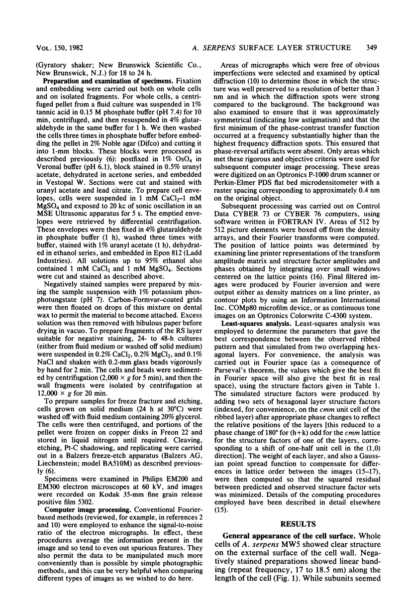
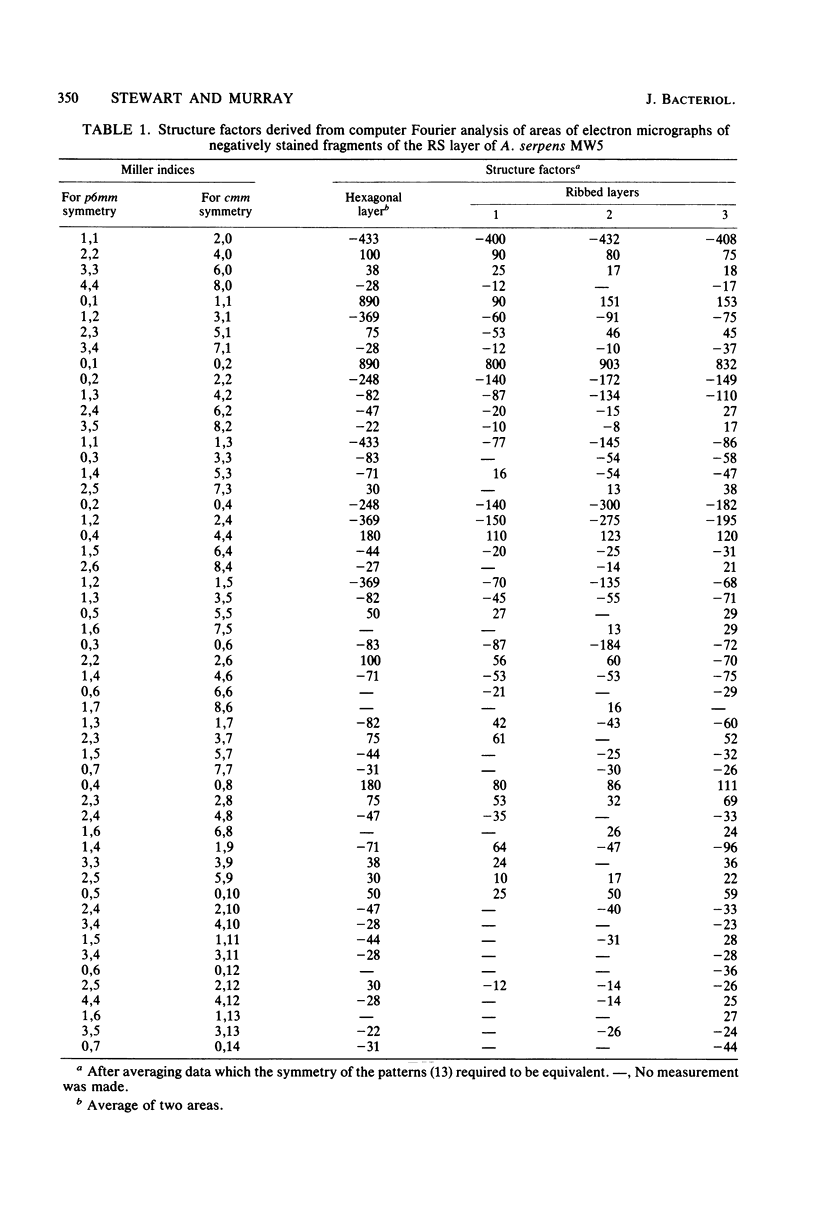
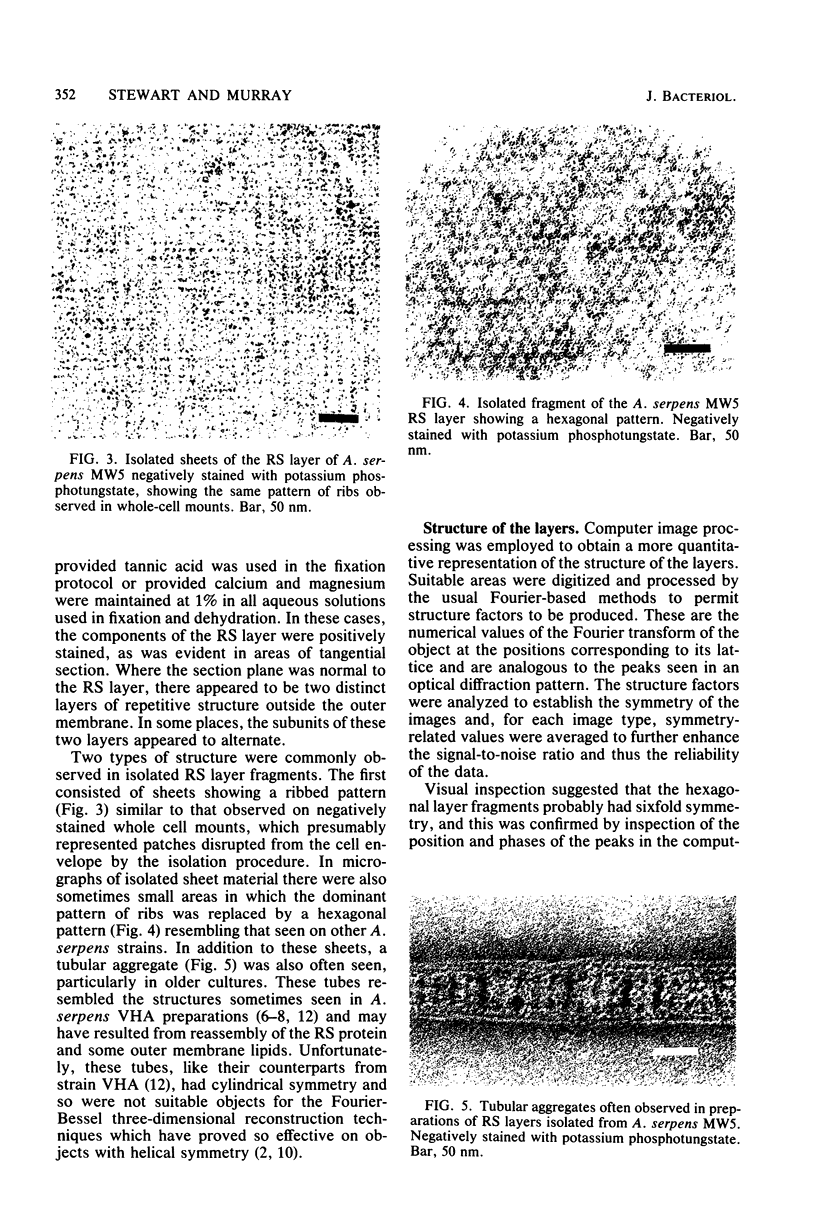
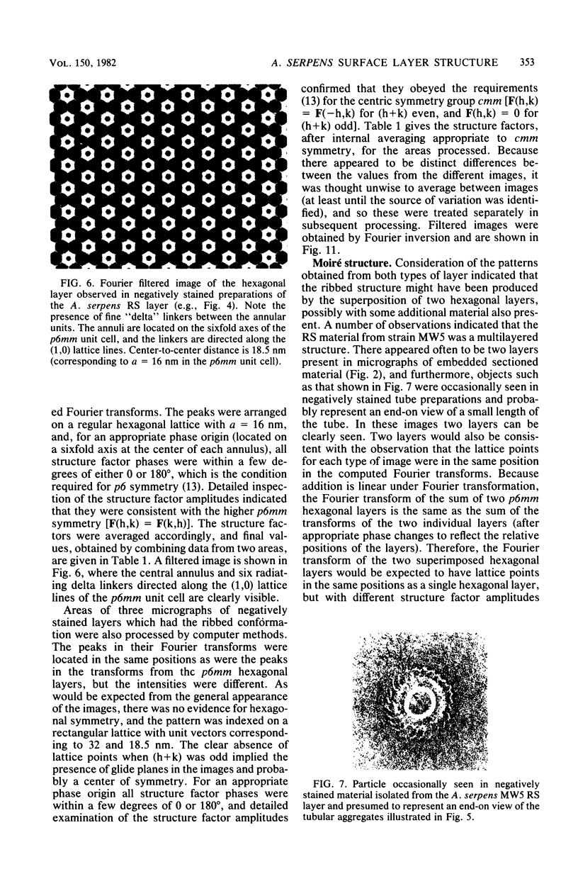
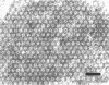
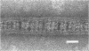
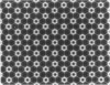
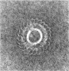
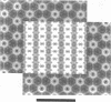
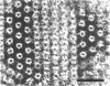
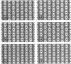
The structure of the regular surface layer of Aquaspirillum serpens MW5 has been investigated by electon microscopy supplemented by computer image processing and least-squares analysis. The layer has a ribbed appearance, both on the bacterium and in isolated, negatively stained fragments. However, detailed analysis indicated that the layer was composed of two hexagonal sheets having p6mm symmetry and a = 16 nm. One sheet was staggered by one half repeat along a (1,0) line of the p6nm lattice relative to the second so that, in projection, the pattern of the composite layer was a translational moiré, characterized by a series of ribs spaced 16 nm apart. The ribbed layer had cmm symmetry with a = 32 nm and b = 18.5 nm. Analysis of this pattern indicated that the two p6nm hexagonal sheets were unevenly stained, and this was confirmed by using least-squares methods to simulate the observed pattern by combining two hexagonal patterns. The general structure of the layer was consistent with a role as a selective and protective barrier on the cell surface.
Full text
PDF
Images in this article

Selected References
These references are in PubMed. This may not be the complete list of references from this article.
- Aebi U., Smith P. R., Dubochet J., Henry C., Kellenberger E. A study of the structure of the T-layer of Bacillus brevis. J Supramol Struct. 1973;1(6):498–522. doi: 10.1002/jss.400010606. [DOI] [PubMed] [Google Scholar]
- Amos L. A. Image analysis of macromolecular structures. J Microsc. 1974 Mar;100(2):143–152. doi: 10.1111/j.1365-2818.1974.tb03924.x. [DOI] [PubMed] [Google Scholar]
- Beveridge T. J., Murray R. G. Surface arrays on the cell wall of Spirillum metamorphum. J Bacteriol. 1975 Dec;124(3):1529–1544. doi: 10.1128/jb.124.3.1529-1544.1975. [DOI] [PMC free article] [PubMed] [Google Scholar]
- Beveridge T. J. Ultrastructure, chemistry, and function of the bacterial wall. Int Rev Cytol. 1981;72:229–317. doi: 10.1016/s0074-7696(08)61198-5. [DOI] [PubMed] [Google Scholar]
- Buckmire F. L., Murray R. G. Studies on the cell wall of Spirillum serpens. 1. Isolation and partial purification of the outermost cell wall layer. Can J Microbiol. 1970 Oct;16(10):1011–1022. doi: 10.1139/m70-171. [DOI] [PubMed] [Google Scholar]
- Buckmire F. L., Murray R. G. Studies on the cell wall of Spirillum serpens. II. Chemical characterization of the outer structured layer. Can J Microbiol. 1973 Jan;19(1):59–66. doi: 10.1139/m73-009. [DOI] [PubMed] [Google Scholar]
- Chester I. R., Murray R. G. Protein-lipid-lipopolysaccharide association in the superficial layer of Spirillum serpens cell walls. J Bacteriol. 1978 Feb;133(2):932–941. doi: 10.1128/jb.133.2.932-941.1978. [DOI] [PMC free article] [PubMed] [Google Scholar]
- Crowther R. A., Klug A. Structural analysis of macromolecular assemblies by image reconstruction from electron micrographs. Annu Rev Biochem. 1975;44:161–182. doi: 10.1146/annurev.bi.44.070175.001113. [DOI] [PubMed] [Google Scholar]
- Finch J. T., Klug A., Nermut M. V. The structure of the macromolecular units on the cell walls of Bacillus polymyxa. J Cell Sci. 1967 Dec;2(4):587–590. doi: 10.1242/jcs.2.4.587. [DOI] [PubMed] [Google Scholar]
- Glaeser R. M., Chiu W., Grano D. Structure of the surface layer protein of the outer membrane of Spirillum serpens. J Ultrastruct Res. 1979 Mar;66(3):235–242. doi: 10.1016/s0022-5320(79)90121-7. [DOI] [PubMed] [Google Scholar]
- Sleytr U. B. Regular arrays of macromolecules on bacterial cell walls: structure, chemistry, assembly, and function. Int Rev Cytol. 1978;53:1–62. doi: 10.1016/s0074-7696(08)62240-8. [DOI] [PubMed] [Google Scholar]
- Stewart M., Beveridge T. J., Murray R. G. Structure of the regular surface layer of Spirillum putridiconchylium. J Mol Biol. 1980 Feb 15;137(1):1–8. doi: 10.1016/0022-2836(80)90153-9. [DOI] [PubMed] [Google Scholar]
- Stewart M., Beveridge T. J. Structure of the regular surface layer of Sporosarcina ureae. J Bacteriol. 1980 Apr;142(1):302–309. doi: 10.1128/jb.142.1.302-309.1980. [DOI] [PMC free article] [PubMed] [Google Scholar]
- Stewart M. Structures of alpha-tropomyosin magnesium paracrystals. II. Stimulation of staining patterns from the sequence and some observations on the mechanism of positive staining. J Mol Biol. 1981 Jun 5;148(4):411–425. doi: 10.1016/0022-2836(81)90184-4. [DOI] [PubMed] [Google Scholar]
- Thorne K. J. Regularly arranged protein on the surfaces of Gram-negative bacteria. Biol Rev Camb Philos Soc. 1977 May;52(2):219–234. doi: 10.1111/j.1469-185x.1977.tb01351.x. [DOI] [PubMed] [Google Scholar]