Streptococcus suis is an important cause of meningitis, arthritis and septicaemia, especially in young pigs. Serotype 2 is the most prevalent type in clinical material from pigs in Europe [9] and the infection causes severe disease outbreaks in swine herds. Different experimental models have been used to elucidate the infection but central parts of the pathogenesis still remain unclear [4]. In spontaneous infection, S. suis is generally believed to invade via the upper respiratory tract [4]. Recently, we described an infection model in minipigs using aerogenous challenge and subsequent steroid treatment [5]. In conventional pigs, reports of models attempting aerosol exposure seem limited to a single study using anaesthetized pigs [2].
In order to evaluate the pathogenesis of S. suis type 2 infection in pigs, we aimed at establishing an aerosol model for the infection using unanaesthetized conventionally reared, weaned pigs. The present report describes the microbiological and pathological findings in a pilot study of an aerosol model for S. suis infection in conventional pigs.
Four clinically healthy, 6-week-old, female, Landrace-Yorkshire crossbred pigs (animals A-D) from the same litter (weaned at app. 4 weeks of age) were included in this study. They were obtained from a herd with no history of disease compatible with S. suis infection and S. suis had never been isolated from the herd. For the aerosol exposure, a 1.5 m3 chamber connected to a nebulizing apparatus was used. The chamber and procedure used were essentially as previously described [5]. Briefly, the animals were in the chamber for 20 min while 40 ml of an aquatic solution of 1% acetic acid (pH 3.4) was dispersed and let into the chamber with atmospheric air. Then the pigs were kept outside the chamber for one hour. Thereafter, 3 of the animals (A-C) were brought back into the chamber and 42.5 ml of a bacterial suspension was dispersed over 20 min in the air supply to the chamber. The bacterial suspension was a 5-h broth culture of S. suis serotype 2 (strain P321/6) concentrated to 1.1 × 10¹⁰ colony-forming units/ml. The fourth pig (D) served as a control to evaluate the immediate effect of acetic acid alone. One h after exposure to acetic acid in the chamber, this animal was euthanized by exsanguination after being anaesthetized by intramuscular injection (1 ml/15 kg) of Zoletil® (25 mg/ml zolezepam, 25 mg/ml tiletamin). The 3 remaining animals were housed in a single pen and observed for clinical signs of disease and rectal temperatures were recorded daily. On the fifth day after exposure, animal A was euthanized due to the severity of clinical signs. The 2 remaining pigs were euthanized on the sixth day.
Following euthanasia, all pigs were necropsied and gross lesions were recorded. Tissues for microscopy as well as swabs for microbiological culturing were collected to cover all parts of the respiratory tract and a range of organs known to be affected in S. suis infection [5]. From each animal, samples were taken from gross lesions as well as a standard of 31 tissues for histopathology and 18 tissues for microbiological culture, which was done aerobically at 37°C on 5% calf blood agar. Morphologically suspect colonies were subcultured and identified biochemically and serologically using standard methods. From aseptically sampled blood as well as from areas with a normal bacterial flora, i.e., nasal cavity, pharyngeal and palatine tonsils, microbiological culture was done as previously described [5].
For histopathology, fixation in 4% neutral buffered formaldehyde, decalcification of relevant tissues, as well as processing of HE stained sections were performed as previously described [6]. The presence of S. suis antigen was examined by immunohistochemistry using a previously published protocol [7].
Clinical signs were only observed in pigs A and C with onset on days 3 and 5 after exposure, respectively. These pigs had loss of appetite, elevated body temperatures (40.0–41.5°C), were reluctant to rise and lame in one or more legs. Gross lesions were noted in animal A, in which a fibrinopurulent polyserositis was seen, and in animal C, which had an exudative meningitis and arthritis.
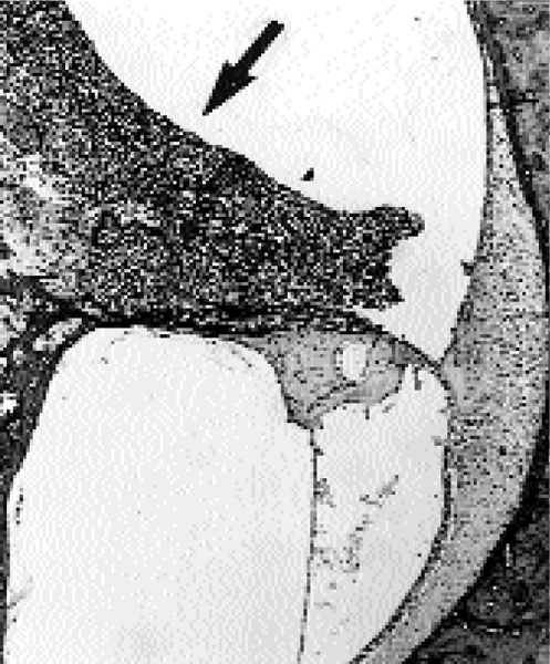
By microscopy, a range of lesions of a generally suppurative nature were noted in the challenged animals (Table 1). Lesions compatible with septicaemia were seen in animal A. In animal C, the histopathological findings included suppurative meningitis, arthritis, and labyrinthitis (Fig. 1). In the third S. suis exposed animal, B, a suppurative otitis media was observed histologically. In the control animal, D, the only changes observed were focal, mild mucopurulent rhinitis and focal, mild, chronic, purulent bronchopneumonia in the right cranial lung lobe.
Table 1.
Distribution of lesions and of S. suis serotype 2 as detected by culture and immunohistochemistry in 3 animals after aerosolic challenge.
| Tissue | Animals positive | Characteristic macroscopic and/or histologic lesions (Animals) | |
| Culture | IHC | ||
| Nasal cavity | C | A,C | Mild, focal mucopurulent rhinitis (A,C) |
| Lung | - | A | Diffuse fibrinopurulent pleuritis (A,B); acute embolic pneumonia (A) |
| Bronchial lnn. | - | A | Follicular hyperplasia and exudation of heterophils (A,B) |
| Middle ear | C | B | Unilateral purulent otitis media and exudate in Eustachian tube (B) |
| Inner ear | n.d. | C | Bilateral purulent labyrinthitis and perineuritis (n. vestibularis) (C) – Fig. 1 |
| Tonsil, pharyngeal | A,C | C | Focal exudation of heterophils (C) |
| Tonsil, soft palate | A,B,C | A,B,C | Heterophils in crypt epithelium and lumen (A,B,C) |
| Brain | C | C | Diffuse fibrinopurulent meningitis with focal submeningeal encephalitis (C) |
| Heart | A | A | Diffuse fibrinopurulent pericarditis (A) |
| Peritoneum | A | A | Diffuse fibrinopurulent peritonitis (A) |
| Mesenteric lnn. | - | A | Follicular hyperplasia and exudation of heterophils (A) |
| Liver | A | A | Multifocal, subcapsular hepatic necrosis and fibrinopurulent perihepatitis (A) |
| Spleen | C | A | - |
| Joints | A,C | A,C | Suppurative arthritis (A,C) |
| Blood | A | n.d. | - |
n.d.: not done; -: negative; IHC: immunohistochemistry for S. suis serotype 2; A,B,C: animal no.
Figure 1.

Inner ear, S. suis infected pig (animal C). Suppurative labyrinthitis with exudate (arrow) in the perilymph of the cochlear scala tympani. HE × 4.
By immunohistochemistry, S. suis serotype 2 antigen was demonstrated in animals A-C in a number of tissues (Table 1). Generally, antigen detection was related to the presence of 1–3 μm structures, resembling coccoid bacteria, located intracellularly or in exudates. Rarely, a more diffuse intracellular immunostaining was observed in phagocytes in inflamed tissues or corresponding lymphoid tissues. S. suis serotype 2 was reisolated from various tissues in animals A, B, and C (Table 1). No bacterial pathogens were isolated from animal D.
The clinical signs as well as the lesions observed in 2 of the 3 animals challenged with S. suis were comparable to spontaneous S. suis serotype 2 infection in pigs [8]. Acute embolic pneumonia was also observed in animal A. Although this observation is not common in S. suis infections, bacterial embolism is not surprising given the bacteraemia present. In animal C, a bilateral labyrinthitis with S. suis antigen present in the exudate was detected. This lesion, which apparently has not been seen previously in experimental pig models of this infection, is a common sequela in porcine meningitis due to spontaneous S. suis serotype 2 infection [7]. Furthermore, hearing loss due to inner ear affection is a major characteristic in humans affected by S. suis meningitis [1]. In animal B, otitis media with S. suis antigen present was observed. As no signs of a systemic infection were observed in this animal, these findings might constitute a local infection after direct spread via the likewise affected Eustachian tube.
In the control animal D, only minor lesions were found. Thus, the observed focal bronchopneumonia is regarded as being insignificant. However, the mucopurulent rhinitis also seen in the infected animals may have been associated with the exposure to acetic acid. This would be in concordance with previous studies, where intranasal instillation of 1% acetic acid was shown to induce loss of cilia and epithelium as well as submucosal oedema and inflammation within 12 h of exposure [3]. However, further studies are required to evaluate the effect of exposure to acetic acid and the interaction with S. suis infection in this model.
In conclusion, by experimental aerosolic exposure, infection with S. suis serotype 2 was established in unanaesthetized conventional pigs, and lesions similar to those seen in spontaneously infected animals were induced.
Acknowledgments
Acknowledgements
We wish to thank Dr. T. Gonzales for her collaboration and appreciate the technical assistance from A. Wamsler-Jensen, L. Johansen, P.R. Mortensen, L. Kioerboe, E. Hjuler and M. Bak. This study was financially supported by the Danish Bacon and Meat Council and by a R N Thomsen grant.
References
- Arends JP, Zanen HC. Meningitis caused by Streptococcus suis in humans. Rev Infect Dis. 1988;10:131–137. doi: 10.1093/clinids/10.1.131. [DOI] [PubMed] [Google Scholar]
- Chengappa MM, Maddux RL, Kadel WL, Greer SC, Herren CE. Proc 29th ann meeting. Amer Assn Vet Lab Diagnosticians, Louisville, Kentucky; 1986. Streptococcus suis infection in pigs: Incidence and experimental reproduction of the syndrome; pp. 25–38. [Google Scholar]
- Gagné S, Martineau-Doizé B. Nasal epithelial changes induced in piglets by acetic acid and by Bordetella bronchiseptica. J Comp Pathol. 1993;109:71–81. doi: 10.1016/s0021-9975(08)80241-1. [DOI] [PubMed] [Google Scholar]
- Gottschalk M, Segura M. The pathogenesis of the meningitis caused by Streptococcus suis: the unresolved questions. Vet Microbiol. 2000;76:259–272. doi: 10.1016/S0378-1135(00)00250-9. [DOI] [PubMed] [Google Scholar]
- Madsen LW, Aalbaek B, Nielsen OL, Jensen HE. Aerogenous infection of microbiologically defined minipigs with Streptococcus suis serotype 2 – a new model. APMIS. 2001. [DOI] [PubMed]
- Madsen LW, Jensen AL, Larsen S. Spontaneous lesions in clinically healthy, microbiologically defined Göttingen minipigs. Scand J Lab Anim Sci. 1998;25:159–166. [Google Scholar]
- Madsen LW, Svensmark B, Elvestad K, Jensen HE. Otitis interna is a frequent sequela to Streptococcus suis meningitis in pigs. Vet Pathol. 2001;38:190–195. doi: 10.1354/vp.38-2-190. [DOI] [PubMed] [Google Scholar]
- Reams RY, Glickman LT, Harrington DD, Thacker HL, Bowersock TL. Streptococcus suis infection in swine: a retrospective study of 256 cases. Part II. Clinical signs, gross and microscopic lesions, and coexisting microorganisms. J Vet Diagn Invest. 1994;6:326–334. doi: 10.1177/104063879400600308. [DOI] [PubMed] [Google Scholar]
- Wisselink HJ, Smith HE, Stockhofe-Zurwieden N, Peperkamp K, Vecht U. Distribution of capsular types and production of muramidase-released protein (MRP) and extracellular factor (EF) of Streptococcus suis strains isolated from diseased pigs in seven European countries. Vet Microbiol. 2000;74:237–248. doi: 10.1016/S0378-1135(00)00188-7. [DOI] [PubMed] [Google Scholar]


