Abstract
Ceramides deriving from sphingomyelin hydrolysis are important mediators of apoptotic signals originating from Fas (APO-1/CD95). However, definitive evidence for the role played by individual sphingomyelinases is still lacking. We have analyzed lymphoblastoid cell lines derived from patients affected by Niemann Pick disease (NPD), an autosomal recessive disorder caused by loss-of-function mutations within the acidic sphingomyelinase (ASM) gene. NPD lymphoblasts, which display normal neutral sphingomyelinase activity, fail to activate ASM in response to Fas cross-linking, unlike normal lymphoblasts. NPD lymphoblasts also fail to accumulate GD3 ganglioside, a downstream mediator of ceramide-induced cell death (De Maria, R., L. Lenti, F. Malisan, F. D'Agostino, B. Tomassini, A. Zeuner, M.R. Rippo, R. Testi. 1997. Science. 277:1652–1655), and display a substantially inefficient apoptosis after Fas cross-linking. Inefficient apoptosis is due to lack of ASM activity, because proximal signaling from Fas in NPD lymphoblasts is not impaired and apoptosis can be efficiently triggered by passing the ASM defect with exogenous ceramides. Moreover, mannose receptor–mediated transfer of ASM into NPD lymphoblasts rescues their ability to transiently activate ASM, accumulate GD3, and rapidly undergo apoptosis after Fas cross-linking. These results provide definitive genetic evidence for the role of ASM in the progression of apoptotic signals originating from Fas.
Cross-linking of the surface receptor Fas (APO-1/ CD95) triggers apoptotic cell death in a variety of cell types (1, 2). Defining the effectors that are involved in the propagation of apoptotic signals originating from Fas bears promises for the possibility to modulate apoptosis in diseases where accelerated Fas-dependent cell death occurs.
Several early events concur in setting the irreversible commitment to cell death after Fas cross-linking. Seconds after receptor engagement, caspase 8 (FLICE/MACH-1/ MCH5), a member of the ICE/CED-3 family of cysteine proteases, is recruited to the receptor “death domain” via the adaptor molecule FADD/MORT1, and is activated (3, 4). Sequential triggering of downstream caspases leads to the hydrolysis of crucial protein substrates within the following hours, contributing to apoptotic cell shape changes (5, 6). Concomitantly, a lipid pathway is initiated by death domain–dependent early caspases (7) and involves the sequential activation of phosphatidylcholine-specific phospholipase C (PC-PLC) and membrane-associated acidic sphingomyelinase (ASM) (8–10). ASM generates transient ceramide accumulation in acidic cellular compartments within minutes. This is followed by enhanced activity of GD3 synthase with accumulation of GD3 ganglioside in the Golgi and early depolarization of the inner mitochondrial membrane within the first 2 h after Fas cross-linking (11).
Mitochondrial damage with induction of membrane permeability transition crucially contributes to the apoptotic program with the release of diffusible factors responsible for the activation of downstream caspases (12–14). More ceramide is then formed by the action of caspase-dependent cytosolic neutral sphingomyelinase (NSM), accumulates beginning within a few hours after Fas cross-linking, and persists throughout the entire apoptotic process (15, 16).
Although ceramides act as important intracellular mediators of apoptotic signals, the relative contribution of individual sphingomyelinases in the apoptotic program is still unclear (17). Here we used lymphoid cell lines derived from patients affected by Niemann Pick disease (NPD) type B, a genetic disorder characterized by deficiency of ASM (18) but not NSM, as a unique model to assess the role of ASM in Fas-mediated apoptosis of human lymphocytes.
Materials and Methods
Cells and Reagents.
EBV-transformed lymphoblasts derived from type B NPD patients (19) or from normal controls were grown in a mixture of RPMI and DMEM media (1:1, vol/vol) supplemented with 15% FCS (Hyclone Laboratories, Hyclone, UT). The parental Chinese hamster ovary (CHO) cell line and the CHO-18 clone overexpressing human ASM were grown in DMEM supplemented with 10% FCS. ASM-enriched supernatant from the CHO-18 line was recovered after 3 d of culture in 35-mm dishes and stored at −20°C until use (20).
Anti-Fas mAb (CH-11 clone) was from Upstate Biotechnology Inc. (Lake Placid, NY). C6-ceramide (N-exanoyl-d-sphyngosine), GD3 ganglioside, and d-mannose-6-phosphate (M6P) were from Sigma Chemical Co. (St. Louis, MO). C2-ceramide and C2-dihydroceramide were from Biomol (Plymouth Meeting, PA). [N-methyl-14C]sphingomyelin and L-3-phosphatidyl[N-methyl-14C]choline,1,2-dipalmitoyl, used for in vitro acidic sphingomyelinase and PC-PLC assays, respectively, were from Amersham International (Little Chalfont, Buckinghamshire, UK). The specific PC-PLC inhibitor D609 (10) was from Kamiya (Thousand Oaks, CA).
Acid and Neutral Sphingomyelinase Assay.
Methyl -14 C–labeled sphingomyelin (Amersham International; specific activity 56 mCi/mmol) was diluted in ethanol containing cold sphingomyelin to a final specific activity of 1 mCi/mmol. The substrate mixture was evaporated, resuspended in ASM or NSM buffer, sonicated to form micelles, and placed on ice until use. Cells were washed twice with ice-cold PBS and dissolved in 0.2% Triton X-100. After 10 min on ice, cell lysates were centrifuged for 5 min at 2,000 rpm, and supernatants were sonicated for 10 min in an ice-cold ultrasound bath. Next, 15 μl of each sample were incubated for 2 h at 37°C with 15 μl of the sphingomyelin mixture resuspended in ASM buffer (250 mM sodium acetate, pH 5.2, and 0.2% Triton X-100), or in NSM buffer (40 mM Hepes, pH 7.4, 5 mM MgCl2, and 0.2% Triton X-100) to determine ASM or NSM activity, respectively. Radioactive phosphocholine (PCho) produced from 14C-labeled sphingomyelin was extracted with 200 μl of chloroform/methanol (1:1 vol/vol) and 90 μl of water. The amount of [14C]PCho produced was quantified by liquid scintillation counting. ASM and NSM activities were expressed as pmol of SM hydrolysed by 106 cells per hour.
Measurement of PC-PLC Activity.
Aliquots of 107 cells were resuspended in 1 ml RPMI medium supplemented with 10% FCS, pretreated for 30 min with zVAD-fmk, and stimulated for the indicated times with anti-Fas mAb (500 ng/ml). Stimulation was stopped by immersion of samples in methanol and dry ice (−70°C) for 10 sec followed by centrifugation at 4°C. Pellets were dissolved in 300 μl of 0.01% Triton X-100 and then were sonicated. 16 μl of lysate were incubated at 37°C for 2 h in PC-PLC buffer (50 mM Tris-HCl, pH 7.3, 6.3 mM CaCl2, 0.13 μg/ ml BSA, 0.15 M ammonium sulfate, and l-3-phosphatidyl[N-methyl-14C]choline, 50 nCi) in 80 μl of total volume. The reaction was stopped and the lipids were extracted by adding CHCl3: CH3OH (1:2, vol/vol; 180 μl), 0.9% NaCl (60 μl), and CHCl3 (60 μl). The aqueous and organic phases containing [14C]PCho and [14C]PC, respectively, were separated and quantitated by liquid scintillation. PC-PLC activation was expressed as picomoles of PCho released per 107 cells.
Immunofluorescence, DNA Labeling, and Flow Cytometry Analysis.
For surface staining, cells were incubated for 30 min at 4°C with saturating amounts of PE-conjugated control IgG1 or anti-Fas antibody (DX2; PharMingen, San Diego, CA), and analyzed by flow cytometry. The cell pellet was gently resuspended in hypotonic fluorochrome solution (propidium iodide 50 μg/ml [Sigma Chemical Co.] in 0.1% sodium citrate plus 0.1% Triton X-100), kept overnight at 4°C in the dark until flow cytometry analysis. Percentage of apoptotic cells was determined by evaluating the number of hypodiploid nuclei.
Detection of GD3 Ganglioside Accumulation.
Cells were suspended in ice-cold deionized water, disrupted by two cycles of freezing and thawing, and extracted twice in water/methanol/chloroform (3: 8:4 vol/vol/vol). Samples were then extracted from the upper-phase of the partition and dried by a rotary evaporator, and salts and other nonlipid contaminants were removed by chromatography over Supelclean L-C18 columns (Supelco, Bellefonte, PA). The eluted gangliosides were spotted on aluminium-backed silica gel 60 high performance thin layer chromatography (HPTLC) plates (Merck, Darmstadt, Germany), developed in chloroform/ methanol/0.25% aqueous KCl (5:4:1 vol/vol/vol). After warm air drying, plates were treated with 0.5% polyisobutylmetacrylate in hexane for 90 s, incubated for 10 min with 2% BSA in PBS and for 1 h with R24 anti-GD3 antibody (mouse monoclonal IgG3, provided by Dr. L.J. Old, Ludwig Institute, New York), and washed three times. Then plates were treated with horseradish peroxidase–conjugated goat anti–mouse IgG, and GD3 was visualized by immunoperoxidase staining.
Results and Discussion
The ASM Defect Impairs the Efficiency of Fas-induced Apoptosis.
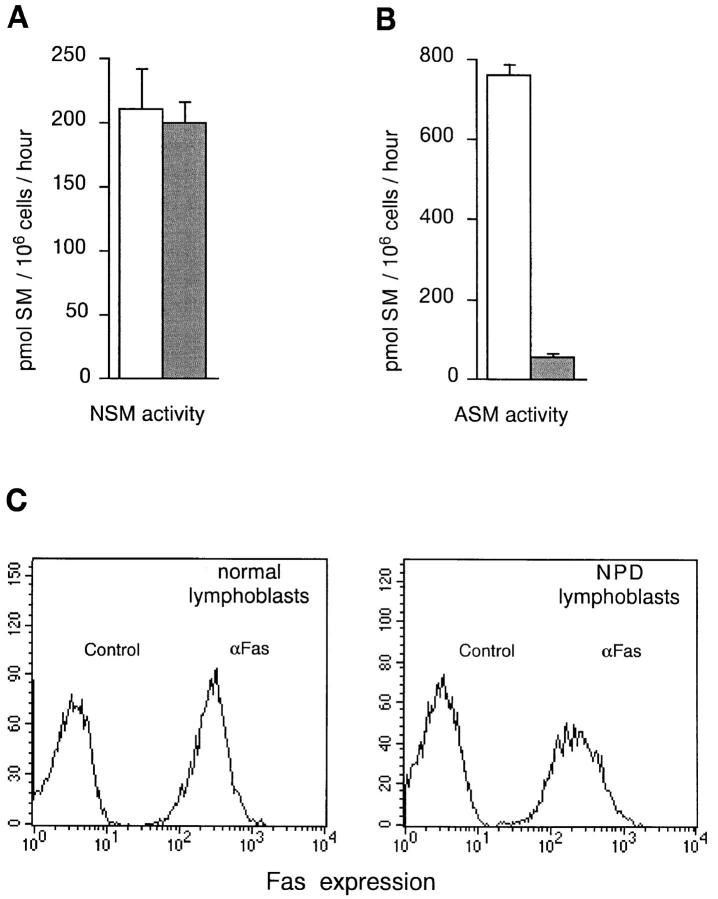
Lymphoblastoid cell lines, derived from peripheral blood lymphocytes of patients affected by NPD, were assessed for basal levels of both NSM and ASM. As shown in Fig. 1, A and B, NPD lymphoblasts and lymphoblasts derived from normal donors display comparable NSM activity, but ASM activity in NPD lymphoblasts is less than 10% of normal levels. Expression of Fas at the cell surface is similar between NPD and normal lymphoblasts (Fig. 1 C). This cellular system therefore represents a powerful model in which to assess the relevance of ASM in Fas-mediated apoptosis of human lymphoid cells.
Figure 1.
NPD lymphoblasts have normal Fas expression and NSM activity, but display a very low ASM activity. Cell lysates from normal (open columns) and NPD (shaded columns) lymphoblasts were incubated with radioactive SM in the presence of NSM (A) or ASM (B) reaction buffer. Data are expressed as mean ± SD from five different experiments. (C) Flow cytometry analysis of normal and NPD lymphoblasts stained with anti-Fas PE or with control isotype-matched IgG. Two NPD and three normal lymphoblastoid cell lines gave comparable results.
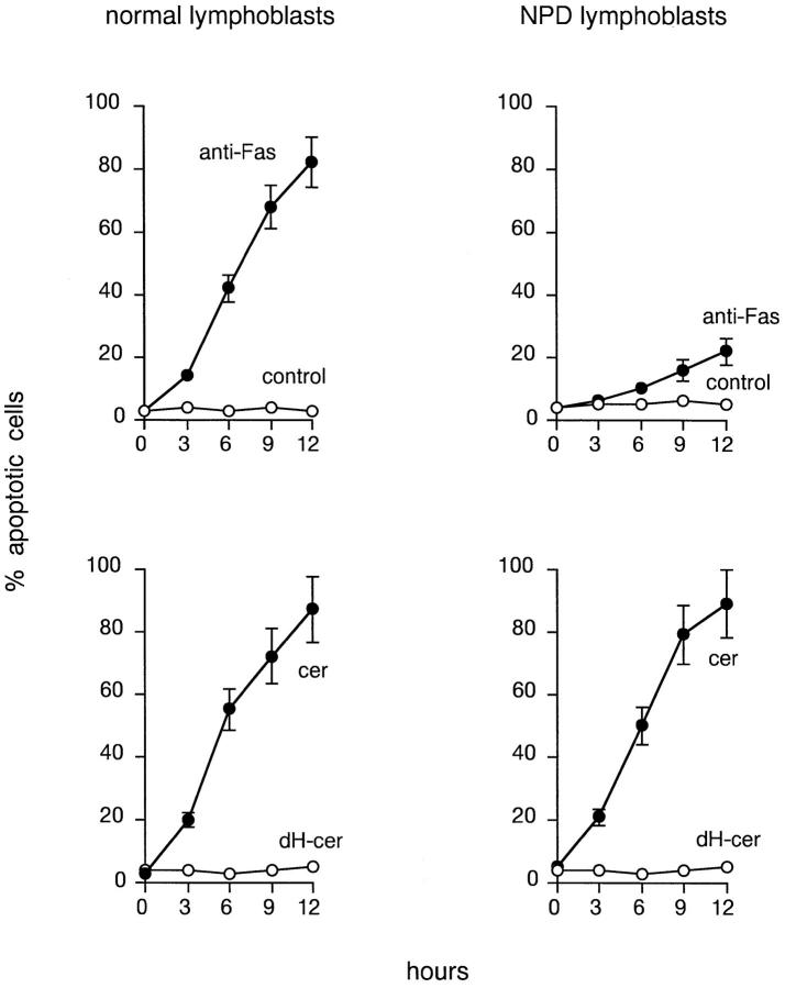
The ability of anti-Fas mAbs to induce apoptosis in NPD lymphoblasts was investigated. Fig. 2 shows that cross-linking of Fas induces apoptosis very inefficiently in NPD lymphoblasts. In fact, the kinetics of both cytosolic changes (data not shown) and nuclear DNA loss (Fig. 2, top) is dramatically delayed compared to normal lymphoblasts. Mitochondrial changes are also delayed in NPD lymphoblasts after Fas cross-linking (Rippo, M.R., et al., manuscript submitted for publication). In contrast, cell permeable C2-ceramide rapidly induces apoptosis in NPD lymphoblasts (Fig. 2, bottom), suggesting that bypassing the genetic defect with exogenous ceramides is sufficient to effectively trigger the apoptotic program.
Figure 2.
Sensitivity to Fas stimulation and exposure to synthetic ceramides. Normal and NPD lymphoblasts were incubated with 200 ng/ml of anti-Fas or control IgM (top), or with 25 μM C2-ceramide or C2-dihydroceramide (bottom). At different time points, the number of apoptotic cells was evaluated by propidium iodide staining and flow cytometry analysis. Results are expressed as mean ± SD of six different experiments.
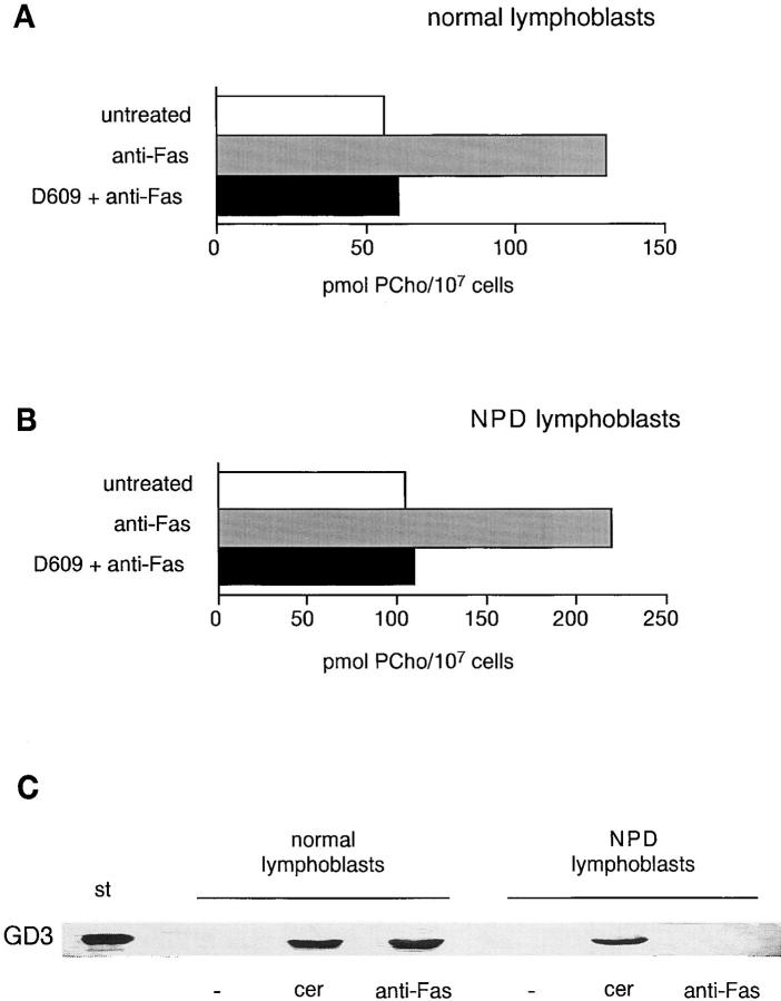
The inefficiency of Fas cross-linking to induce apoptosis in NPD lymphoblasts could be due to impaired signal generation. To rule out this possibility, we investigated the ability of Fas-derived signals to induce PC-PLC activity, as this is immediately upstream from and required for ASM triggering. Fas cross-linking resulted in levels of PC-PLC induction comparable to those observed in normal lymphoblasts (Fig. 3, A and B), suggesting that early signal generation from Fas occurs correctly in ASM-deficient cells.
Figure 3.
Fas stimulation in NPD lymphoblasts efficiently induces PC-PLC activity, but fails to generate GD3 ganglioside accumulation. Cell extracts from 107 normal (A) and NPD (B) lymphoblasts treated for 15 min with control IgM or 500 ng/ml anti-Fas mAb, preceded or not by preincubation with 50 μg/ml D609 for 30 min, were reacted with radiolabeled PC, and the released PCho was quantitated. (C) 107 normal and NPD lymphoblasts were left untreated or were stimulated with 100 μM C6-ceramide (cer) or 500 ng/ml anti-Fas for 5 or 15 min, respectively. Then polar lipids were extracted and run on HPTLC plates along with GD3 standard (st), and GD3 was detected by anti-GD3 immunostaining. Four experiments gave comparable results.
We recently showed that the synthesis and transient accumulation of GD3 ganglioside is required for the progression of apoptotic signals induced by Fas cross-linking and ceramides (11). The ability of Fas-delivered signals to induce GD3 accumulation was therefore investigated in NPD lymphoblasts. Fig. 3 C shows that, unlike normal lymphoblasts, NPD lymphoblasts are unable to induce GD3 accumulation after Fas cross-linking. They can, however, induce GD3 by direct exposure to exogenous ceramides. Together, these data indicated that GD3 accumulation that follows Fas cross-linking requires ceramides derived from the ASM, and strongly suggested that the ASM defect is responsible for the inability to accumulate GD3 and undergo apoptosis efficiently after Fas cross-linking.
Restoring the ASM Defect Reinstates an Efficient Apoptotic Program.
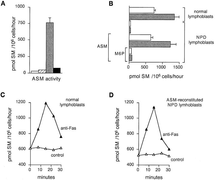
To prove that the ASM defect was indeed responsible for the inefficient apoptotic program, we attempted to reconstitute both basal and inducible ASM activities in NPD lymphoblasts. To accomplish this aim we took advantage of a clone of CHO cells stably expressing a secreted functional ASM (20). CHO-18 cells secrete ASM in the supernatant, which is therefore enriched in functional ASM. NPD lymphoblasts cultured for 24 h in the presence of 25% CHO-18 culture supernatant, but not wild-type CHO culture supernatant, could essentially recover a “normal” basal ASM activity (Fig. 4 A). NPD lymphoblasts internalize ASM through M6P receptor–mediated endocytosis, since reconstitution is completely inhibited by excess M6P (Fig. 4 A). ASM-treated NPD lymphoblasts have basal ASM levels comparable to those of normal lymphoblasts, and can also respond to Fas cross-linking with an ∼100% increase in ASM activity, as do normal lymphoblasts (Fig. 4 B). No induction of ASM activity in response to Fas cross-linking can be measured in untreated NPD lymphoblasts or in NPD lymphoblasts treated with ASM in the presence of excess M6P (Fig. 4 B). Also, the kinetics of ASM induction in response to Fas cross-linking in reconstituted NPD lymphoblasts is comparable to that one observed in normal lymphoblasts (Fig. 4 C).
Figure 4.
Exposure to exogenous ASM reconstitutes ASM activity in NPD lymphoblasts. (A) ASM activity in NPD lymphoblasts cultivated in complete medium alone (open columns), medium containing 25% of parental CHO cell supernatant (hatched columns), 25% CHO-18 supernatant (shaded columns), or 25% CHO-18 supernatant in the presence of 3 mM M6P (closed columns). (B) ASM activity in normal or NPD lymphoblasts treated for 15 min with control IgM (open bars) or 500 ng/ml anti-Fas (shaded bars). NPD cells were also preincubated for 24 h with 25% CHO-18 supernatant in the absence or in the presence of 3 mM M6P. Results represent mean ± SD from four different experiments. Time course of ASM activity in control (open triangles), anti-Fas stimulated (closed triangles) normal (C) or ASM-reconstituted NPD (D) lymphoblasts. A representative experiment out of three performed is shown.
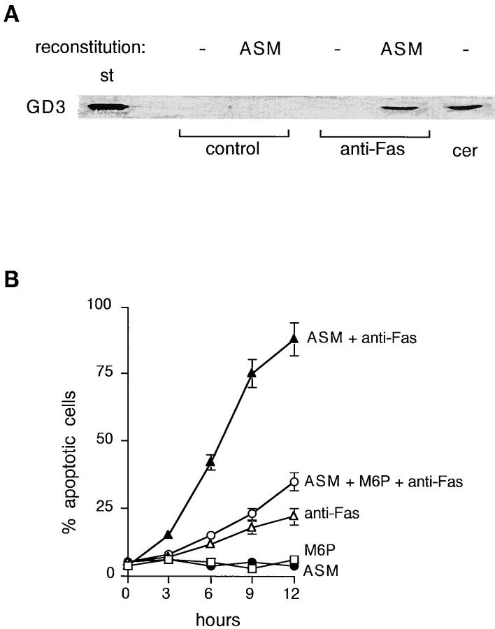
We therefore investigated whether Fas-deriving signals in ASM-reconstituted NPD lymphoblasts were now able to progress up to GD3 accumulation and efficiently trigger apoptosis. Indeed, Fas cross-linking led to normal GD3 accumulation in ASM-reconstituted NPD lymphoblasts (Fig. 5 A). Accordingly, ASM-reconstituted lymphoblasts recovered the ability to undergo apoptosis in response to Fas cross-linking with a kinetics comparable to that observed in normal lymphoblasts. Functional recovery was largely prevented by excess M6P, which also prevented the reconstitution of ASM activity (Fig. 5 B).
Figure 5.
Reconstitution of ASM restores Fas-induced GD3 accumulation and efficient apoptosis in NPD lymphoblasts. (A) NPD lymphoblasts were preincubated for 24 h with CHO (−) or CHO-18 (ASM) supernatant and stimulated for 15 min with control IgM or 500 ng/ml anti-Fas, or for 5 min with 100 μM C6-ceramide (cer). Then polar lipids were extracted, run on HPTLC plates along with GD3 standard (st), and GD3 was detected by anti-GD3 immunostaining. Three experiments gave comparable results. (B) NPD lymphoblasts were incubated for 24 h with or without ASM-containing medium, in the presence or in the absence of 3 mM M6P, and stimulated for different time points with 200 ng/ml anti-Fas or control IgM. The percentage of apoptotic cells was evaluated by propidium iodide staining and flow cytometry analysis of cell nuclei. Mean values and standard deviations at each time point, from four different experiments are shown.
Concluding Remarks.
Here we show that ASM is required for GD3 ganglioside accumulation in response to Fas cross-linking and efficient apoptosis in lymphoid cells. Previous observations indicated that ASM was part of a lipid pathway originating from the “death domains” of receptors belonging to the TNF-R family (10, 21). This pathway was shown to be responsible for the generation of ceramides relevant for Fas-induced cell death (8, 10, 22). Direct genetic evidence also indicated that the ASM defect is responsible for significant protection from radiation-induced apoptosis in NPD-derived human fibroblasts or in mice where the ASM gene has been disrupted by gene targeting (19). Here we reconstituted an efficient Fas-induced apoptotic program in ASM-deficient lymphoblasts by direct ASM replacement. Our data suggest that ASM-derived ceramides are used for ganglioside biosynthesis, up to the generation of GD3 ganglioside. GD3 is a key effector molecule of the lipid pathway, being extremely effective in inducing membrane permeability changes in mitochondria (11). In fact, preventing ganglioside biosynthesis and GD3 accumulation in lymphocytes significantly protects from Fas-derived apoptotic signals (11). ASM activation and GD3 accumulation are therefore tightly linked steps along the same pathway. Since ASM is activated within minutes from Fas cross-linking, ASM-deriving ceramides, and the resultant GD3, probably play a role in the initial stages of the apoptotic program, most likely in the early damage to mitochondria (Rippo, M.R., et al., manuscript submitted for publication). Subsequent activation of cytosolic NSM is then responsible for massive ceramide accumulation, crucially contributing to the apoptotic process during the following hours (15, 16). The data presented here indicate that ASM activation is critical for the optimal progression of early apoptotic signals originating from Fas in lymphoid cells.
Acknowledgments
This work has been supported by grants from Associazione Italiana Ricerca sul Cancro, Istituto Superiore di Sanita' Progetto Sclerosi Multipla, Associazione Spaziale Italiana, Ministero Universita' Ricerca Scientifica e Tecnologica and European Commission Biomed2 Program to R. Testi, and by National Institutes of Health grant HD-28607 and March of Dimes grant I-224 to E.H. Schuchmann.
Abbreviations used in this paper
- ASM
acidic sphingomyelinase
- CHO
Chinese hamster ovary
- HPTLC
high performance thin layer chromatography
- M6P
d-mannose-6-phosphate
- NPD
Niemann Pick disease
- NSM
neutral sphingomyelinase
- PCho
phosphocholine
- PC-PLC
phosphatidylcholine-specific phospholipase C
References
- 1.Nagata S, Golstein P. The Fas death factor. Science. 1995;267:1449–1456. doi: 10.1126/science.7533326. [DOI] [PubMed] [Google Scholar]
- 2.Krammer PH. The CD95(APO-1/Fas) receptor/ ligand system: death signals and disease. Cell Death and Differentiation. 1996;3:159–160. [PubMed] [Google Scholar]
- 3.Muzio M, Chinnaiyan AM, Kischkel FC, O'Rourke K, Shevchenko A, Carsten-Scaffidi JN, Bretz JD, Zhang M, Gentz R, Mann M, et al. FLICE, a novel FADD homologous ICE/CED-3–like protease, is recruited to the CD95 (Fas/Apo-1) death-inducing signaling complex. Cell. 1996;85:817–827. doi: 10.1016/s0092-8674(00)81266-0. [DOI] [PubMed] [Google Scholar]
- 4.Boldin MP, Goncharov TM, Goltsev YV, Wallach D. Involvement of MACH, a novel MORT1/ FADD-interacting protease, in Fas/APO-1- and TNF receptor–induced cell death. Cell. 1996;85:803–815. doi: 10.1016/s0092-8674(00)81265-9. [DOI] [PubMed] [Google Scholar]
- 5.Los M, Van de Craen M, Penning LC, Schenk H, Westendorp M, Bauerle PA, Dröge W, Krammer PH, Fiers W, Schulze-Osthoff K. Requirement of an ICE/CED-3 protease for Fas/APO-1-mediated apoptosis. Nature. 1995;375:81–83. doi: 10.1038/375081a0. [DOI] [PubMed] [Google Scholar]
- 6.Enari M, Hug H, Nagata S. Involvement of an ICE-like protease in Fas-mediated apoptosis. Nature. 1995;375:78–81. doi: 10.1038/375078a0. [DOI] [PubMed] [Google Scholar]
- 7.Testi R. Sphingomyelin breakdown and cell fate. Trends Biochem Sci. 1996;21:468–471. doi: 10.1016/s0968-0004(96)10056-6. [DOI] [PubMed] [Google Scholar]
- 8.Cifone MG, De Maria R, Roncaioli P, Rippo MR, Azuma M, Lanier LL, Santoni A, Testi R. Apoptotic signaling through CD95 (Fas/APO-1) activates an acidic sphingomyelinase. J Exp Med. 1994;180:1547–1552. doi: 10.1084/jem.180.4.1547. [DOI] [PMC free article] [PubMed] [Google Scholar]
- 9.Gulbins E, Bissonette R, Mahboubi A, Martin S, Nishioka W, Brunner T, Baier G, Baier-Bitterlich G, Byrd C, Lang F, et al. Fas-induced apoptosis is mediated via a ceramide-initiated RAS signaling pathway. Immunity. 1995;2:341–351. doi: 10.1016/1074-7613(95)90142-6. [DOI] [PubMed] [Google Scholar]
- 10.Cifone MG, Roncaioli P, De Maria R, Camarda G, Santoni A, Ruberti G, Testi R. Multiple signaling originate at the Fas/Apo-1 (CD95) receptor: sequential involvement of phosphatidylcholine-specific phospholipase C and acidic sphingomyelinase in the propagation of the apoptotic signal. EMBO (Eur Mol Biol Organ) J. 1995;14:5859–5868. doi: 10.1002/j.1460-2075.1995.tb00274.x. [DOI] [PMC free article] [PubMed] [Google Scholar]
- 11.De Maria R, Lenti ML, Malisan F, d'Agostino F, Tomassini B, Zeuner A, Rippo MR, Testi R. Requirement for GD3 ganglioside in CD95- and ceramide-induced apoptosis. Science. 1997;277:1652–1655. doi: 10.1126/science.277.5332.1652. [DOI] [PubMed] [Google Scholar]
- 12.Liu X, Kim CN, Yang J, Jemmerson R, Wang X. Induction of apoptotic program in cell-free extracts: requirement for cytochrome c. Cell. 1996;86:147–157. doi: 10.1016/s0092-8674(00)80085-9. [DOI] [PubMed] [Google Scholar]
- 13.Susin SA, Zamzami N, Castedo M, Hirsch T, Marchetti P, Macho A, Daugas E, Geuskens M, Kroemer G. Bcl-2 inhibits the mitochondrial release of an apoptogenic protease. J Exp Med. 1996;184:1331–1341. doi: 10.1084/jem.184.4.1331. [DOI] [PMC free article] [PubMed] [Google Scholar]
- 14.Kroemer G, Zamzami N, Susin SA. Mitochondrial control of apoptosis. Immunol Today. 1997;18:44–51. doi: 10.1016/s0167-5699(97)80014-x. [DOI] [PubMed] [Google Scholar]
- 15.Tepper CG, Jayadev S, Liu B, Bielawska A, Wolff R, Yonehara S, Hannun YA, Seldin M. Role for ceramide as an endogenous mediator of Fas-induced cytotoxicity. Proc Natl Acad Sci USA. 1995;92:8443–8447. doi: 10.1073/pnas.92.18.8443. [DOI] [PMC free article] [PubMed] [Google Scholar]
- 16.Hannun YA. Functions of ceramide in coordinating cellular responses to stress. Science. 1996;274:1855–1859. doi: 10.1126/science.274.5294.1855. [DOI] [PubMed] [Google Scholar]
- 17.Kolesnick R, Fuks Z. Ceramide: a signal for apoptosis or mitogenesis? . J Exp Med. 1995;181:1949–1952. doi: 10.1084/jem.181.6.1949. [DOI] [PMC free article] [PubMed] [Google Scholar]
- 18.Schuchman EH, Suchi M, Takahashi T, Sandhoff K, Desnick R. Human acidic sphingomyelinase. Isolation, nucleotide sequence and expression of the full length and alternatively spliced cDNA. J Biol Chem. 1991;266:8531–8539. [PubMed] [Google Scholar]
- 19.Santana P, Peña L, Haimovitz-Friedman A, Martin S, Green D, McLaughlin M, Cordon-Cardo C, Schuchman EH, Fuks Z, Kolesnick R. Acid sphingomyelinase–deficient human lymphoblasts and mice are defective in radiation-induced apoptosis. Cell. 1996;86:189–199. doi: 10.1016/s0092-8674(00)80091-4. [DOI] [PubMed] [Google Scholar]
- 20.Schissel SL, Schuchman EH, Williams KJ, Tabas I. Zn2+-stimulated sphingomyelinase is secreted by many cell types and is the product of the acidic sphingomyelinase gene. J Biol Chem. 1996;271:18431–18436. doi: 10.1074/jbc.271.31.18431. [DOI] [PubMed] [Google Scholar]
- 21.Wiegmann K, Schütze S, Machleidt T, Witte D, Krönke M. Functional dichotomy of neutral and acidic sphingomyelinases in tumor necrosis factor signaling. Cell. 1994;78:1005–1015. doi: 10.1016/0092-8674(94)90275-5. [DOI] [PubMed] [Google Scholar]
- 22.De Maria R, Boirivant M, Cifone MG, Roncaioli P, Hahne M, Tschopp J, Pallone F, Santoni A, Testi R. Functional expression of Fas and Fas ligand on human gut lamina propria lymphocytes. A potential role for the acidic sphingomyelinase pathway in normal immunoregulation. J Clin Invest. 1996;97:316–322. doi: 10.1172/JCI118418. [DOI] [PMC free article] [PubMed] [Google Scholar]


