

Vol. 201, No. 4, February 21, 2005. Pages 627–636.
The authors regret that an inaccurate version of Fig. 4 appears in the original article. The correct version of Fig. 4 and its legend appear below.
Figure 4.
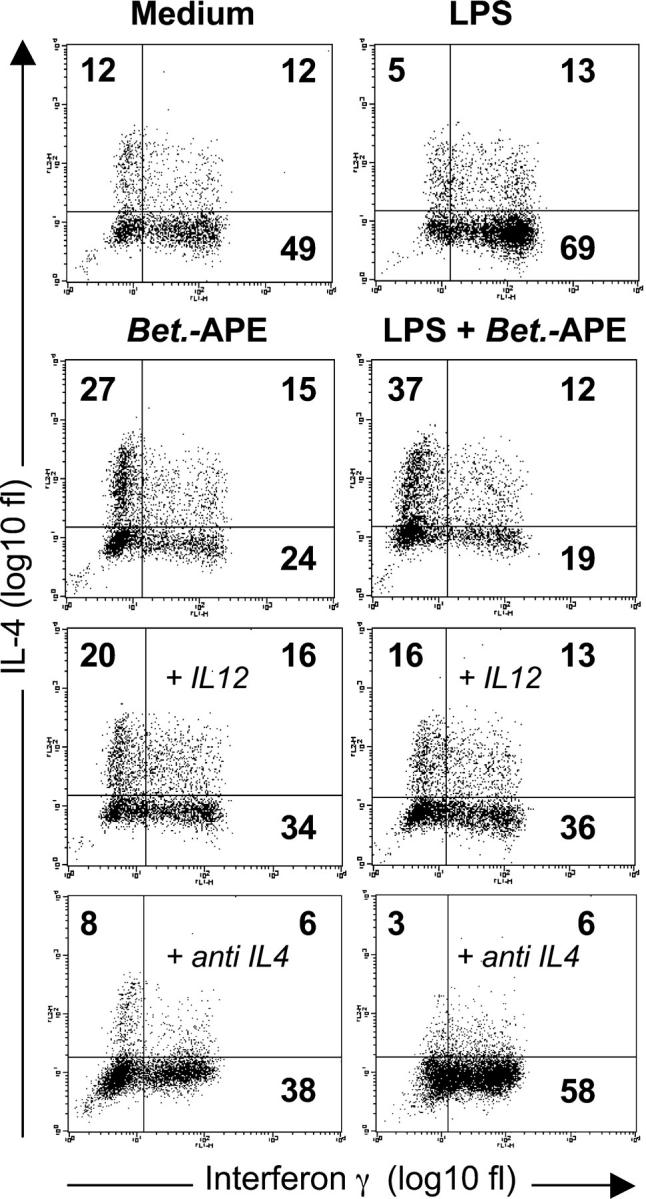
DCs matured in the presence of Bet.-APE display reduced Th1- and increased Th2-polarizing capacity. DCs were left untreated or stimulated with Bet.-APE (3 mg/ml) in the presence or absence of LPS (100 ng/ml). After 24 h DCs were washed and cocultured with CD4+CD45RA+ allogenic T cells (DC/T cell ratio 1:4) that were expanded for 12 d in the presence of IL-2. T cell polarization was determined by analyzing intracellular IFN-γ and IL-4 accumulation via flow cytometry after restimulation with PMA and ionomycin in the presence of brefeldin A during the last 2 h of stimulation. To address, if the Bet.-APE–dependent Th2 polarization could be reverted by exogenous IL-12, hrIL-12 (10 ng/ml) was added at the beginning of the coculture of Bet.-APE/LPS-treated DCs and T cells. Representative experiment of n = 3–6 (compare Table I). To explore the role of IL-4 in the Th2 polarization induced by Bet.-APE–treated DCs, IL-4–neutralizing antibodies (10 μg/ml) were added at the beginning of the DC/T cell coculture. Representative experiment of n = 3.