Abstract
The B cell antigen receptor (BCR) consists of the membrane-bound immunoglobulin (Ig) molecule as antigen-binding subunit and the Ig-α/Ig-β heterodimer as signaling subunit. BCR signal transduction involves activation of protein tyrosine kinases (PTKs) and phosphorylation of several proteins, only some of which have been identified. The phosphorylation of these proteins can be induced by exposure of B cells either to antigen or to the tyrosine phosphatase inhibitor pervanadate/H2O2. One of the earliest substrates in B cells is a 65-kD protein, which we identify here as a B cell adaptor protein. This protein, named SLP-65, is part of a signaling complex involving Grb-2 and Vav and shows homology to SLP-76, a signaling element of the T cell receptor. In pervanadate/H2O2-stimulated cells, SLP-65 becomes phosphorylated only upon expression of the BCR. These data suggest that SLP-65 is part of a BCR transducer complex.
Keywords: antigen receptor, tyrosine phosphorylation, Src homology 2 domain, SLP-65
Engagement of the B cell antigen receptor (BCR)1 triggers the phosphorylation of proteins on tyrosine and the concomitant activation of a variety of signaling pathways. The tyrosine phosphorylation of phospholipase C-γ leads to hydrolysis of phosphoinositides which produces second messengers able to elevate intracellular [Ca2+] and activate protein kinase C (1–3). The BCR-mediated activation of the ras/Raf/Map kinase pathway is less well understood but is likely to involve p120rasGAP and the Grb-2–Sos complex (1–3). Tyrosine-phosphorylated Vav exhibits GDP/ GTP exchange activity for Rac-1, a small G protein implicated in cell proliferation and cytoskeletal organization (4). The activation of these and other signaling cascades is not restricted to BCR-stimulated B cells, and some signaling elements are used by many receptors. The stimulated BCR must both maintain signal specificity and avoid interference with other types of receptor that are not stimulated. A preformed BCR transducer complex would make this possible by allowing the rapid and selective activation of BCR-specific signaling pathways, even in those cases where some signaling elements are common to the BCR and another receptor. The analysis of substrate phosphorylation in J558L B cells treated with pervanadate/H2O2 suggests that as soon as it is expressed, the BCR indeed recruits and organizes intracellular signaling elements into a signaling complex (5). The existence of preformed signaling complexes has been directly demonstrated for the photoreceptor system in drosophila (6) and for the osmoadaptation–pheromone response system in yeast (7). In both cases, specific adaptor proteins were found to organize the physical and functional coupling of intracellular signaling proteins to receptors in the plasma membrane. We have tried to identify protein tyrosine kinase (PTK) substrate proteins that become tyrosine phosphorylated early after BCR stimulation. Here we report on the identification and cloning of such a protein, which we named SLP-65 (Src homology [SH2] domain–containing leukocyte protein of 65 kD). This B cell–specific adaptor protein contains a number of protein– protein interaction domains and is constitutively associated with Grb-2 and Vav. In the absence of a BCR, treatment of the cells with pervanadate/H2O2 does not induce phosphorylation of SLP-65. Our results suggest a scaffolding function for SLP-65 which would couple BCR activation to intracellular signaling pathways.
Materials and Methods
Materials.
Immobilized PT66 antiphosphotyrosine-agarose beads (Sigma-Aldrich, Deisenhofen, Germany) and soluble 4G10 antiphosphotyrosine antibodies (Upstate Biotechnology Inc., Lake Placid, NY) were used for precipitation and immunoblot analysis of phosphotyrosine-containing proteins, respectively. Anti-Vav and anti–Grb-2 rabbit polyclonal antibodies were purchased from Santa Cruz Biotechnology Inc. (Santa Cruz, CA). Polyclonal anti–SLP-65 antibodies were produced by immunizing rabbits with KLH-coupled SLP-65 peptides encompassing amino acids 148–161 (KARLA peptide; see Fig. 1). The glutathione S-transferase (GST)–Grb-2 fusion proteins contain amino acids 1–168 (SH3-SH2) and 49–168 (SH2) of the human Grb-2 protein, respectively. Primer sequences used for amplifications of the respective Grb-2 cDNA fragments are available upon request. The plasmid encoding the GST–Grb-2 (SH3) fusion protein was a gift of Dr. William Wishart, Novartis (Basel, Switzerland). The GST fusion protein encompassing the complete murine Grb-2 was obtained from Upstate Biotechnology Inc.
Figure 1.
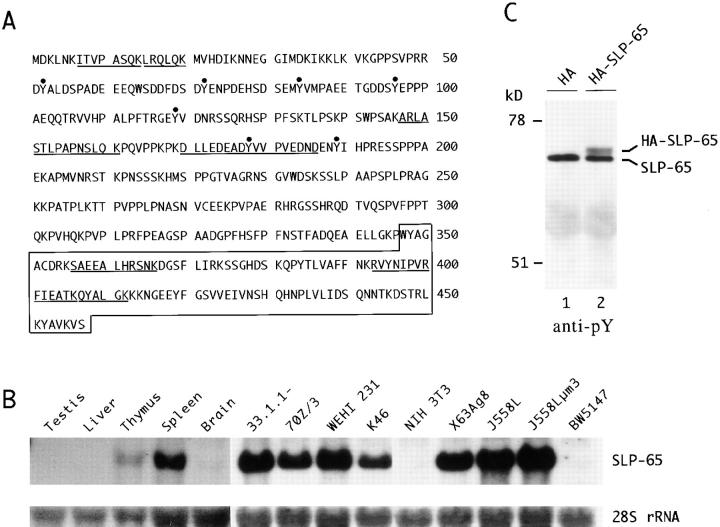
The SLP-65 gene encodes a protein with several regions of similarity to SLP-76 and is transcribed in lymphoid tissues. (A) Deduced amino acid sequence of murine SLP-65. Dots, Potential tyrosine-phosphorylation sites are marked. Box, The COOH-terminal SH2 domain. Underline, Peptide sequences obtained from affinity-purified SLP-65. (B) SLP-65 transcripts of ∼2 kb were detected by Northern blot analysis of RNA from the indicated tissues and cell lines. The SLP-65 probe used was derived from the ORF, and a reference signal for ribosomal 28S RNA was used as a loading control. (C) Anti–SLP-65 precipitates were prepared from postnuclear supernatants of pervanadate/H2O2-stimulated J558Lμm3 cells, transiently transfected either with the parental vector (lane 1) or an expression vector encoding SLP-65 with an NH2-terminal HA tag (lane 2). Precipitated proteins were analyzed by antiphosphotyrosine (anti-pY) immunoblotting.
Protein Analysis.
J558L cell lines, pervanadate/H2O2 stimulations, immunoprecipitations, affinity purifications with GST fusion proteins, and Western blot analysis were described previously (5, 8).
Large-scale Preparation and Amino Acid Sequencing of PTK Substrate Proteins.
In total, 1010 J558Lδm7.1 cells were stimulated in aliquots of 3 × 10⁷ cells per ml RPMI 1640 medium for 3 min with 50 μM pervanadate/H2O2 at 37°C (5). After lysis of the cells in 600 μl of 1% NP-40 lysis buffer (5), postnuclear supernatants were collected and precleared for 3 h with 7.5 ml of mouse IgG agarose (Sigma-Aldrich). Subsequently, the lysate was subjected to antiphosphotyrosine immunoprecipitation with 6 ml of agarose-conjugated PT66 mAb (Sigma-Aldrich). Settled beads were extensively washed with 1% NP-40 lysis buffer, and bound proteins were eluted eight times with 7.5 ml 50 mM phenyl phosphate/200 mM NaCl. Proteins were concentrated with centrifugal filter devices (microsep 10K; Pall Filtron Corp., East Hills, NY), separated by 10% SDS-PAGE, and stained with Coomassie blue. The 65-kD protein band was excised, subjected to in-gel digestion with lysyl endopeptidase C (Lys-C), and the resulting HPLC-purified peptides were sequenced (TOPLAB, Munich, Germany; see Fig. 1).
cDNA Isolation.
Several independent clones of a 2-kb cDNA (GenBank accession no. Y17159) were isolated using a combination of standard PCR with primers derived from a partial cDNA sequence in GenBank (accession no. AJ222814) and 5′ rapid amplification of cDNA ends (5′RACE; GIBCO BRL, Gaithersburg, MD) and completely sequenced.
Northern Blot Analysis.
As described previously (9), total cellular RNA was isolated, and 15 μg per sample was separated in a denaturing agarose gel. After blotting, hybridization was performed with a randomly primed 795-bp PstI fragment from nucleotide 650 to 1445 (the open reading frame [ORF] extends from 478 to 1851).
Transient Protein Expression.
For transient expression of SLP-65 in J558Lμm3 cells, a 1490-bp PCR fragment (amplified with PfuI [Stratagene Inc., La Jolla, CA]) containing the entire ORF was cloned downstream of the CMV promoter in pCGN (10). The resulting expression vector, pCGN-p65, encodes a SLP-65 fusion protein containing an NH2-terminal, 20 amino acid peptide tag which is derived from the hemagglutinin protein of human influenza virus (HA tag [11]). The conditions for electroporation have been described previously (9).
Results and Discussion
To identify PTK substrate proteins in B cells, we affinity purified these proteins from the lysate of pervanadate/ H2O2-stimulated J558Lδm7.1 cells via antiphosphotyrosine antibody columns and determined their peptide sequence. We focused on a 65-kD protein which is the most rapidly phosphorylated substrate after exposure of J558Lδm7.1 cells to antigen or to pervanadate. Several of the sequenced peptides matched with a partial sequence in GenBank (accession no. AJ222814). Using mouse splenic cDNA as template, we used a combination of conventional and 5′RACE PCR to isolate ∼2 kb of the corresponding cDNA (accession no. Y17159). The cDNA we obtained has an ORF encoding a protein of 457 amino acids (Fig. 1 A). Seven of the peptides sequenced from the purified protein were found in the predicted sequence (Fig. 1 A, underline). The NH2-terminal portion of the protein contains seven tyrosine residues which are putative targets for phosphorylation (Fig. 1 A, dots). Five of these are found in a YxxP context otherwise found in the PTK substrates Cas, p62dok, Cbl, and SLP-76 (12–15). The central part of the protein is rich in prolines and contains several SH3 domain–binding motifs. The COOH-terminal part contains an SH2 domain which, when compared with the database, is most similar to the SH2 domain of SLP-76, a recently identified PTK substrate in T lymphocytes (15). Both proteins share the same overall structure. A comparison with human and mouse SLP-76 also reveals a modest similarity in the NH2 terminus (not shown). We conclude that the 65-kD protein substrate is the B cell analogue of SLP-76, and call it SLP-65. In a Northern blot analysis, the 2-kb SLP-65 transcript is found only in spleen and weakly in thymus, but not in liver, testis, or brain (Fig. 1 B). The transcript is also found in B cell lines representing different developmental stages from the pre-B to the plasma cell stage, but not in a T cell or a fibroblast line (Fig. 1 B). These results suggest that SLP-65 is primarily expressed in B lymphocytes but perhaps to some extent also in thymocytes.
SLP-65 has a predicted pI of 8.2, in good agreement with our experimental data showing a pI of 8.5 (data not shown). The predicted molecular mass of 50.7 kD is considerably smaller than the apparent size of 65 kD in SDS-PAGE. To test whether this could be due to aberrant migration, and to obtain further evidence that we had indeed isolated the cDNA encoding the tyrosine-phosphorylated 65-kD substrate, we transiently expressed SLP-65 in J558Lμm3 cells as a fusion protein with an NH2-terminal HA tag (see Materials and Methods). Anti–SLP-65 peptide antibodies (see Materials and Methods) precipitated the HA-tagged fusion protein from pervanadate/H2O2-stimulated J558Lμm3 transfectants but not from control cells. The HA-tagged SLP-65 migrates slightly above the endogenous SLP-65 as detected by antiphosphotyrosine antibodies (Fig. 1 C) and anti-HA antibodies (data not shown). We conclude that the cDNA we isolated encodes the 65-kD substrate for activated PTKs.
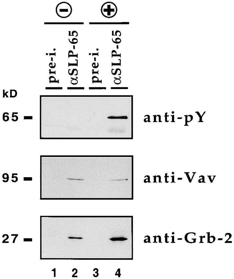
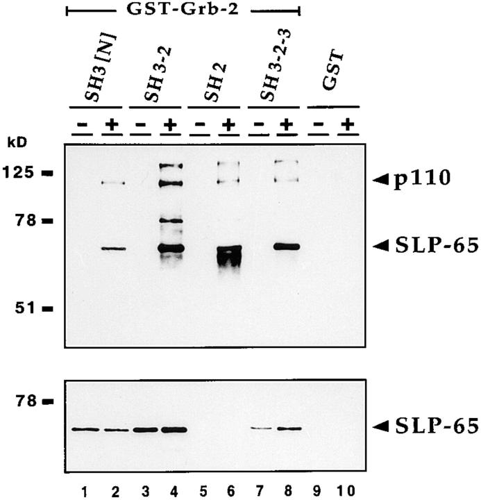
In T cells, the SLP-76 adaptor protein is bound by Grb-2 and Vav (16–18). Anti–SLP-65 antibodies, but not the preimmune serum, coprecipitate Grb-2 and Vav from lysates of unstimulated and pervanadate/H2O2-stimulated J588Lμm3 cells (Fig. 2, lanes 2 and 4). The phosphorylated form of SLP-65 is only found in the lysates of pervanadate/ H2O2-stimulated cells (Fig. 2, top, lane 4). These data show that SLP-65 is constitutively associated with Grb-2 and Vav, supporting the notion that SLP-65 is the B cell analogue of SLP-76. Using different GST–Grb-2 fusion proteins, SLP-65 could be purified from lysates of unstimulated and pervanadate/H2O2-stimulated J558Lμm3 cells with the NH2-terminal SH3 domain of Grb-2 (Fig. 3, bottom, lanes 1 and 2) and to an even stronger extent with the combined SH3 and SH2 domains (Fig. 3, lanes 3 and 4). SLP-65 bound only weakly to the SH2 domain of Grb-2 and not at all to GST alone (Fig. 3, lanes 5, 6, 9, and 10). Probing the same blot with antiphosphotyrosine antibodies showed that, in addition to SLP-65, the Grb-2 SH3 domain coprecipitates a phosphoprotein of 110 kD. This protein may be identical or similar to the SLP-76–associated protein SLAP130/FYB (19, 20). These proteins, as well as other phosphoproteins, were more efficiently purified by the combination of the SH3 and SH2 domains of Grb-2 but not by GST alone (lanes 4 and 10). Two proteins with characteristics similar to SLP-65 have been also found in a human B cell line (21).
Figure 2.
SLP-65 is constitutively associated with Vav and Grb-2. J558Lμm3 cells were left unstimulated (lanes 1 and 2) or stimulated for 2 min with 50 μM pervanadate/ H2O2 (lanes 3 and 4). Cleared cellular lysates were subjected to immunoprecipitation with preimmune serum (pre-i., lanes 1 and 3) or anti–SLP-65 antibodies (lanes 2 and 4). Tyrosine-phosphorylated SLP-65 was detected by antiphosphotyrosine immunoblotting (anti-pY, top). The Vav and Grb-2 proteins were detected by probing the same filter with anti-Vav (middle) and anti–Grb-2 antibodies (bottom).
Figure 3.
The NH2-terminal SH3 domain of Grb-2 is sufficient to bind SLP-65. Grb2-binding proteins were purified from lysates of unstimulated (lanes 1, 3, 5, and 7) and pervanadate/H2O2-stimulated J558Lμm3 cells (lanes 2, 4, 6, and 8) using GST fusion proteins containing different domains of Grb-2: NH2-terminal SH3 domain (lanes 1 and 2), NH2-terminal SH3 plus SH2 domain (lanes 3 and 4), SH2 domain (lanes 5 and 6), and complete Grb-2 (lanes 7 and 8). GST was used as a control (lanes 9 and 10). Proteins were detected by antiphosphotyrosine and anti–SLP-65 immunoblotting (top and bottom, respectively).
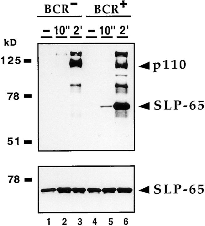
We have previously shown that after exposure to pervanadate, a 65-kD protein is rapidly phosphorylated only in BCR-positive cells (5). To verify that SLP-65 is identical to this early PTK substrate, we stimulated the BCR-negative J558L line and its BCR-positive transfectant J558Lμm3 for different times with pervanadate/H2O2. Similar amounts of SLP-65 were purified from these cells with the GST–Grb-2[SH3-SH2] fusion protein (Fig. 4, bottom, lanes 1–6). However, the SLP-65 protein remained completely unphosphorylated in the BCR-negative line, even after 2 min of stimulation (Fig. 4, top, lane 3). In the BCR-positive transfectant, SLP-65 is rapidly phosphorylated, 10 s after stimulation (lane 5). Other PTK substrate proteins in the BCR-negative and -positive lines require longer times of stimulation for detection with antiphosphotyrosine antibodies (Fig. 4, top, lanes 3 and 6). Thus, the rapid phosphorylation of SLP-65 in J558Lμm3 B cells is dependent on the BCR surface expression.
Figure 4.
Tyrosine phosphorylation of SLP-65 is dependent on BCR expression. BCR-negative J558L cells (lanes 1–3) and BCR-positive J558Lμm3 cells (lanes 4–6) were unstimulated (lanes 1 and 4) or stimulated with 25 μM pervanadate/H2O2 either for 10 s (lanes 2 and 5) or 2 min (lanes 3 and 6). Proteins complexed with GST–Grb-2[SH3-SH2] fusion proteins were detected by antiphosphotyrosine and anti–SLP-65 immunoblotting (top and bottom, respectively).
Our data suggest that SLP-65 is part of a BCR transducer complex comprising not only kinases, phosphatases, and their substrates, but also adaptor proteins which are necessary to physically and functionally connect these elements. This may be a common theme in signal transduction (6, 7). The early and BCR-dependent phosphorylation of SLP-65, together with its restricted expression pattern, suggest that SLP-65 is critically involved in coupling intracellular signaling elements specifically to the BCR. This may contribute to a basal level of signaling in the absence of antigen, a mechanism that may be necessary for B cell maintenance (22, 23). Such a central role in signal transduction is also suggested for SLP-76. Overexpression of SLP-76 in T cells can augment signal transduction from the TCR, leading to increased nuclear factor of activated T cells (NFAT) activation and IL-2 secretion (24, 25). Furthermore, mice deficient for SLP-76 contained no peripheral T cells as a result of an early block in T cell development, whereas B cells develop normally (26). To better understand the function of the SLP protein family, it will be necessary to reveal exactly how they are connected to the antigen receptors.
Acknowledgments
We thank Dr. Lise Leclercq for critical reading of the manuscript.
This work is supported by the Deutsche Forschungsgemeinschaft through SFB 388, and by the Leibniz program.
Abbreviations used in this paper
- BCR
B cell antigen receptor
- GST
glutathione S-transferase
- HA
hemagglutinin
- ORF
open reading frame
- PTK
protein tyrosine kinase
- RACE
rapid amplification of cDNA ends
- SH2 and SH3 domain
Src homology 2 and 3 domain, respectively
- SLP-65
SH2 domain–containing leukocyte protein of 65 kD
References
- 1.DeFranco AL. The complexity of signaling pathways activated by the BCR. Curr Opin Immunol. 1997;9:296–308. doi: 10.1016/s0952-7915(97)80074-x. [DOI] [PubMed] [Google Scholar]
- 2.Kurosaki T. Molecular mechanisms in B cell antigen receptor signaling. Curr Opin Immunol. 1997;9:309–318. doi: 10.1016/s0952-7915(97)80075-1. [DOI] [PubMed] [Google Scholar]
- 3.Reth M, Wienands J. Initiation and processing of signals from the B cell antigen receptor. Annu Rev Immunol. 1997;15:453–479. doi: 10.1146/annurev.immunol.15.1.453. [DOI] [PubMed] [Google Scholar]
- 4.Crespo P, Schuebel KE, Ostrom AA, Gutkind JS, Bustelo XR. Phosphotyrosine-dependent activation of Rac-1 GDP/GTP exchange by the vav proto-oncogene product. Nature. 1997;385:169–172. doi: 10.1038/385169a0. [DOI] [PubMed] [Google Scholar]
- 5.Wienands J, Larbolette O, Reth M. Evidence for a preformed transducer complex organized by the B cell antigen receptor. Proc Natl Acad Sci USA. 1996;93:7865–7870. doi: 10.1073/pnas.93.15.7865. [DOI] [PMC free article] [PubMed] [Google Scholar]
- 6.Tsunoda S, Sierralta J, Sun Y, Bodner R, Suzuki E, Becker A, Socolich M, Zuker CS. A multivalent PDZ-domain protein assembles signalling complexes in a G-protein-coupled cascade. Nature. 1997;388:243–249. doi: 10.1038/40805. [DOI] [PubMed] [Google Scholar]
- 7.Posas F, Saito H. Osmotic activation of the HOG MAPK pathway via Ste11p MAPKKK: scaffold role of PBS2p MAPKK. Science. 1997;276:1702–1705. doi: 10.1126/science.276.5319.1702. [DOI] [PubMed] [Google Scholar]
- 8.Baumann G, Maier D, Freuler F, Tschopp C, Baudisch K, Wienands J. In vitro characterization of major ligands for Src homology 2 domains derived from tyrosine kinases, from the adaptor protein SHC and from GTPase- activating protein in Ramos B cells. Eur J Immunol. 1994;42:1799–1807. doi: 10.1002/eji.1830240812. [DOI] [PubMed] [Google Scholar]
- 9.Jumaa H, Nielsen PJ. The splicing factor SRp20 modifies splicing of its own mRNA and ASF/F2 antagonizes this regulation. EMBO (Eur Mol Biol Organ) J. 1997;16:5077–5085. doi: 10.1093/emboj/16.16.5077. [DOI] [PMC free article] [PubMed] [Google Scholar]
- 10.Tanaka M, Herr W. Differential transcriptional activation by Oct-1 and Oct-2: independent activation domains induce Oct-2 phosphorylation. Cell. 1997;60:375–386. doi: 10.1016/0092-8674(90)90589-7. [DOI] [PubMed] [Google Scholar]
- 11.Wilson IA, Niman HL, Houghten RA, Cherenson AR, Connolly ML, Lerner RA. The structure of an antigenic determinant in a protein. Cell. 1984;37:767–778. doi: 10.1016/0092-8674(84)90412-4. [DOI] [PubMed] [Google Scholar]
- 12.Sakai R, Iwamatsu A, Hirano N, Ogawa S, Tanaka T, Mano H, Yazaki Y, Hirai H. A novel signaling molecule, p130, forms stable complexes in vivo with v-Crk and v-Src in a tyrosine phosphorylation-dependent manner. EMBO (Eur Mol Biol Organ) J. 1994;13:3748–3756. doi: 10.1002/j.1460-2075.1994.tb06684.x. [DOI] [PMC free article] [PubMed] [Google Scholar]
- 13.Yamanashi Y, Baltimore D. Identification of the Abl- and rasGAP-associated 62 kDa protein as a docking protein, Dok. Cell. 1997;88:205–211. doi: 10.1016/s0092-8674(00)81841-3. [DOI] [PubMed] [Google Scholar]
- 14.Langdon WY, Hartley JW, Klinken SP, Ruscetti SK, Morse HCD. v-cbl, an oncogene from a dual-recombinant murine retrovirus that induces early B-lineage lymphomas. Proc Natl Acad Sci USA. 1989;86:1168–1172. doi: 10.1073/pnas.86.4.1168. [DOI] [PMC free article] [PubMed] [Google Scholar]
- 15.Jackman JK, Motto DG, Sun Q, Tanemoto M, Turck CW, Peltz GA, Koretzky GA, Findell PR. Molecular cloning of SLP-76, a 76-kDa tyrosine phosphoprotein associated with Grb2 in T cells. J Biol Chem. 1995;270:7029–7032. doi: 10.1074/jbc.270.13.7029. [DOI] [PubMed] [Google Scholar]
- 16.Tuosto L, Michel F, Acuto O. p95vav associates with tyrosine-phosphorylated SLP-76 in antigen-stimulated T cells. J Exp Med. 1996;184:1161–1166. doi: 10.1084/jem.184.3.1161. [DOI] [PMC free article] [PubMed] [Google Scholar]
- 17.Wu J, Motto DG, Koretzky GA, Weiss A. Vav and SLP-76 interact and functionally cooperate in IL-2 gene activation. Immunity. 1996;4:593–602. doi: 10.1016/s1074-7613(00)80485-9. [DOI] [PubMed] [Google Scholar]
- 18.Onodera H, Motto DG, Koretzky GA, Rothstein DM. Differential regulation of activation-induced tyrosine phosphorylation and recruitment of SLP-76 to Vav by distinct isoforms of the CD45 protein-tyrosine phosphatase. J Biol Chem. 1996;271:22225–22230. doi: 10.1074/jbc.271.36.22225. [DOI] [PubMed] [Google Scholar]
- 19.Musci MA, Hendricks-Taylor LR, Motto DG, Paskind M, Kamens J, Turck CW, Koretzky GA. Molecular cloning of SLAP-130, an SLP-76-associated substrate of the T cell antigen receptor-stimulated protein tyrosine kinases. J Biol Chem. 1997;272:11674–11677. doi: 10.1074/jbc.272.18.11674. [DOI] [PubMed] [Google Scholar]
- 20.da Silva AJ, Li Z, de Vera C, Canto E, Findell P, Rudd CE. Cloning of a novel T-cell protein FYB that binds FYN and SH2-domain-containing leukocyte protein 76 and modulates interleukin 2 production. Proc Natl Acad Sci USA. 1997;94:7493–7498. doi: 10.1073/pnas.94.14.7493. [DOI] [PMC free article] [PubMed] [Google Scholar]
- 21.Fu C, Chan AC. Identification of two tyrosine phosphoproteins, pp70 and pp68, which interact with phospholipase C-γ, Grb2, and Vav after B cell antigen receptor activation. J Biol Chem. 1997;272:27362–27368. doi: 10.1074/jbc.272.43.27362. [DOI] [PubMed] [Google Scholar]
- 22.Lam KP, Kuhn R, Rajewsky K. In vivo ablation of surface immunoglobulin on mature B cells by inducible gene targeting results in rapid cell death. Cell. 1997;90:1073–1083. doi: 10.1016/s0092-8674(00)80373-6. [DOI] [PubMed] [Google Scholar]
- 23.Neuberger M. Antigen receptor signaling gives lymphocytes a long life. Cell. 1997;90:971–973. doi: 10.1016/s0092-8674(00)80362-1. [DOI] [PubMed] [Google Scholar]
- 24.Motto DG, Ross SE, Wu J, Hendricks-Taylor LR, Koretzky GA. Implication of the GRB2-associated phosphoprotein SLP-76 in T cell receptor–mediated interleukin 2 production. J Exp Med. 1996;183:1937–1943. doi: 10.1084/jem.183.4.1937. [DOI] [PMC free article] [PubMed] [Google Scholar]
- 25.Fang N, Motto DG, Ross SE, Koretzky GA. Tyrosines 113, 128, and 145 of SLP-76 are required for optimal augmentation of NFAT promoter activity. J Immunol. 1996;157:3769–3773. [PubMed] [Google Scholar]
- 26.Clements JL, Yang B, Ross-Barta SE, Eliason SL, Hrstka RF, Williamson RA, Koretzky GA. Requirement for the leukocyte-specific adaptor protein SLP-76 for normal T cell development. Science. 1998;281:416–419. doi: 10.1126/science.281.5375.416. [DOI] [PubMed] [Google Scholar]


